2. Introduction
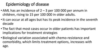
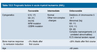
•Acute myeloid leukemia (AML) is a heterogeneous
clonal disorder of hematopoietic stem/progenitor
cells.
•Characterized by the rapid growth of abnormal white
blood cells (blasts) that accumulate in the bone
marrow and interfere with the production of normal
blood cells
3. Learning outcome
•Describe the epidemiology of disease
•Discus the pathophysiology of AML
•Discuss the clinical presentation of AML
•Discuss the laboratory findings in AML
•Discuss the common Acute myeloid leukaemia with
recurrent genetic Abnormalities
4. Epidemiology of disease
•AML has an incidence of 2 – 3 per 100 000 per annum in
children, rising to 15 per 100 000 in older adults.
•It can occur at all ages but has its peak incidence in the seventh
decade
•The fact that most cases occur in older patients has important
implications for treatment strategies
•Biological variation associated with chemo-resistance and
comorbidity, which limits treatment options, increases with
age.
7. M1 Acute myeloblastic leukaemia without
maturation
• Medium-sized blasts with high (n:c)ratio,
rounded nuclei with immature, dispersed
chromatinwith one or more prominent
nucleoli.
• Blasts can showfine azurophilic
granulation or isolated Auer rods inthe
cytoplasm in 5% to 10% of cases
Immunophenotype
•MPO +
•CD13 +
•CD33 +
•CD117+
•CD34 +/-
8. M2 Acute myeloblastic leukaemiawith
maturation
• Small to medium-sized blasts with high (n:c)
• ratio and rounded nuclei sometimes located in a
corner of the cytoplasm.
• The nucleus shows dispersed, immature chromatin
with one or more nucleoli.
• The cytoplasm is basophilic and can containtraces
of primary azurophilic granulation or isolated Auer
rods.
Immunophenotype
•MPO +
•CD34 +/-
•CD13 +
•CD15 +
•HLA-DR +/-
•Sudan black +
9. M3 Promyelocytic leukaemia
• Abundant, intensely azurophilic granulation.
• The nucleus is usually monocytic
inappearance(reniform) and is either irregular or
bilobed with adeep cleft.
• Scarcely basophilic cytoplasm due to the proliferation
of azurophilic granulation.
• Some atypicalpromyelocytes also contain elongated or
splinter-shaped crystalline cytoplasmic inclusions
specific tothis type of leukaemia.
• These usually form clumps,but differ from Auer rods in
that they show a tubularsubstructure on electronic
microscopy.
Immunophenotype
•CD13 +
•CD33 +
•HLA-DR –
•CD34
11. Pathophysiology
•AML is a malignant clonal disorder of immature cells in the
haemopoietic hierarchical system.
•Leukaemic transformation is assumed to occur in many cases
at, or near, the level of the haemopoietic stem cell before it
has embarked on any lineage commitment.
•Some cases may originate at a slightly later stage in cells that
are committed to lineage differentiation
•There is considerable heterogeneity between cases with
respect to morphology, immunological phenotype, associated
cytogenetic and molecular abnormalities and, more recently,
patterns of gene expression.
12. Pathophysiology
•AML cells have abnormal function characterized by failure to
progress through the expected differentiation programme
and/or to die by the process of apoptosis.
• Associated with this may be retention of the stem cell
characteristic of self - renewal.
•This leads to the accumulation of a clone of cells, which
dominates bone marrow activity and leads to marrow failure.
•Adenopathy or organomegaly can occur but are not usual
features.
•Extramedullary disease can occur, including cerebrospinal
13. Clinical Presentation
• Most patients with AML have a total WBC count between 5 and
• 30 × 109/L, although the WBC count may range from 1 to 200 × 109/L.
• Myeloblasts are present in the peripheral blood in 90% of patients.
• Anemia, thrombocytopenia, and neutropenia give rise to the clinical
findings of pallor, fatigue, fever with infections, bruising, and
bleeding.
• In addition, disseminated intravascular coagulation and other
bleeding abnormalities can be significant.
• Infiltration of malignant cells into the gums and other mucosal sites
and skin also can be seen.
• Bone and joint pain are the first symptoms in only 25% of patients
14. Common abnormalities in laboratory test
•hyperuricemia (caused by increased cellular turnover),
hyperphosphatemia (due to cell lysis), and hypocalcemia (the
latter two are also involved in progressive bone destruction).
•Hypokalemia is also common at presentation.
•, especially when the WBC is quite elevated, tumor lysis
syndrome may occur
•Tumor lysis syndrome may occur due to induction
chemotherapy
• by hyperkalemia, hyperphosphatemia, hyperuricemia and
hyperuricosuria, and hypocalcemia.
• The hyperkalemia alone can be life threatening
16. Acute Myeloid Leukemia with t(8;21) (q22;q22);
RUNX1/RUNX1T1
•mutation is found in about 5% of AML cases.
•Seen predominantly in children and young adults, AML with
this translocation has myeloblasts with dysplastic granular
cytoplasm, Auer rods, and some maturation .
•similar to the FAB M2 classification Various anomalies, such as
pseudo–Pelger-Huët cells and hypogranulation, can be seen.
•Eosinophilia is possible.
•The diagnosis of this subtype is based on the genetic
abnormality, regardless of blast count
17. Acute Myeloid Leukemia with inv(16)(p13.1q22) or
t(16;16)(p13.1;q22);CBFB-MYH11.
• Accounting for approximately 5% to 8% of all AML cases, core-binding
factor (CBF)
• AML occurs at all ages, but is found predominantly in younger
patients.
• Cytogenetic hallmarks of CBF AML may represent cooperative events
in CBF AML leukemogenesis and include mutations in the KIT, FLT3,
JAK2, and RAS genes.
• The genetic aberration is sufficient for diagnosis regardless of blast
count.
• Myeloblasts, monoblasts, and promyelocytes are seen in the
peripheral blood and bone marrow
18. Acute Myeloid Leukemia with t(15;17)
(q22;q12);PML-RARA.
• Also known as acute promyelocytic leukemia (APL),
• mutation comprises 5% to 10% of AML cases.
• It occurs in all age groups but is seen most commonly in young adults.
• This disorder is characterized by a differentiation block at the promyelocytic stage.
• Detection of the 15;17 translocation is sufficient for diagnosis regardless of blast
count.
• Characteristic of this presentation are the abnormal hypergranular promyelocytes,
some with Auer rods
• When Auer rod bundles are found, the cell is called a faggot cell.
• When promyelocytes release primary granule contents, their procoagulant activity
initiates disseminated intravascular coagulation