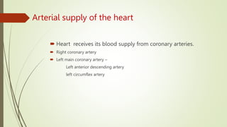
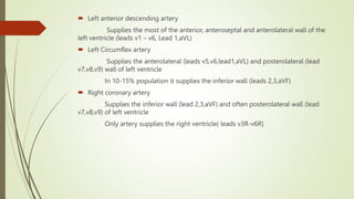
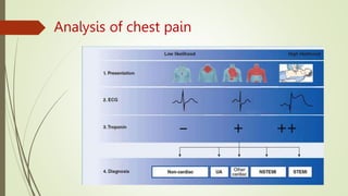
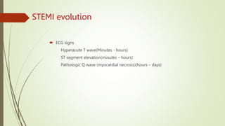
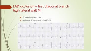
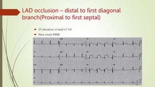
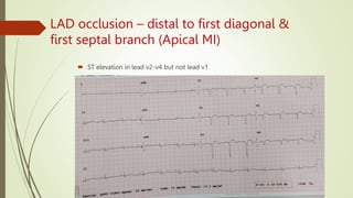
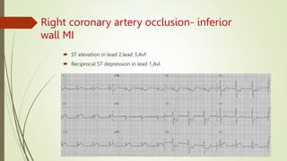
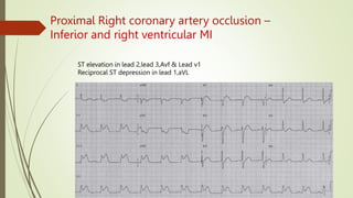
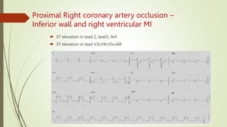
LOCALIZATION OF
MYOCARDIAL INFARCTION
UNIT 2
CHIEF
DR. D.SOJI MD GEN MED
PRESENTER
Rajamuthukrishnan PG
 A 46 Years old male presented with complaints of sudden onset of chest pain which
is retrosternal, compressive type radiating to left side of the chest and left arm for
the past 1 hour
 C/o shortness of breath present
 C/o excessive sweating present
 C/o dizziness
 No other complaints
 Not a k/c/o DM,HTN,CAD,CKD,TB,BA,Epilepsy,other illness
 Chronic smoker
 Vitals – PR- 70/Mt BP- 114/70 mm of Hg Spo2- 96% in RA
 CVS-s1s2 heard, no murmur
 RS- bilateral air entry present, no added sounds
 Abdomen- soft, no organomegaly
 CNS- NFND
What next?
Arterial supply of the heart
 Heart receives its blood supply from coronary arteries.
 Right coronary artery
 Left main coronary artery –
Left anterior descending artery
left circumflex artery
 Left anterior descending artery
Supplies the most of the anterior, anteroseptal and anterolateral wall of the
left ventricle (leads v1 – v6, Lead 1,aVL)
 Left Circumflex artery
Supplies the anterolateral (leads v5,v6,lead1,aVL) and posterolateral (lead
v7,v8,v9) wall of left ventricle
In 10-15% population it supplies the inferior wall (leads 2,3,aVF)
 Right coronary artery
Supplies the inferior wall (lead 2,3,aVF) and often posterolateral wall (lead
v7,v8,v9) of left ventricle
Only artery supplies the right ventricle( leads v3R-v6R)
ECG leads representation on heart
 Lead v1,v2 – ventricular septum
 Lead v2-v4 – anterior wall of the left ventricle
 Lead v5,v6,1,aVL - lateral wall of the left ventricle
 Lead 2,3,aVF – inferior wall of left ventricle
 Lead v7-v9 (special leads) – posterolateral wall of left ventricle
 Lead v3R-v6R (special right pre cordial leads) – right ventricle
Analysis of chest pain
Acute coronary syndrome
 Myocardial infarction with ST segment elevation(STEMI)
 Non ST segment elevation acute coronary syndrome
NSTEMI
Unstable angina
STEMI evolution
 ECG signs
Hyperacute T wave(Minutes - hours)
ST segment elevation(minutes – hours)
Pathologic Q wave (myocardial necrosis)(hours – days)
Left anterior descending artery
 Proximal occlusion
 First diagonal branch
 First septal branch
 Distal occlusion
Proximal occlusion of LAD – Extensive Anterior
wall MI
 ST elevation lead v1- v6,aVL , lead 1
 Reciprocal ST depression In lead 3,aVF
LAD occlusion – first diagonal branch
high lateral wall MI
 ST elevation in lead 1,Avl
 Reciprocal ST depression in lead 3,aVF
LAD occlusion – distal to first diagonal
branch(Proximal to first septal)
 ST elevation in lead v1-V4
 New onset RBBB
LAD occlusion – distal to first diagonal &
first septal branch (Apical MI)
 ST elevation in lead v2-v4 but not lead v1
LAD Wrap around occlusion
 ST elevation & Q wave in lead 2,3,Avf in addition to lead V2-V4
Left circumflex artery occlusion –
anterolateral MI
 ST segment elevation in lead v5,v6,lead1,Avl
 Reciprocal ST depression in Lead 3,aVF
Left circumflex artery occlusion –
posterolateral MI
 ST depression with tall R wave in lead v1-v3
 ST elevation in lead v5,v6
 ST elevation in posterior leads(v7-v9)
Left circumflex artery occlusion vs right
coronary artery occlusion
 Inferior wall MI due to left circumflex artery occlusion
ST elevation in lead 2,3 avF (Lead 2 > lead 3)
ST Segment isoelectric or elevated in lead 1,Avl
Reciprocal ST depression in lead v2,v3
 Inferior wall MI due to right coronary artery occlusion
ST segment elevation in lead 3 > lead 2
Reciprocal ST depression in lead Avl > lead 1
Features suggestive of Right ventricle MI(Proximal RCA occlusion) like ST
elevation in lead v1, V3r – v6R, AV nodal block
Right coronary artery occlusion- inferior
wall MI
 ST elevation in lead 2,lead 3,Avf
 Reciprocal ST depression in lead 1,Avl
Right coronary artery- inferolateral wall MI
 ST elevation in lead 2,lead 3,Avf with ST elevation in lead v5,v6
Right coronary artery occlusion –
inferoposterior wall MI
 ST elevation in lead 2, lead 3, Avf
 Reciprocal ST depression with or without tall R wave in lead v1-v3
 ST Elevation in lead v7-v9
Right coronary artery occlusion-
inferoposterior wall MI
Proximal Right coronary artery occlusion –
Inferior and right ventricular MI
ST elevation in lead 2,lead 3,Avf & Lead v1
Reciprocal ST depression in lead 1,aVL
Proximal Right coronary artery occlusion –
Inferior wall and right ventricular MI
 ST elevation in lead 2, lead3, Avf
 ST elevation in lead V3r,V4r,V5r,v6R
ST depression in lead v1- v3
Posterior wall MI vs Sub endocardial ischemia of anterior wall
 Necessity of thrombolysis
 Confirm with posterior leads( leads v7-v9)
 In Some people left circumflex artery is small and its occlusion does not cause any
changes in ECG( bio markers & angiography needed)
Differential diagnosis of ST segment
elevation
Thank you

MI LOCALISATION.pptx

  • 1.
    LOCALIZATION OF MYOCARDIAL INFARCTION UNIT2 CHIEF DR. D.SOJI MD GEN MED PRESENTER Rajamuthukrishnan PG
  • 2.
     A 46Years old male presented with complaints of sudden onset of chest pain which is retrosternal, compressive type radiating to left side of the chest and left arm for the past 1 hour  C/o shortness of breath present  C/o excessive sweating present  C/o dizziness  No other complaints  Not a k/c/o DM,HTN,CAD,CKD,TB,BA,Epilepsy,other illness  Chronic smoker  Vitals – PR- 70/Mt BP- 114/70 mm of Hg Spo2- 96% in RA  CVS-s1s2 heard, no murmur  RS- bilateral air entry present, no added sounds  Abdomen- soft, no organomegaly  CNS- NFND
  • 4.
  • 6.
    Arterial supply ofthe heart  Heart receives its blood supply from coronary arteries.  Right coronary artery  Left main coronary artery – Left anterior descending artery left circumflex artery
  • 7.
     Left anteriordescending artery Supplies the most of the anterior, anteroseptal and anterolateral wall of the left ventricle (leads v1 – v6, Lead 1,aVL)  Left Circumflex artery Supplies the anterolateral (leads v5,v6,lead1,aVL) and posterolateral (lead v7,v8,v9) wall of left ventricle In 10-15% population it supplies the inferior wall (leads 2,3,aVF)  Right coronary artery Supplies the inferior wall (lead 2,3,aVF) and often posterolateral wall (lead v7,v8,v9) of left ventricle Only artery supplies the right ventricle( leads v3R-v6R)
  • 8.
    ECG leads representationon heart  Lead v1,v2 – ventricular septum  Lead v2-v4 – anterior wall of the left ventricle  Lead v5,v6,1,aVL - lateral wall of the left ventricle  Lead 2,3,aVF – inferior wall of left ventricle  Lead v7-v9 (special leads) – posterolateral wall of left ventricle  Lead v3R-v6R (special right pre cordial leads) – right ventricle
  • 9.
  • 10.
    Acute coronary syndrome Myocardial infarction with ST segment elevation(STEMI)  Non ST segment elevation acute coronary syndrome NSTEMI Unstable angina
  • 11.
    STEMI evolution  ECGsigns Hyperacute T wave(Minutes - hours) ST segment elevation(minutes – hours) Pathologic Q wave (myocardial necrosis)(hours – days)
  • 12.
    Left anterior descendingartery  Proximal occlusion  First diagonal branch  First septal branch  Distal occlusion
  • 13.
    Proximal occlusion ofLAD – Extensive Anterior wall MI  ST elevation lead v1- v6,aVL , lead 1  Reciprocal ST depression In lead 3,aVF
  • 14.
    LAD occlusion –first diagonal branch high lateral wall MI  ST elevation in lead 1,Avl  Reciprocal ST depression in lead 3,aVF
  • 15.
    LAD occlusion –distal to first diagonal branch(Proximal to first septal)  ST elevation in lead v1-V4  New onset RBBB
  • 16.
    LAD occlusion –distal to first diagonal & first septal branch (Apical MI)  ST elevation in lead v2-v4 but not lead v1
  • 17.
    LAD Wrap aroundocclusion  ST elevation & Q wave in lead 2,3,Avf in addition to lead V2-V4
  • 18.
    Left circumflex arteryocclusion – anterolateral MI  ST segment elevation in lead v5,v6,lead1,Avl  Reciprocal ST depression in Lead 3,aVF
  • 19.
    Left circumflex arteryocclusion – posterolateral MI  ST depression with tall R wave in lead v1-v3  ST elevation in lead v5,v6  ST elevation in posterior leads(v7-v9)
  • 20.
    Left circumflex arteryocclusion vs right coronary artery occlusion  Inferior wall MI due to left circumflex artery occlusion ST elevation in lead 2,3 avF (Lead 2 > lead 3) ST Segment isoelectric or elevated in lead 1,Avl Reciprocal ST depression in lead v2,v3  Inferior wall MI due to right coronary artery occlusion ST segment elevation in lead 3 > lead 2 Reciprocal ST depression in lead Avl > lead 1 Features suggestive of Right ventricle MI(Proximal RCA occlusion) like ST elevation in lead v1, V3r – v6R, AV nodal block
  • 21.
    Right coronary arteryocclusion- inferior wall MI  ST elevation in lead 2,lead 3,Avf  Reciprocal ST depression in lead 1,Avl
  • 22.
    Right coronary artery-inferolateral wall MI  ST elevation in lead 2,lead 3,Avf with ST elevation in lead v5,v6
  • 23.
    Right coronary arteryocclusion – inferoposterior wall MI  ST elevation in lead 2, lead 3, Avf  Reciprocal ST depression with or without tall R wave in lead v1-v3  ST Elevation in lead v7-v9
  • 24.
    Right coronary arteryocclusion- inferoposterior wall MI
  • 25.
    Proximal Right coronaryartery occlusion – Inferior and right ventricular MI ST elevation in lead 2,lead 3,Avf & Lead v1 Reciprocal ST depression in lead 1,aVL
  • 26.
    Proximal Right coronaryartery occlusion – Inferior wall and right ventricular MI  ST elevation in lead 2, lead3, Avf  ST elevation in lead V3r,V4r,V5r,v6R
  • 27.
    ST depression inlead v1- v3 Posterior wall MI vs Sub endocardial ischemia of anterior wall  Necessity of thrombolysis  Confirm with posterior leads( leads v7-v9)  In Some people left circumflex artery is small and its occlusion does not cause any changes in ECG( bio markers & angiography needed)
  • 28.
    Differential diagnosis ofST segment elevation
  • 29.