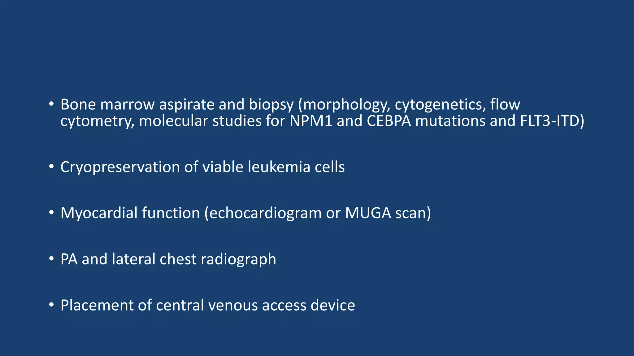
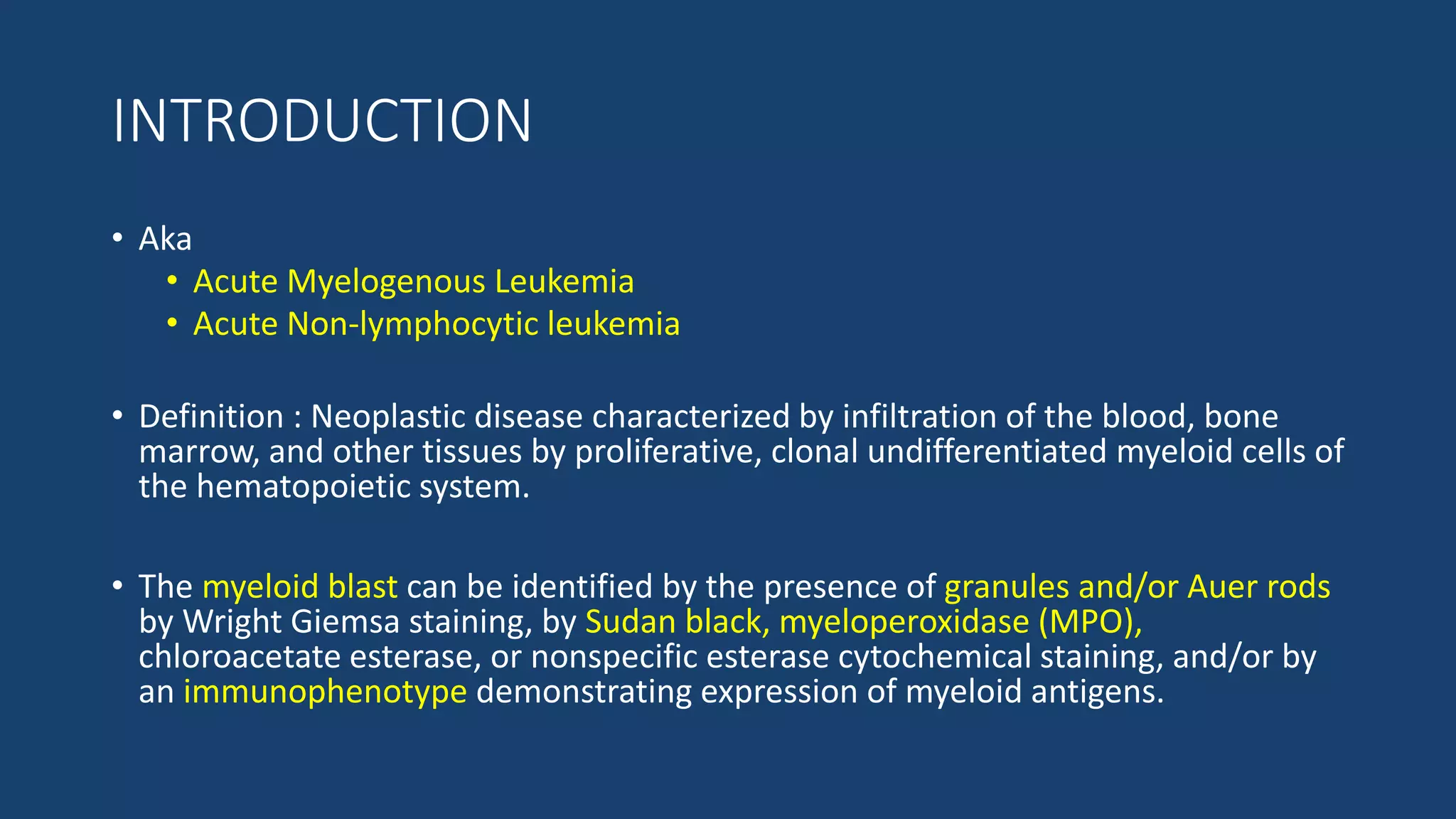
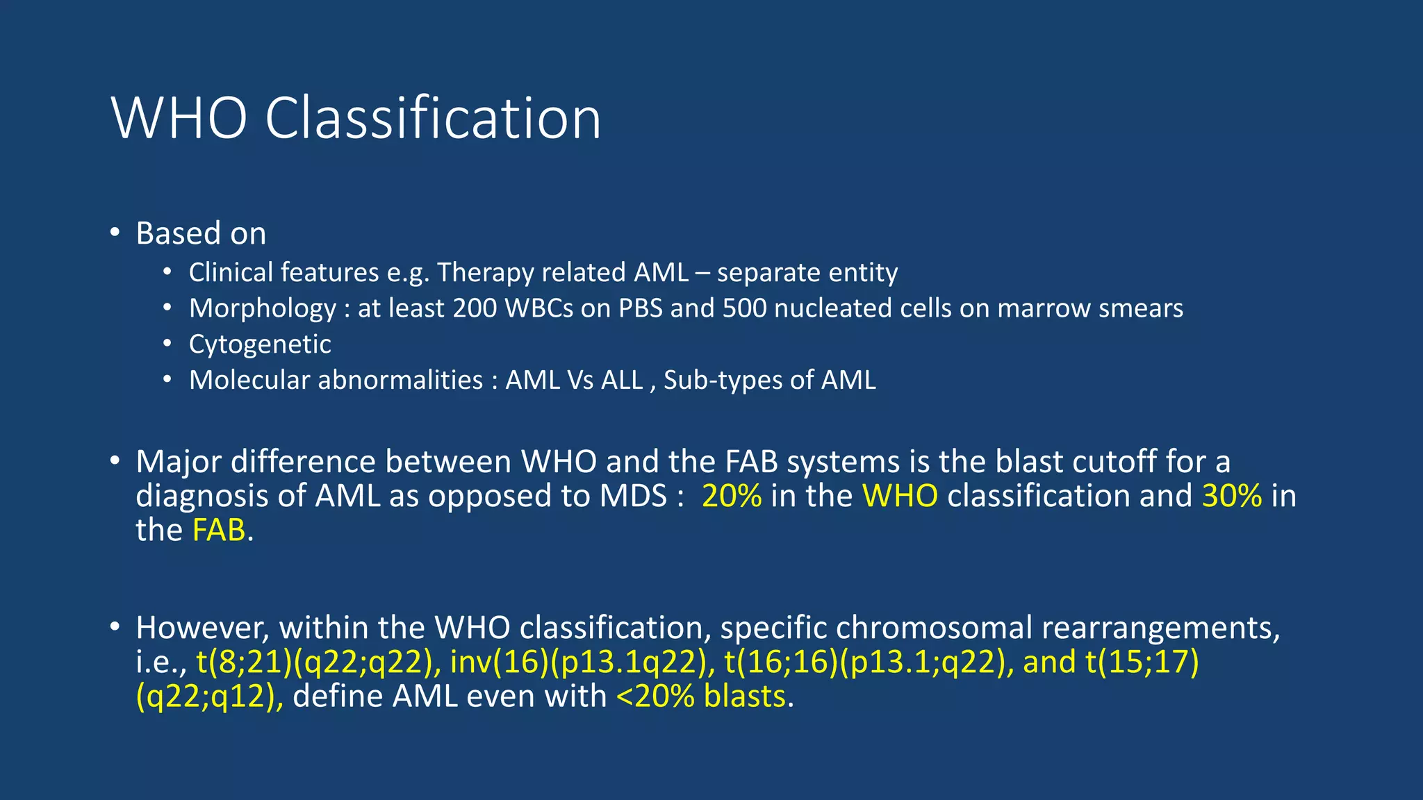
3. INTRODUCTION
• Aka
• Acute Myelogenous Leukemia
• Acute Non-lymphocytic leukemia
• Definition : Neoplastic disease characterized by infiltration of the blood, bone
marrow, and other tissues by proliferative, clonal undifferentiated myeloid cells of
the hematopoietic system.
• The myeloid blast can be identified by the presence of granules and/or Auer rods
by Wright Giemsa staining, by Sudan black, myeloperoxidase (MPO),
chloroacetate esterase, or nonspecific esterase cytochemical staining, and/or by
an immunophenotype demonstrating expression of myeloid antigens.
4. Cont..
• Incidence : ~3.5 per 100,000 people per year
• M:F = 4.5 : 3.1
• Age :
• AML incidence increases with age; it is 1.7 in individuals age <65 years and
15.9 in those age >65 years.
• The median age at diagnosis is 67 years.
5. HISTORICAL BACKGROUND
Hilstad described APL
in 1957
Von Boros and
Karenyi described
acute megakaryocytic
leukemia in 1931
DiGugliemo described
acute
erythroleukemia in
1917
Reschad and Schilling
described acute
monoblastic leukemia
in 1913;
Naegeli identified the
myeloblast in
leukemia in 1900
Ebstein introduced
the term “acute
leukemia” in 1889
6. RISK FACTORS
• OCCUPATIONAL EXPOSURES
• Exposure to rubber, paint, embalming fluids, pesticides, ethylene oxide,
petroleum, poultry, automobiles, nuclear power, plastics and electrical wiring,
as well as gasoline station attendants, beauticians, barbers, and
cosmetologists.
• The most fully characterized occupational exposure associated with AML is to
the aromatic hydrocarbon benzene.
• Average latency was 11.4 years
7. • ENVIRONMENTAL FACTORS
• Ionizing radiation is carcinogenic primarily via induction of double-strand DNA
breaks.
• The risk of leukemia correlates with radiation dosage and age at exposure.
• LIFESTYLE RELATED FACTORS
• Smoking : 2-3 times higher in male smokers > 20 pack years.
• Obesity
8. • THERAPY-RELATED ACUTE MYELOID LEUKEMIA
AML diagnosed following cytotoxic therapy for a prior malignant or benign
condition is considered to be t-AML.
Most commonly develops following t/t with alkylating agents(4-6 years) or
topoisomerase II inhibitors(1-3 years).
Pts with chromosome 5 and 7 abnormalities have the worst prognosis.
Low complete remission rate and short disease-free and overall survival.
11. WHO Classification
• Based on
• Clinical features e.g. Therapy related AML – separate entity
• Morphology : at least 200 WBCs on PBS and 500 nucleated cells on marrow smears
• Cytogenetic
• Molecular abnormalities : AML Vs ALL , Sub-types of AML
• Major difference between WHO and the FAB systems is the blast cutoff for a
diagnosis of AML as opposed to MDS : 20% in the WHO classification and 30% in
the FAB.
• However, within the WHO classification, specific chromosomal rearrangements,
i.e., t(8;21)(q22;q22), inv(16)(p13.1q22), t(16;16)(p13.1;q22), and t(15;17)
(q22;q12), define AML even with <20% blasts.
12. CYTOGENETICS
• The diagnosis of APML is based on the presence of either the t(15;17)(q22;q12)
cytogenetic rearrangement or the PML-RARA fusion product of the translocation.
• More commonly associated with younger age are t(8;21) and t(15;17), and with
older age, del(5q) and del(7q).
• Myeloid sarcomas are a/w t(8;21), and DIC is a/w t(15;17).
• AML with recurrent genetic abnormalities are those characterized by the
leukemogenic fusion genes RUNX1-RUNX1T1, CBFB-MYH11, MLLT3-MLL, and
DEK-NUP214, resulting, respectively, from t(8;21), inv(16) or t(16;16), t(9;11), and
t(6;9)(p23;q34).
13. MDS-AML OVERLAP/SECONDARY AML
• Mutations a/w secondary AML occur in genes encoding SRSF2, SF3B1, U2AF1,
and ZRSR2 (splicing factors); ASXL1, EZH2, and BCOR (epigenetic regulators); and
STAG2 (a member of the cohesin complex).
• In contrast, NPM1 mutations, and CBF and KMT2A rearrangements are highly
specific for de novo AML.
• Genetic features in MDS that are associated with prognosis and progression to
AML include mutations in TP53, RUNX1, ETV6, EZH2, and ASXL.
• TP53 mutations are associated with a particularly poor survival, including
following allogeneic HCT.
14.
15.
16.
17.
18.
19. PROGNOSTICATION IN AML- Significance ?
FACTOR FAVOURABLE UNFAVOURABLE
CLINICAL
1. Age <45 y <2 y, >60 y
2. ECOG performance status 0-1 >1
3. Leukemia De novo Antecedent hematologic disorder;
MDS ,MPN
4. Infection Absent Present
5. Prior chemotherapy No Yes
6. WBC count <25,000/cu mm >100,000/cu mm
24. HISTORY
• BM suppression cytopenia
• Fatigue and malaise : M/C symptom (50%)
• Anorexia , LOW
• Increasing fatigue or decreased exercise tolerance (anemia) : 30-40%
• Excess bleeding or bleeding from unusual sites (DIC,
thrombocytopenia) : 5%
• Fevers or recurrent infections (neutropenia) : 10%
• Headache, vision changes, non-focal neurologic abnormalities (CNS
leukemia or bleed)
25. • Early satiety (splenomegaly)
• Family history of AML (Fanconi, Bloom, or Kostmann syndromes or
ataxia-telangiectasia)
• History of cancer (exposure to alkylating agents, radiation,
topoisomerase II inhibitors)
• Occupational exposures (radiation, benzene, petroleum products,
paint, smoking, pesticides)
26. PHYSICAL EXAMINATION
• Performance status (prognostic factor)
• Ecchymosis and oozing from IV sites (DIC, possible APML)
• Fever and tachycardia (signs of infection)
• Papilledema, retinal infiltrates, cranial nerve abnormalities (CNS leukemia)
• Poor dentition, dental abscesses
27. • Gum hypertrophy (leukemic infiltration, most common in monocytic
leukemia)
• Skin infiltration or nodules (leukemia infiltration, most common in
monocytic leukemia)
• Lymphadenopathy, splenomegaly, hepatomegaly
• Back pain, lower extremity weakness [spinal granulocytic sarcoma,
most likely in t(8;21) patients]
30. • Bone marrow aspirate and biopsy (morphology, cytogenetics, flow
cytometry, molecular studies for NPM1 and CEBPA mutations and FLT3-ITD)
• Cryopreservation of viable leukemia cells
• Myocardial function (echocardiogram or MUGA scan)
• PA and lateral chest radiograph
• Placement of central venous access device
31. HEMATOLOGY FINDINGS
• Anemia :
• Usually present at diagnosis
• Usually Normocytic Normochromic.
• The median presenting leukocyte count is about 15,000/μL.
• Between 25 and 40% of patients have counts <5000/μL, and
• 20% have counts >100,000/μL.
• Platelet counts <100,000/μL are found at diagnosis in ~75% of pts,
and about 25% have counts <25,000/μL.
• Both morphologic and functional platelet abnormalities are found.
32.
33. INTERVENTIONS FOR SPECIFIC PATIENTS
• Dental evaluation (for those with poor dentition)
• Lumbar puncture (for those with symptoms of CNS involvement)
• Screening spine MRI (for patients with back pain, lower extremity
weakness,
34. BIOBANKING
• Pretreatment leukemic marrow and blood s/b stored within a biobank.
• Pretreatment samples should include nucleic acid (DNA and RNA, stored
at280°C) and viable cells (stored at2196°C).
• Skin fibroblasts may be the preferred tissue source.
• Skin biopsy can be performed using a punch biopsy or by taking a small biopsy at the
site of skin incision during bone marrow A/B.
• Other sources include finger nails and hair follicles, although the amount of
DNA that can be extracted may be limited.
38. SUPPORTIVE CARE
• Multi-lumen right atrial catheters should be inserted as soon as pts with
newly diagnosed AML have been stabilized.
• Platelet transfusions : to maintain platelet count ≥10,000/μL.
• The platelet count should be kept at higher levels in febrile pts and during episodes
of active bleeding or DIC.
• RBC transfusions : to maintain Hb >8 g/dL in the absence of active
bleeding, or CCF , which require higher hemoglobin levels(9-10 g/dL)
• Blood products leuko-depleted by filtration should be used to avert or
delay alloimmunization as well as febrile reactions.
39. • CMV-negative blood products should be used for CMV-seronegative
patients who are potential candidates for allogeneic HSCT.
• Antibacterial (i.e., quinolones) and antifungal (i.e., posaconazole)
prophylaxis in the absence of fever is likely to be beneficial.
• For pts who are herpes simplex virus or varicella zoster seropositive,
antiviral prophylaxis should be initiated (e.g., acyclovir, valacyclovir).
• Manage for febrile neutropenia , TLS , Leucostasis, DIC.
40.
41. MONITORING OF MRD
• Minimal Residual Disease monitoring : 2 approaches
• Multiparameter Flow Cytometry (MFC )
• Molecular techniques
• Real Time quantitative PCR(RT-qPCR)
• Next generation sequencing based techniques
• MRD can be assessed at
• Early time points
• Sequentially based consolidation to detect impending morphologic relapse
52. ROLE OF OTHER DRUGS
• FLT3 INHIBITORS
• Pts. with FLT3-mutated AML may be considered to receive intensive
chemotherapy in combination with midostaurin.
• GEMTUZUMAB OZOGAMICIN (GO)
• Antibody-toxin (calicheamicin) conjugate that targets CD331 AML
• Studies have suggested that the addition of GO was associated with longer
relapse-free survival (RFS) in the favorable-risk subset of CBF-AML.
53. • CPX-351
• CPX-351 may improve therapy of older pts. with high-risk features.
• PURINE ANALOGUES( CLADRIBINE)
• Results awaited
54. MYELOABLATIVE CONDITIONING VS RIC
Current recommendations
• Myeloablative regimens(busulfan/cyclophosphamide)
• For healthy younger patients.
• RIC(Reduced Intensive Conditioning )
• For elderly patients or in younger pts. with severe comorbidities.
60. APML
• Highly curable subtype of AML : approx. 85% of pts. achieve long-term
survival with current approaches.
• Initial regimen : Cytarabine + Daunorubicin increased incidence of DIC
abandoned.
• Current regimen : Tretinoin
• An oral drug that induces the differentiation of leukemic cells bearing the t(15;17),
where disruption of the RARA gene encoding a retinoid acid receptor occurs.
• Tretinoin decreases the frequency of DIC but produces another complication called
the APL differentiation syndrome.
61. APL DIFFERENTIATION SYNDROME
• Occurs within the first 3 weeks of t/t.
• Characterized by fever, fluid retention, dyspnea, chest pain, pulmonary infiltrates,
pleural and pericardial effusions, and hypoxemia.
• Pathogenesis : adhesion of differentiated neoplastic cells to the pulmonary
vasculature endothelium endothelial dysfunction pulmonary hemorrhage.
62. • Management
Glucocorticoids, chemotherapy, and/ or supportive measures
Temporary discontinuation of tretinoin in cases of severe ds. (i.e., patients
developing renal failure or requiring admission to ICU due to respiratory distress).
The mortality rate of this syndrome is about 10%.
63. • Tretinoin (45 mg/m2 per day orally until remission is documented) plus
concurrent anthracycline-based (i.e., idarubicin or daunorubicin) chemotherapy
appears to be among the most effective treatment for APL, leading to CR rates of
90–95%.
• Following achievement of CR, patients should receive at least two cycles of
anthracycline based chemotherapy
64. • Arsenic trioxide
In a RCT arsenic trioxide improved outcome if used after achievement of CR and
before consolidation therapy with anthracycline- based chemotherapy.
Patients receiving arsenic trioxide are at risk of APL differentiation syndrome,
especially when it is administered during induction or salvage treatment after
disease relapse.
In addition, arsenic trioxide may prolong the QT interval, increasing the risk of
cardiac arrhythmias.
65. • A study compared the gold standard (tretinoin plus chemotherapy) in newly
diagnosed non-high-risk APL with a chemotherapy-free combination of tretinoin
and arsenic trioxide.
• An equivalent outcome was demonstrated between the two arms, and the chemotherapy-
free regimen will likely become a new standard for non-high-risk APL patients.
• Combinations of tretinoin, arsenic trioxide, and/or chemotherapy and/or
gemtuzumab ozogamicin have shown favorable responses in high-risk APL pts. at
diagnosis
66. • Assessment of MRD by RT-PCR amplification of the t(15;17) chimeric
gene product PML-RARA following the final cycle of chemotherapy
• Disappearance of the signal is associated with long-term disease-free survival
• Persistence documented by two consecutive tests performed 2 weeks apart
invariably predicts relapse.
• Pts in molecular, cytogenetic, or clinical relapse should be salvaged
with arsenic trioxide with or without tretinoin.
• Produces meaningful responses in up to 85% of pts
• Can be followed by autologous or, less frequently, especially if RT-PCR positive
for PML-RARA, allogeneic HSCT.
67. REFERENCE
• Harrison 19th edition
• Wintrobe’s clinical hematology 13th edition
• UpToDate 2018
• How I treat AML in adults blood journal 2017