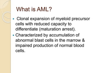
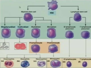
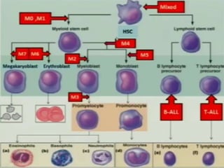
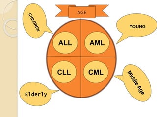
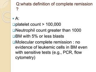
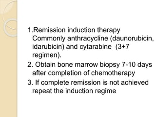
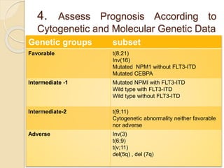
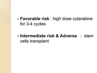
What is AML?
 Clonal expansion of myeloid precursor
cells with reduced capacity to
differentiate (maturation arrest).
 Characterized by accumulation of
abnormal blast cells in the marrow &
impaired production of normal blood
cells.
Causes of leukemia
 No single causative agent
 Most form combination of factors:
 Genetic predisposition
 Down syndrome
 Bloom syndrome.
 Klinefelter syndrome
 Turner syndrome
 Neurofibromatosis
 Environmental factors:
Chemical agents (benzene exposure)
Chemotherapeutic agents
(topoisomerase inhibitors , alkylating
agents)
Radiation
Immunological deficiencies
ALL AML
CMLCLL
AGE
YOUNG
Elderly
Clinical symptoms
Due to the excessive proliferation of myeloid
precursor cells in bone marrow, certain symptoms &
lab findings are expected
• Neutropenia – fever, chills
•Thrombocytopenia – bleeding, bruising
•Anemia – weakness, fatigue
• Bone pain (sternum, lower extremities) – secondary
to expansion of medullary cavity
Physical Findings
 Gum hypertrophy especially in the
monocytic variants (M4 or M5)
 Pallor, petechiae , ecchymoses
 Bone tenderness
 Retinal hemorrhage
 CNS infiltration (more common in M4 and
M5)
 Skin, soft tissue infiltration
 Extramedullary disease (ie, myeloid
sarcoma)
Work up
1.CBC & blood film
2. PT , PTT & fibrinogen
3. B.M aspiration, biopsy , cytogenetic &
molecular analysis
4.Immunophenotyping and
cytochemistry
5.Human leukocyte antigen (HLA) for
sibling or unrelated donor
6. Evaluate myocardial function before
chemotherapy
Diagnosis
1. 20% blasts on BM aspirate ( WHO
criteria)
2.patients with certain cryptogenic
abnormalities are considered to have
AML regardless of blast percentage
 t(8;21)(q22;q22)
 Inversion (16)(p13q22)
 t(16;16)(p13;q22)
 t(15;17)(q22;q12)
WHO Classification
 AML with certain genetic abnormalities
 t(8;21), t(16), inv(16), chromosome 11 changes
 t(15;17) as usually seen with AML M3
 AML with multilineage dysplasia (followed MDS or MPD)
 AML related to previous chemotherapy or radiation
 AML not otherwise specified
 Undifferentiated AML (M0)
 AML with minimal maturation (M1)
 AML with maturation (M2)
 Acute myelomonocytic leukemia (M4)
 Acute monocytic leukemia (M5)
 Acute erythroid leukemia (M6)
 Acute megakaryoblastic leukemia (M7)
 Acute basophilic leukemia
 Acute panmyelosis with fibrosis
 Myeloid sarcoma (also known as granulocytic sarcoma or
chloroma)
AML with minimal maturation
(M1)
AML with maturation (M2)
Acute promyelocytic leukemia
Acute myelomonocytic leukemia
(M4)
Acute monocytic leukemia
(M5)
Acute erythroid leukemia
(M6)
Acute megakaryoblastic
leukemia
CASE 1
 52 year M no significant PMH presents with
general malaise for several days, headache and
shortness of breath. Exam reveals pallor and
scattered petechiae. Found to have 95,000 WBC
with HCT of 22 and platelet count of 22,000.
biochemistry panel unremarkable. Treatment of
this condition would involve:
A. Induction chemotherapy with
anthracycline/cytarabine
B. Leukapheresis
C. Transfusion of platelets, PRBCs followed by
leukapheresis
D. Referring to ICU
Leukostasis
 Leukostasis – predominantly in those
with WBC counts > 100,000
 Common symptoms :
Pulmonary: dyspnea, chest pain
CNS: headaches, visual changes,
confusion , coma
 Other symptoms :Priapism , MI ,renal
insufficiency
What's treatment of
leukostasis?
1. Aggressive hydration
2. Leukapheresis
3. Induction Chemotherapy or high
dose hydroxyurea
 Avoid transfusion due to increase
blood viscosity.
 Monitoring for tumor lysis syndrome or
DIC
Case 2
 72 y women initiating chemotherapy
with intermediate dose cytarabine.
Develops progressive nausea and
lethargy with generalized muscle
cramps. An ECG is obtained. What
diagnosis is concerning?
Tumor lysis syndrome
Tumor Lysis Syndrome
 Characterized by metabolic
derangements caused by massive
release of cellular components following
lysis of malignant cells
 Commonly seen in malignancies with
high rates of cell proliferation (esp. ALL,
Burkitt’s lymphoma & AML).
 Preexisting hyperuricemia , renal
insufficiency , hypevolemia is considered
as a risk factors.
What's laboratory finding in
TLS?
 HIGH: potassium , phosphate and
uric acid.
 LOW: calicium
 Metabolic acidosis & renal impairment
Clinical feature
 Hyperuircemia: Crystallization of uric
acid and in renal tubules -> impaired
renal function.
 Hyperphosphatemia: nausea, vomiting,
diarrhea, seizures, lethargy
 Hypercalcemia: arrhythmia, hypotension,
tetany, cramps
 Hyperkalemia: arrhythmia, cramps &
paresthesia
What's is the treatment of
TLS?
1.A aggressive IV fluid hydration with
diuresis to promotes excretion of uric
acid and phosphate; improves renal
blood flow/GFR
2.Hpouricemic agents
A. Allopurinol:
• Inhibit xanthine oxidase
• Decrease formation of new uric acid
NOT preexisting level
• Need to be renally dosed
B. Rasburicase
 Recombinant urate oxidase
 Decreases serum uric acid
 Canot be given in pregnant women &
G6PD
3.Dialysis can be used in severe
cases
What about urinary
alkalinization?
 Urine alkalization is NOT recommended
because its increased propensity to
develop xanthine-obstructive uropathies
( with Allopurinol use).
Case 3
 30-year-old nulliparous female suffered
from placental abruption at the 25th
week of pregnancy, and emergent C/S
was done immediately with termination
of the pregnancy. Labs were significant
for a prolonged PTT/INR, anemia,
thrombocytopenia. What is the likely
diagnosis?
DIC
DIC
 Common symptoms/findings
Weakness (anemia), infections/fever
(malfunctioning WBCs)
Petechiae, ecchymoses, hematuria,
bleeding from venipuncture sites
Migratory thrombophlebitis (Trousseau’s
syndrome)
Nonbacterial thrombotic (marantic)
endocarditis
DVT/PE
What's are lab finding of DIC?
 Lab findings
Prolonged PT/INR, PTT
Microangiopathic anemia
(schistocytes)
Thrombocytopenia
Elevated fibrin split products
Elevated D-dimer
Low fibrinogen
What's treatment of DIC ?
 Supportive therapy
Platelets
Cryoprecipitate (fibrinogen)
FFP
 Treatment for obvious thrombosis (e.g
thrombophlebitis, mural thrombus)
UFH or LMWH; often resistant to
warfarin
Activated protein C
 Treatment of underlying malignancy
Treatment of AML
Q:whats definition of complete remission
?
 A:
platelet count > 100,000
Neutrophil count greater than 1000
BM with 5% or less blasts
Molecular complete remission : no
evidence of leukemic cells in BM even
with sensitive tests (e.g., PCR, flow
cytometry)
1.Remission induction therapy
Commonly anthracycline (daunorubicin,
idarubicin) and cytarabine (3+7
regimen).
2. Obtain bone marrow biopsy 7-10 days
after completion of chemotherapy
3. If complete remission is not achieved
repeat the induction regime
4. Assess Prognosis According to
Cytogenetic and Molecular Genetic Data
subsetGenetic groups
t(8;21)
Inv(16)
Mutated NPM1 without FLT3-ITD
Mutated CEBPA
Favorable
Mutated NPMI with FLT3-ITD
Wild type with FLT3-ITD
Wild type without FLT3-ITD
Intermediate -1
t(9;11)
Cytogenetic abnormality neither favorable
nor adverse
Intermediate-2
Inv(3)
t(6;9)
t(v;11)
del(5q) , del (7q)
Adverse
 Favorable risk : high dose cytarabine
for 3-4 cycles
 Intermediate risk & Adverse : stem
cells transplant
Acute myeloid leukemia

Acute myeloid leukemia

  • 2.
    What is AML? Clonal expansion of myeloid precursor cells with reduced capacity to differentiate (maturation arrest).  Characterized by accumulation of abnormal blast cells in the marrow & impaired production of normal blood cells.
  • 5.
    Causes of leukemia No single causative agent  Most form combination of factors:  Genetic predisposition  Down syndrome  Bloom syndrome.  Klinefelter syndrome  Turner syndrome  Neurofibromatosis
  • 6.
     Environmental factors: Chemicalagents (benzene exposure) Chemotherapeutic agents (topoisomerase inhibitors , alkylating agents) Radiation Immunological deficiencies
  • 7.
  • 8.
    Clinical symptoms Due tothe excessive proliferation of myeloid precursor cells in bone marrow, certain symptoms & lab findings are expected • Neutropenia – fever, chills •Thrombocytopenia – bleeding, bruising •Anemia – weakness, fatigue • Bone pain (sternum, lower extremities) – secondary to expansion of medullary cavity
  • 9.
    Physical Findings  Gumhypertrophy especially in the monocytic variants (M4 or M5)  Pallor, petechiae , ecchymoses  Bone tenderness  Retinal hemorrhage  CNS infiltration (more common in M4 and M5)  Skin, soft tissue infiltration  Extramedullary disease (ie, myeloid sarcoma)
  • 12.
    Work up 1.CBC &blood film 2. PT , PTT & fibrinogen 3. B.M aspiration, biopsy , cytogenetic & molecular analysis 4.Immunophenotyping and cytochemistry 5.Human leukocyte antigen (HLA) for sibling or unrelated donor 6. Evaluate myocardial function before chemotherapy
  • 13.
    Diagnosis 1. 20% blastson BM aspirate ( WHO criteria) 2.patients with certain cryptogenic abnormalities are considered to have AML regardless of blast percentage  t(8;21)(q22;q22)  Inversion (16)(p13q22)  t(16;16)(p13;q22)  t(15;17)(q22;q12)
  • 14.
    WHO Classification  AMLwith certain genetic abnormalities  t(8;21), t(16), inv(16), chromosome 11 changes  t(15;17) as usually seen with AML M3  AML with multilineage dysplasia (followed MDS or MPD)  AML related to previous chemotherapy or radiation  AML not otherwise specified  Undifferentiated AML (M0)  AML with minimal maturation (M1)  AML with maturation (M2)  Acute myelomonocytic leukemia (M4)  Acute monocytic leukemia (M5)  Acute erythroid leukemia (M6)  Acute megakaryoblastic leukemia (M7)  Acute basophilic leukemia  Acute panmyelosis with fibrosis  Myeloid sarcoma (also known as granulocytic sarcoma or chloroma)
  • 15.
    AML with minimalmaturation (M1)
  • 16.
  • 17.
  • 18.
  • 19.
  • 20.
  • 21.
  • 22.
    CASE 1  52year M no significant PMH presents with general malaise for several days, headache and shortness of breath. Exam reveals pallor and scattered petechiae. Found to have 95,000 WBC with HCT of 22 and platelet count of 22,000. biochemistry panel unremarkable. Treatment of this condition would involve: A. Induction chemotherapy with anthracycline/cytarabine B. Leukapheresis C. Transfusion of platelets, PRBCs followed by leukapheresis D. Referring to ICU
  • 23.
    Leukostasis  Leukostasis –predominantly in those with WBC counts > 100,000  Common symptoms : Pulmonary: dyspnea, chest pain CNS: headaches, visual changes, confusion , coma  Other symptoms :Priapism , MI ,renal insufficiency
  • 24.
    What's treatment of leukostasis? 1.Aggressive hydration 2. Leukapheresis 3. Induction Chemotherapy or high dose hydroxyurea  Avoid transfusion due to increase blood viscosity.  Monitoring for tumor lysis syndrome or DIC
  • 25.
    Case 2  72y women initiating chemotherapy with intermediate dose cytarabine. Develops progressive nausea and lethargy with generalized muscle cramps. An ECG is obtained. What diagnosis is concerning? Tumor lysis syndrome
  • 26.
    Tumor Lysis Syndrome Characterized by metabolic derangements caused by massive release of cellular components following lysis of malignant cells  Commonly seen in malignancies with high rates of cell proliferation (esp. ALL, Burkitt’s lymphoma & AML).  Preexisting hyperuricemia , renal insufficiency , hypevolemia is considered as a risk factors.
  • 27.
    What's laboratory findingin TLS?  HIGH: potassium , phosphate and uric acid.  LOW: calicium  Metabolic acidosis & renal impairment
  • 28.
    Clinical feature  Hyperuircemia:Crystallization of uric acid and in renal tubules -> impaired renal function.  Hyperphosphatemia: nausea, vomiting, diarrhea, seizures, lethargy  Hypercalcemia: arrhythmia, hypotension, tetany, cramps  Hyperkalemia: arrhythmia, cramps & paresthesia
  • 29.
    What's is thetreatment of TLS? 1.A aggressive IV fluid hydration with diuresis to promotes excretion of uric acid and phosphate; improves renal blood flow/GFR 2.Hpouricemic agents A. Allopurinol: • Inhibit xanthine oxidase • Decrease formation of new uric acid NOT preexisting level • Need to be renally dosed
  • 30.
    B. Rasburicase  Recombinanturate oxidase  Decreases serum uric acid  Canot be given in pregnant women & G6PD 3.Dialysis can be used in severe cases
  • 31.
    What about urinary alkalinization? Urine alkalization is NOT recommended because its increased propensity to develop xanthine-obstructive uropathies ( with Allopurinol use).
  • 32.
    Case 3  30-year-oldnulliparous female suffered from placental abruption at the 25th week of pregnancy, and emergent C/S was done immediately with termination of the pregnancy. Labs were significant for a prolonged PTT/INR, anemia, thrombocytopenia. What is the likely diagnosis? DIC
  • 33.
    DIC  Common symptoms/findings Weakness(anemia), infections/fever (malfunctioning WBCs) Petechiae, ecchymoses, hematuria, bleeding from venipuncture sites Migratory thrombophlebitis (Trousseau’s syndrome) Nonbacterial thrombotic (marantic) endocarditis DVT/PE
  • 34.
    What's are labfinding of DIC?  Lab findings Prolonged PT/INR, PTT Microangiopathic anemia (schistocytes) Thrombocytopenia Elevated fibrin split products Elevated D-dimer Low fibrinogen
  • 35.
    What's treatment ofDIC ?  Supportive therapy Platelets Cryoprecipitate (fibrinogen) FFP  Treatment for obvious thrombosis (e.g thrombophlebitis, mural thrombus) UFH or LMWH; often resistant to warfarin Activated protein C  Treatment of underlying malignancy
  • 36.
  • 37.
    Q:whats definition ofcomplete remission ?  A: platelet count > 100,000 Neutrophil count greater than 1000 BM with 5% or less blasts Molecular complete remission : no evidence of leukemic cells in BM even with sensitive tests (e.g., PCR, flow cytometry)
  • 38.
    1.Remission induction therapy Commonlyanthracycline (daunorubicin, idarubicin) and cytarabine (3+7 regimen). 2. Obtain bone marrow biopsy 7-10 days after completion of chemotherapy 3. If complete remission is not achieved repeat the induction regime
  • 39.
    4. Assess PrognosisAccording to Cytogenetic and Molecular Genetic Data subsetGenetic groups t(8;21) Inv(16) Mutated NPM1 without FLT3-ITD Mutated CEBPA Favorable Mutated NPMI with FLT3-ITD Wild type with FLT3-ITD Wild type without FLT3-ITD Intermediate -1 t(9;11) Cytogenetic abnormality neither favorable nor adverse Intermediate-2 Inv(3) t(6;9) t(v;11) del(5q) , del (7q) Adverse
  • 40.
     Favorable risk: high dose cytarabine for 3-4 cycles  Intermediate risk & Adverse : stem cells transplant