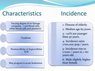
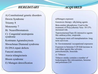
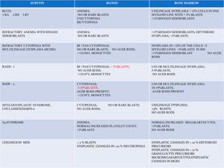
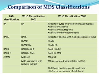
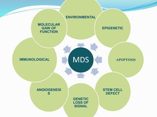
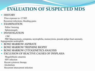
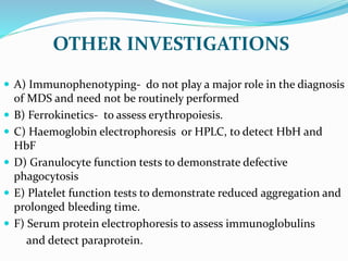
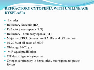
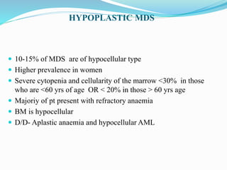
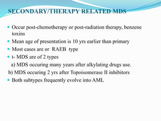
Myelodysplastic syndrome (MDS) is a heterogeneous group of clonal stem cell disorders where the bone marrow cannot produce blood cells effectively. Patients present with varying degrees of cytopenias. MDS may progress to acute leukemia. The WHO classification system recognizes subtypes based on blast percentage and cytogenetics that provide prognostic information. MDS results from genetic mutations and epigenetic changes in hematopoietic stem cells. Presenting signs include anemia, infections, and bleeding due to cytopenias. Evaluation involves blood tests, bone marrow aspiration/biopsy, and cytogenetics. Refractory cytopenia with unilineage dysplasia is the most common subtype.
acute leukemia
For More Medicine Free PPT - http://playnever.blogspot.com/
For Health benefits and medicine videos Subscribe youtube channel - https://www.youtube.com/playlist?list=PLKg-H-sMh9G01zEg4YpndngXODW2bq92w
acute leukemia
For More Medicine Free PPT - http://playnever.blogspot.com/
For Health benefits and medicine videos Subscribe youtube channel - https://www.youtube.com/playlist?list=PLKg-H-sMh9G01zEg4YpndngXODW2bq92w
Myelodesplastic syndrome by haider zamanHaider zaman
Myelodesplastic syndrome presentation may easy for Medical student to know about Myelodesplastic syndrome & its classification and Sign and Symptoms,Genetically Abnormalities in MDS,Diagnosing,Morphological abnormalities in MDSs
AML:ACUTE MYELOID LEUKAEMIA
for medical colleges teaching faculty and students as well. it includes AML causes , histopathological slides of subclasses of Acute myeloid leukemia, classification , diagnosis, management modalities, complications .Acute leukemias are stem cell disorders characterized by malignant neoplastic proliferation and accumulation of immature and non functional hematopoietic cells in the bone marrow.
The neoplastic cells show increased proliferation and/or decreased apoptosis.
If the defect primarily affects the common myeloid progenitor (CMP) then it is called Acute myeloid leukemia.
Acute myeloid leukemia (AML) is a neoplastic disease characterized by infiltration of the blood, bone marrow, and other tissues by proliferative, clonal undifferentiated cells of the hematopoietic system.
AML is the result of a sequence of somatic mutations in a multipotential primitive hematopoietic cell or, in some cases, a more differentiated progenitor cell.
It can be slow growing or rapidly fatal.
AML is the predominant form of leukemia during the neonatal period
Incidence : 1.5/100,000/year in infants decreases to approximately 0.4 per 100,000 children ages 5 to 9 years, increases gradually to 1.0 persons per 100,000 until age 25 years, and thereafter increases exponentially until the rate reaches approximately 25/100,000 persons.
AML accounts for 15 to 20 percent of the acute leukemias in children and 80 percent of the acute leukemias in adults.
Men > Women (4.5 : 3)
HEREDITY
1) Chromosomal aneuploidy like Trisomy 21 noted in Down syndrome
2) Defective DNA repair, e.g., Fanconi anemia, Bloom syndrome, and Ataxia telangiectasia
3) Congenital neutropenia ie Kostmann syndrome
4) Germline mutations of CCAAT/enhancer-binding protein α (CEBPA), runt-related transcription factor 1 (RUNX1), and tumor protein p53 (TP53) have also been associated with a higher predisposition to AML
RADIATION
Peaks after 5 to 7 yrs of exposure.
Therapeutic radiation alone seems to add little risk of AML but can increase the risk in people also exposed to alkylating agents.
CHEMICAL AND OTHER EXPOSURES
Exposure to benzene, plastic, rubber, petroleum products, paint, ethylene oxide, herbicides and pesticides can increase the risk.
Smoking can also increase the risk
DRUGS
Anticancer drugs are the leading cause of therapy-associated AML.
Alkylating agent–associated leukemias occur on average 4–6 years after exposure, and affected individuals have aberrations in chromosomes 5 and 7.
Topoisomerase II inhibitor–associated leukemias occur 1–3 years after exposure, and affected individuals often have aberrations involving chromosome 11q23.
Other agents like Chloramphenicol, phenylbutazone, and, less commonly, chloroquine and methoxypsoralen.
SYMPTOMS :
Present with nonspecific symptoms initially.
Fatigue is the first symptom
Fever with or without infection will be present in approximately 10% patients
Bleeding, easy bruising
occasional
Couples presenting to the infertility clinic- Do they really have infertility...Sujoy Dasgupta
Dr Sujoy Dasgupta presented the study on "Couples presenting to the infertility clinic- Do they really have infertility? – The unexplored stories of non-consummation" in the 13th Congress of the Asia Pacific Initiative on Reproduction (ASPIRE 2024) at Manila on 24 May, 2024.
Title: Sense of Taste
Presenter: Dr. Faiza, Assistant Professor of Physiology
Qualifications:
MBBS (Best Graduate, AIMC Lahore)
FCPS Physiology
ICMT, CHPE, DHPE (STMU)
MPH (GC University, Faisalabad)
MBA (Virtual University of Pakistan)
Learning Objectives:
Describe the structure and function of taste buds.
Describe the relationship between the taste threshold and taste index of common substances.
Explain the chemical basis and signal transduction of taste perception for each type of primary taste sensation.
Recognize different abnormalities of taste perception and their causes.
Key Topics:
Significance of Taste Sensation:
Differentiation between pleasant and harmful food
Influence on behavior
Selection of food based on metabolic needs
Receptors of Taste:
Taste buds on the tongue
Influence of sense of smell, texture of food, and pain stimulation (e.g., by pepper)
Primary and Secondary Taste Sensations:
Primary taste sensations: Sweet, Sour, Salty, Bitter, Umami
Chemical basis and signal transduction mechanisms for each taste
Taste Threshold and Index:
Taste threshold values for Sweet (sucrose), Salty (NaCl), Sour (HCl), and Bitter (Quinine)
Taste index relationship: Inversely proportional to taste threshold
Taste Blindness:
Inability to taste certain substances, particularly thiourea compounds
Example: Phenylthiocarbamide
Structure and Function of Taste Buds:
Composition: Epithelial cells, Sustentacular/Supporting cells, Taste cells, Basal cells
Features: Taste pores, Taste hairs/microvilli, and Taste nerve fibers
Location of Taste Buds:
Found in papillae of the tongue (Fungiform, Circumvallate, Foliate)
Also present on the palate, tonsillar pillars, epiglottis, and proximal esophagus
Mechanism of Taste Stimulation:
Interaction of taste substances with receptors on microvilli
Signal transduction pathways for Umami, Sweet, Bitter, Sour, and Salty tastes
Taste Sensitivity and Adaptation:
Decrease in sensitivity with age
Rapid adaptation of taste sensation
Role of Saliva in Taste:
Dissolution of tastants to reach receptors
Washing away the stimulus
Taste Preferences and Aversions:
Mechanisms behind taste preference and aversion
Influence of receptors and neural pathways
Impact of Sensory Nerve Damage:
Degeneration of taste buds if the sensory nerve fiber is cut
Abnormalities of Taste Detection:
Conditions: Ageusia, Hypogeusia, Dysgeusia (parageusia)
Causes: Nerve damage, neurological disorders, infections, poor oral hygiene, adverse drug effects, deficiencies, aging, tobacco use, altered neurotransmitter levels
Neurotransmitters and Taste Threshold:
Effects of serotonin (5-HT) and norepinephrine (NE) on taste sensitivity
Supertasters:
25% of the population with heightened sensitivity to taste, especially bitterness
Increased number of fungiform papillae
Factory Supply Best Quality Pmk Oil CAS 28578–16–7 PMK Powder in Stockrebeccabio
Factory Supply Best Quality Pmk Oil CAS 28578–16–7 PMK Powder in Stock
Telegram: bmksupplier
signal: +85264872720
threema: TUD4A6YC
You can contact me on Telegram or Threema
Communicate promptly and reply
Free of customs clearance, Double Clearance 100% pass delivery to USA, Canada, Spain, Germany, Netherland, Poland, Italy, Sweden, UK, Czech Republic, Australia, Mexico, Russia, Ukraine, Kazakhstan.Door to door service
Hot Selling Organic intermediates
Prix Galien International 2024 Forum ProgramLevi Shapiro
June 20, 2024, Prix Galien International and Jerusalem Ethics Forum in ROME. Detailed agenda including panels:
- ADVANCES IN CARDIOLOGY: A NEW PARADIGM IS COMING
- WOMEN’S HEALTH: FERTILITY PRESERVATION
- WHAT’S NEW IN THE TREATMENT OF INFECTIOUS,
ONCOLOGICAL AND INFLAMMATORY SKIN DISEASES?
- ARTIFICIAL INTELLIGENCE AND ETHICS
- GENE THERAPY
- BEYOND BORDERS: GLOBAL INITIATIVES FOR DEMOCRATIZING LIFE SCIENCE TECHNOLOGIES AND PROMOTING ACCESS TO HEALTHCARE
- ETHICAL CHALLENGES IN LIFE SCIENCES
- Prix Galien International Awards Ceremony
The prostate is an exocrine gland of the male mammalian reproductive system
It is a walnut-sized gland that forms part of the male reproductive system and is located in front of the rectum and just below the urinary bladder
Function is to store and secrete a clear, slightly alkaline fluid that constitutes 10-30% of the volume of the seminal fluid that along with the spermatozoa, constitutes semen
A healthy human prostate measures (4cm-vertical, by 3cm-horizontal, 2cm ant-post ).
It surrounds the urethra just below the urinary bladder. It has anterior, median, posterior and two lateral lobes
It’s work is regulated by androgens which are responsible for male sex characteristics
Generalised disease of the prostate due to hormonal derangement which leads to non malignant enlargement of the gland (increase in the number of epithelial cells and stromal tissue)to cause compression of the urethra leading to symptoms (LUTS
Pulmonary Thromboembolism - etilogy, types, medical- Surgical and nursing man...VarunMahajani
Disruption of blood supply to lung alveoli due to blockage of one or more pulmonary blood vessels is called as Pulmonary thromboembolism. In this presentation we will discuss its causes, types and its management in depth.
Explore natural remedies for syphilis treatment in Singapore. Discover alternative therapies, herbal remedies, and lifestyle changes that may complement conventional treatments. Learn about holistic approaches to managing syphilis symptoms and supporting overall health.
MANAGEMENT OF ATRIOVENTRICULAR CONDUCTION BLOCK.pdfJim Jacob Roy
Cardiac conduction defects can occur due to various causes.
Atrioventricular conduction blocks ( AV blocks ) are classified into 3 types.
This document describes the acute management of AV block.
These simplified slides by Dr. Sidra Arshad present an overview of the non-respiratory functions of the respiratory tract.
Learning objectives:
1. Enlist the non-respiratory functions of the respiratory tract
2. Briefly explain how these functions are carried out
3. Discuss the significance of dead space
4. Differentiate between minute ventilation and alveolar ventilation
5. Describe the cough and sneeze reflexes
Study Resources:
1. Chapter 39, Guyton and Hall Textbook of Medical Physiology, 14th edition
2. Chapter 34, Ganong’s Review of Medical Physiology, 26th edition
3. Chapter 17, Human Physiology by Lauralee Sherwood, 9th edition
4. Non-respiratory functions of the lungs https://academic.oup.com/bjaed/article/13/3/98/278874
Report Back from SGO 2024: What’s the Latest in Cervical Cancer?bkling
Are you curious about what’s new in cervical cancer research or unsure what the findings mean? Join Dr. Emily Ko, a gynecologic oncologist at Penn Medicine, to learn about the latest updates from the Society of Gynecologic Oncology (SGO) 2024 Annual Meeting on Women’s Cancer. Dr. Ko will discuss what the research presented at the conference means for you and answer your questions about the new developments.
2. What Is Myelodysplastic Syndrome?
Heterogeneous group of clonal hematopoietic stem cell
disorders.
In MDS the bone marrow cannot produce blood cells
effectively, and many of the blood cells formed are
defective.
MDS is best considered a preleukemic disorder in which
the neoplastic clone that has been established may or may
not fully progress to acute leukemia.
3. Characteristics
Varying degree of tri-lineage
cytopenia ( red blood cells,
white blood cells and platelets).
Dysplasia
Normocellular or hypercellular
B.M
May progress to acute leukaemia
Incidence
1- Disease of elderly.
2- Median age 65 years.
3- <10% are younger
than 50 years.
4- Incidence rates
1/100,000 pop./ years.
5- Incidence rise to
1/1000 / years in > 60
years old.
6- Male slightly higher
than female
5. HEREDITARY ACQUIRED
A) Constitutional genetic disorders
Downs Syndrome
Trisomy 8
Monosomy 7
B) Neurofibromatosis
C) Congenital neutropenia
syndrome
Kostmann Agranulocytosis
Shwachman Diamond syndrome
D) DNA repair defects
Fanconi anemia,
Ataxia telangiectasia
Bloom syndrome
E) Mutagen detoxification
a)Mutagen exposure
Genotoxic therapy- alkylating agents
Beta-emitter phosphorus; Used in the
treatment of Polycythemia Vera- 10-15%
increased risk.
Topoisomerase(Topo-II) interactive agents
like anthracycline, etoposide.
Autologous stem cell transplantation- long
term survivors
b) Environmental /occupational exposures
Exposure to benzene-5-20 fold increase in
risk.Other agents like solvents,
petrochemicals, Insectide.
c) Tobacco
Tobacco smoke contains a number of
leukemogens like nitrosamines, benzene
and polonium-210
6. Morphological dysplasia in MDS
Dyserythropoiesis
Dysmyelopoiesis
Dysmegakaryopoiesis
Ring Sideroblast
Type I, II & III blasts
11. Dysmegakaryocytopoiesis
Peripheral Blood:
Thrombocytopenia
Large platelets with abnormal or decreased granularity
Abnormal platelet function
Bone Marrow:
Reduced numbers of megakaryocytes
Micromegakaryocytes
Megakaryocytes with large, single nuclei or multiple small
nuclei
15. MDS: Limitations of FAB Classification
Multilineage cytopenia with <5% BM blasts
Rough prediction of prognosis
Cytogenetics not given importance
Ill defined entities: childhood MDS, T-MDS & other
secondary MDS
Immunophenotyping and genetic techniques not included
16. WHO Classification System( 2001)
Refractory Anemia (RA)
Refractory Anemia with ringed sideroblasts
Refractory Cytopenia (MDS) with Multilineage Dysplasia
(RCMD) – with & without sideroblasts
Refractory Anemia with Excess Blasts (RAEB)
5q- syndrome
Myelodysplastic syndrome, unclassifiable
18. SUBTYPE BLOOD BONE MARROW
RCUD:
1.RA 2.RN 3.RT
ANEMIA;
NO OR RARE BLASTS
UNICYTOPENIA
BICYTOPENIA
UNILINEAGE DYSPLASIA > 10% CELLS IN ONE
MYELOID LINE WITH < 5% BLASTS
<15%RINGED SIDEROBLASTS
REFRACTORY ANEMIA WITH RINGED
SIDEROBLASTS
ANEMIA;
NO OR RARE BLASTS
>15%RINGED SIDEROBLASTS; ERYTHROID
DYSPLASIA; <5%BLASTS;
REFRACTORY CYTOPENIA WITH
MULTILINEAGE DYSPLASIA (RCMD)
BI / PAN CYTOPENIAS;
NO OR RARE BLASTS; NO AUER RODS;
<1X10⁹/L MONOCYTES
DYSPLASIA IN >10% OF THE CELLS >2
MYELOID LINES <5%BLASTS IN BM
>15%RINGED SIDEROBLAST NO AUER
RODS
RAEB - 1 BI / PAN CYTOPENIAS; < 5%BLASTS;
NO AUER RODS;
<1X10⁹/L MONOCYTES
UNI OR MULTILINEAGE DYSPLASIA;
5-9%BLASTS;
NO AUER RODS
RAEB – 2 CYTOPENIAS;
5-19%BLASTS;
AUER RODS PRESENT;
<1X10⁹/L MONOCYTES
UNI OR MULTILINEAGE DYSPLASIA;
10-19%BLASTS;
AUER RODS PRESENT
MYELODYSPLASTIC SYNDROME,
UNCLASSIFIED(MDS-u
CYTOPENIAS; NO OR RARE BLASTS;
NO AUER RODS;
UNILINEAGE DYSPLASIA;
<5% BLASTS;
NO AUER RODS
5q-SYNDROME ANEMIA;
NORMAL/INCREASED PLATELET COUNT;
<5%BLASTS
NORMAL/INCREASED MEGAKARYOCYTES;
<5%BLASTS;
NO AUER RODS
CHILDHOOD MDS < 2 % BLASTS
DYSPLASTIC CHANGES IN >10 % NEUTROPHILS
DYSPLASTIC CHANGES IN > 10 % ERYTHROID
PRECURSORS
DYSPLASTIC CHANGES IN > 10 %
GRANULOCYTE PRECURSORS
MICROMEGAKARYOCYTES,DYSPLASTIC
CHANGES IN MGKS
19. Comparison of MDS Classifications
FAB
classification
WHO Classsification
2001
WHO Classification 2008
RA RA Refractory cytopenia with unilineage dyplasia
• Refractory anemia
• Refractory neutropenia
• Refractory thrombocytopenia
RARS RARS Refractory anemia with ring sideroblasts (RARS)
RCMD RCMD
RCMD-RS RCMD-RS
RAEB RAEB I and 2 RAEB I and 2
RAEB-T RAEB II/ AML RAEB II/ AML
CMML MDS-UC MDS-UC
MDS associated with
isolated del(5q)
MDS associated with isolated del(5q)
Childhood myelodysplastic syndrome
• Refractory cytopenia of childhood
20. DIFFERENCES BETWEEN WHO AND FAB
The WHO system…
Makes use of cytogenetic findings.
The category of RAEB-t was eliminated as it got included
within AML(>20%blasts).
CMML was removed and put in a new category of
myelodysplastic/ myeloproliferative diseases.
Adds the subtypes 5q syndrome and unclassifiable MDS.
Recognizes the prognostic importance of % of bone marrow
blasts
22. MDS : a stem cell disorder
The abnormal cells in MDS are clones derived from an abnormal stem cell
Apoptosis in MDS
Mechanism appears to be one of increased apoptosis of haemopoietic precursors in
the marrow,
For those patients undergoing leukaemic transformation,the cytopenias arise due to
maturation block of the malignant cells
Apoptosis is more prominent in early MDS, such as RA and RARS, than in
advanced MDS with excess myeloblasts
Ineffective Hematopoiesis
Colony forming capacities of pleuripotent stem cells and their progeny are low or
absent
Lower level of GM-CSF, M-CSF,IL 6 .IL 3,
CFU- GM less responsive to both G-CSF & GM-CSF
More dramatic in pts with RAEB or RAEB –t
23. Immunological abnormalities in MDS
Commonly encountered in MDS, suggesting that they may play a role in the
aetiology and pathogenesis of the disease.
Particularly apparent in cases of hypoplastic MDS that share a number of features in
common with aplastic anaemia, notably clinical presentation with macrocytosis and
varying levels of dyserythropoiesis
Acquired mutations in the PIG-A gene characteristic of paroxysmal nocturnal
haemoglobinuria (PNH) are also encountered
Angiogenesis
Autocrine production of angiogenic molecules promotes expansion of leukemic
clone
Vascular endothelial growth factor(VEGF) and its receptor VEGFR-1 And VEGFR-2
is overexpressed
24. Molecular basis of MDS
The common chromosomal abnormalities
found in MDS include loss of Y,
monosomy 5, monosomy 7,trisomy 8,
20q – , abnormalities of 11q23, and
deletions of 17p, 12p, 13q and 11q among
others.
Genetic abnormalities in MDS
Mutations of the AML1 gene (also known as RUNX1 ) have recently been recognized to occur
in MDS, particularly where it is treatment - related or radiation - induced.
Activating mutations of RAS , usually involving NRAS , are found in up to 20% of cases of
MDS
Class 1 mutation-mutation involving Tyrosine kinase GATA1 , PU.1 ( SPI1 ), CEBPA , MLL
and TP53 .
Class 2 mutation –mutation involving Transcription factors FMS (now called CSF1R ), KIT ,
FLT3 , PDGFRB and GCSFR
Association of both Class 1 &2 – highly predisposed to MDS & AML
25. Epigenetic abnormalities
Refers to alteration of gene expression without altering the DNA sequence
Two important epigenetic modifications relevant to MDS, are DNA methylation and
histone modification.
Promoter methylation of p15INK4B – t-MDS
methylation of p15INK4B also seen in loss of Chr 7 and in pts who progresses from
RA to RAEB
27. Signs and Symptoms
Excessive tiredness, shortness of breath, and pale skin
can be caused by anemia.
Serious infections with high fevers can be caused by
leukopenia and, in particular, by having neutropenia or
granulocytopenia.
Excessive bruising and bleeding, for example, frequent
or severe nosebleeds and/or bleeding from the gums,
can be due to thrombocytopenia.
29. EVALUATION OF SUSPECTED MDS
HISTORY
Prior exposure to CT/RT
Recurrent infections, bleeding gums
EXAMINATION
Pallor/ bruising
Splenomegaly
INVESTIGATION
CBC
PBF:Macrocytosis, cytopenia, neytrophilia, monocytosis, pseudo-pelger huet anomaly,
hypogranular neutrophils
BONE MARROW ASPIRATE
BONE MARROW TREPHINE BIOPSY
BONE MARROW CYTOGENETICS ANALYSIS
EXCLUSION OF REACTIVE CAUSES OF DYSPLASIA
Megaloblastic anaemia
HIV infection
Recent cytotoxic therapy
Alcoholism
Recurrent intercurrent infection
30. BONE MARROW ASPIRATE
Well stained BM aspirate smears
At least 500 cells are to be counted
At least 30 megakaryocyte to be evaluated
Dysplastic features should be present in > 10 % cells
TREPHINE BIOPSY IN MDS
Useful for determining
Cellularity of marrow
Abnormal localization of immature precursors (ALIP)
Reticulin fibrosis, Megakaryocytic dysplasia, Lymphoid aggregate
Hypoplastic MDS
Increases the diagnostic accuracy & helps in refining the IPSS score
Flowcytometry
Erythroid abnormalities detected by
H- ferritin , CD71 ,CD105 in Glycophorin A
Abnormal maturation pattern in Granulocytes
For borderline dysplasia ,FC is highly suggestive for MDS
only if aberrant features are present in all three lineages
31. OTHER INVESTIGATIONS
A) Immunophenotyping- do not play a major role in the diagnosis
of MDS and need not be routinely performed
B) Ferrokinetics- to assess erythropoiesis.
C) Haemoglobin electrophoresis or HPLC, to detect HbH and
HbF
D) Granulocyte function tests to demonstrate defective
phagocytosis
E) Platelet function tests to demonstrate reduced aggregation and
prolonged bleeding time.
F) Serum protein electrophoresis to assess immunoglobulins
and detect paraprotein.
32. REFRACTORY CYTOPENIA WITH UNILINEAGE
DYSPLASIA
Includes
Refractory Anaemia (RA),
Refractory neutropenia (RN)
Refractory Thrombocytopenia (RT)
Majority of RCUD cases are RA. RN and RT are rare
10-20 % of all cases of MDS
Older age 65-70 yrs
M:F equal prediliction
C/F due to type of cytopenia
Cytopenia refractory to hematinics , but respond to growth
factors
33. RCUD: Refractory Anaemia
Dyplasia of the erythroid series only.
Clinically, anaemia is refractory to hematinic therapy
Myeloblasts < 1% blood and < 5% marrow
No Auer rods
Other etiologies of erythroid abnormalities must be excluded. These
include:
drug/toxin exposure -vitamin deficiency
viral infection -congenital disease
Epidemiology:
5-10% of MDS cases.
Older patients
Morphology:
Anisopoikilocytosis on peripheral smears
Dyserythropoiesis with nuclear abnormalities (megaloblastoid change)
< 15% ringed sideroblasts
Genetics:
25% may have genetic abnormalities
Prognosis:
Median survival is 66 months
6% rate of progression to acute leukemia
34. RCUD: Refractory Neutropenia
Most important to exclude secondary causes eg. drugs ,toxins
Characteristics of Dysgranulopoiesis
Nuclear: hypolobation (pseudo-Pelger Huet), irregular
hypersegmentation
Cytoplasmic: hypogranularity, Auer rods, small or abnormally
large size
36. Refractory Anemia with Ringed Sideroblasts
Dyplasia of the erythroid series only.
Clinically, anemia is refractory to hematinic therapy
Myeloblasts < 5% in marrow, absent in blood
>15% ringed sideroblasts in marrow
No Auer rods
Other etiologies of ringed sideroblasts must be excluded. These
include:
Anti- tuberculosis drugs
Alcoholism
Epidemiology:
10-12% of MDS cases.
Older patients
Males > females
Morphology:
Dimorphic pattern on peripheral smears
Majority RBC’s normochromic, 2nd population hypochromic
Dyserythropoiesis with nuclear abnormalities (megaloblastoid
change)
37. Refractory Anemia with Ringed Sideroblasts
Genetics:
Clonal chromosomal abnormalities in <10%; in fact,
development of such an abnormality should prompt
reassessment of diagnosis.
Prognosis:
Median survival 6 years (72 months)
1-2% rate of progression to acute leukemia
Contd…
Ring sideroblast
Megaloblastoid Change
38. REFRACTORY CYTOPENIA WITH MULTILINEAGE
DYSPLASIA
MDS with one or more cytopenias and dysplastic changes in two or more of
the myeloid lineage
≤ 1% blasts in PBS and ≤ 5% in the BM
Poor prognosis if even 1% blasts in peripheral blood
Proposed modified criteria are refractory anemia, >10% pseudo-Pelger-Huet
anomalies, dysmegakaryopoiesis in ≥40% or micromegakaryocytes in
≥10%, and no 5q- syndrome
Termed RCMD with ringed sideroblasts if ≥15% ringed sideroblasts
Epidemiology
30 % of cases of MDS
Slight predominance in males
Age 70- 79
39. RCMD: contd...
Genetics
Cytogenetic abnormalities include Trisomy
8,Monosomy 7,del 7q , del 20q as well as complex
karyotype
Prognosis
Frequency of AML development at 2 yrs – 10 %
Overall survival – 30 months
Pts with complex karyotype have survival rate similar
to RAEB
40. Refractory Anemia with Excess Blasts
Refractory anemia with 5-19% myeloblasts in the bone marrow.
RAEB-1:
5-9% blasts in bone marrow and <5% blasts in blood.
RAEB-2:
10-19% blasts in the bone marrow
Auer rods present
Epidemiology: 40% of MDS cases.
Older patients (over 50 years)
Morphology:
Dysplasia of all three cell lines often present
Neutrophil abnormalities may include:
Hypogranulation
Pseudo-Pelger-huet (hyposegmentation/barbells)
Megkaryocyte abnormalities may include
Hypolobation -Micromegakaryocytes
41. Refractory Anemia with Excess Blasts:
Contd…
Morphology (con’t.)
Erythroid precursor abnormalities may include:
Abnormal lobulation -megaloblastoid change
Multinucleation
0-19% myeloblasts in the blood
5-19% in the marrow
Bone marrow:
Usually hypercellular (10-15% hypocellular)
Abnormal localization of immature precursors (ALIP) may be present
Immunophenotype:
Blasts express CD 13, CD33 or CD117
Genetics:
Clonal chromosomal abnormalities found in 30% - 50% of RAEB cases. The
abnormalities include:
+8 – -5 – del(5q)
– -7 – del(7q) – Complex karyotypes
Prognosis:
Median survival, RAEB-1 = 18 months
Median survival, RAEB-2 = 10 months
RAEB-1 = 25% rate of progression to acute leukemia
RAEB-2 = 33% rate of progression to acute leukemia
42. MDS WITH ISOLATED del 5q
Anaemia with or without other cytopenia and/or thrombocytosis
in which the sole genetic abnormality is del 5q
Myeloblasts ≤ 5% of nucleated BM cells and ≤ 1% of PB
leucocytes
Auer rods are absent
More in women
Median age 67 yrs
Anaemia is often severe and usually macrocytic
Thrombocytosis is seen in majority of cases while
thrombocytopenia is uncommon
BM is usually hypercellular or normocellular and frequently
exhibits erythroid hyperplasia
Megakaryocytes are increased in no. and are normal to slightly
decreased in size with conspicuously hypolobated and nonlobated
nuclei
43. Contd…
Genetic abnormality
Sole cytogenetic abnormality interstitial deletion of Chr 5
Recent report a small subset of patients with isolated del 5q may
show a concomitant JAK2 V617F mutation.
Subtype of refractory anemia with good prognosis
Stable clinical course but often transfusion dependent causing
frequent hemochromatosis
10% progress to AML
lenalidomide, a thalidomide analogue and immunomodulating
drug, has high response rate
44. MDS UNCLASSIFIABLE
Subtype of MDS which lacks findings appropriate for classification
into any other MDS category
3 possible instances for MDS-U
1. Patients with findings of refractory cytopenia with unilineage
dysplasia (RCUD) or refractory cytopenia with multilineage
dysplasia (RCMD) but with 1% blasts in PB
2. Cases of MDS with unilineage dysplasia which are associated with
pancytopenia
3. Patients with persistent cytopenia with 1 % or fewer blasts in the
blood and fewer than 5% in BM , unequivocal dysplasia in less than
10% of cells in one or more of the myeloid lineage and who have
cytogenetic abnormalities considered as presumptive evidence of
MDS
Some cases associated with prior aplastic anemia and monosomy 7
45. CHILDHOOD MYELODYSPLASTIC SYNDROME
MDS in children is very uncommon ,accounting less than 5%
of all hematopoietic neoplasms in patients less than 14 yrs
This entity should be distinguished from “ secondary MDS”
that follow congenital or acquired BM failure syndromes and
from MDS that follows cytotoxic therapy for a previous
neoplastic or non neoplastic condition
This entity should be distinguished from MDS with Down
Syndrome
46. Childhood MDS: Contd...
Most of childhood MDS become symptomatic rather early
and transform to AML in a very short span
Has an aggressive clinical couse irrespective of WHO
subtype
Often associated with preexisting BM failure syndromes or
congenital abnormalities like Kostmann Syndrome
Schwachmann Diamond syndrome, Fanconi anaemia, NF 1
down syndrome, juvenile xanthogranuloma
JMML is the commonest
Cytogenetic abnormalities- occurs in 60-70% of primary
MDS in children. Monosomy 7 is the most common
47.
48. DIFFERENCE BETWEEN ADULT AND CHILDHOOD MDS
Pts may not have increased blasts in their PB or BM
RARS and MDS with del 5q are exceedingly rare in children
Neutropenia or Thrombocytopenia is more likely seen
Hypocellular bone marrow is more commonly observed in childhood MDS
REFRACTORY CYTOPENIA OF CHILDHOOD (RCC)
It’s a type of MDS characterized by persistent cytopenia with <5% blasts in BM
and < 2% blasts in PB
BM trephine biopsy specimen is indispensable
75% of children with RCC shows BM hypocellularity
Down syndrome related myeloid neoplasms are excluded
RCC is the most common MDS in childhood accounting for 50% of the cases
Equal incidence in both sexes
49. MINIMAL DIAGNOSTIC CRITERIA FOR MDS IN
CHILDREN
At least two of the following
Sustained unexplained cytopenia( neutropenia,
thrombocytopenia , anemia)
At least bilineage morphological myelodysplasia
Acquired clonal cytogenetic abmormality in hematopoietic
cells
Increased blasts > 5%
50. DIFFERENTIAL DIAGNOSIS
1. Vitamin B 12 and folic acid deficiency
2. AML M6
3. HIV infection
4. Parvo virus B 19 infection
5. Exposure to arsenic and other heavy metals
6. Congenital Dyserythropoietic anemia
7. G- CSF Therapy
51. HYPOPLASTIC MDS
10-15% of MDS are of hypocellular type
Higher prevalence in women
Severe cytopenia and cellularity of the marrow <30% in those
who are <60 yrs of age OR < 20% in those > 60 yrs age
Majoriy of pt present with refractory anaemia
BM is hypocellular
D/D- Aplastic anaemia and hypocellular AML
52. MDS-F (MDS with Myelofibrosis)
Significant marrow fibrosis in 10-15% MDS
Most cases: excess blasts, aggressive course
Unclear whether fibrosis has independent prognostic value
Blast % from aspirate smears alone may understage the disease
CD34 on BMB may help
Cytogenetic abnormalities +ve
JAK2 - negative
53. SECONDARY/THERAPY RELATED MDS
Occur post-chemotherapy or post-radiation therapy, benzene
toxins
Mean age of presentation is 10 yrs earlier than primary
Most cases are or RAEB type
t- MDS are of 2 types
a) MDS occuring many years after alkylating drugs use.
b) MDS occuring 2 yrs after Topoisomerase II inhibitors
Both subtypes frequently evolve into AML
54. IPSS: Prognostic Variables
0 0.5 1.0 1.5 2.0
Marrow blasts % <5 5-10 — 11-20 21-30
Karyotype Good Intermediate Poor
Cytopenias 0/1 2/3 - - -
Overall score is the sum of the scores for following parameters:
BM blasts %: score 0 =< 5%; 0.5=5-10%; 1.5=11-19%; 2.0=20-30%.
Cytopenias: score 0 = no/ one cytopenia; 0.5 = 2 or 3 cytopenias.
Cytogenetics: score 0 (good)= Normal karyotype, -Y, 5q- or 20q-;
score 1.0 (poor)= 7q- or -7, complex translocations;
score 0.5 (intermediate)= all others.
Risk group Overall score Median survival (years)
Low 0 5.7
Intermediate 1 0.5 or 1.0 3.5
Intermediate 2 1.5 or 2.0 1.2
Poor >/= 2.5 0.4
Greenberg P et al. Blood 1997;89:2079-2088.
55. 2012 Revised IPSS
Schanz J, et al. J Clin Oncol. 2012;30:820-829. Greenberg PL, et al. Blood. 2012;120:2454-2465.
Prognostic
Subgroup
Cytogenetic Abnormality Median OS,
Mos
Median
Time to
AML, Mos
Very good del(11q), -Y 60.8 NR
Good Normal, del(20q), del(5q) alone or double, del(12p) 48.6 NR
Intermediate
+8, del(7q), i(17q), +19, any other single or double,
independent clones
26.0 78.0
Poor
inv(3)/t(3q)/del(eq), -7, double including del(7q),
complex (3)
15.8 21.0
Very poor complex (≥ 3) 5.9 8.2
Fine tune the prognostic impact of
•Cytogenetic abnormalities
•Depth of cytopenia
56. IPSS-R
Risk Category Risk Score
Very low ≤ 1.5
Low >1.5 - 3
Intermediate >3 – 4.5
High >4.5 - 6
Very High >6
Variable 0 0.5 1 1.5 2 3 4
Cytogenetics V. good - Good - Int Poor V. poor
BM blast% ≤2 - >2 - <5 - 5-10 >10 -
Hb ≥10 - 8-<10 <8 - - -
Platelets ≥100 50-100 <50 - - - -
ANC ≥0.8 <0.8 - - - - -
59. Treatment considerations
Treatment considerations must take into account many
factors, including the
Pathologic diagnosis
The prognosis based on the IPSS-R or WPSS
Cell line /s affected
Feasibility of performing a clinical trial
61. Role of Growth Factors
GCSF Support improves ANC (75% patients)
Has no impact on overall survival.
Not recommended for routine infection prophylaxis
Thrombopoietic
agents
Most have no significant impact on transfusion needs:
Main utility
–Fewer dose modifications of disease modifying agents
–Romiplostim: 500/750mcg weekly
–Eltrombopag: under study
Erythropoiesis
stimulating agents
(ESA)
–First line therapy for IPSS low or Int-1 risk MDS with
EPO <500U/L (NCCN guidelines)
–Response rates; 20-30%, durability:2 years
–Epoeitin alpha: 60,000-80,000 U once per week
–Darbopoietin alpha: 500mcg once 3 weekly
Most widely prescribed class of medications for MDS (55%)
62.
63. Immunosuppressants
Immunologic suppression of normal BM function, similar to the situation in
aplastic anemia, has been postulated to account for cytopenias in some MDS
patients
Specific candidates- Refractory anemia with relatively
hypoplastic marrow
Predictor of Response to Immunosuppressant
HLA-DR-15-positivity
RA (<5% blasts)
IPSS Low/Int-1
Age <60 years
Brief transfusion history
Trisomy 8 abnormality
Normal cytogenetics
Marrow cellularity <30%
65. Study Design
Dose Reduction
5 mg qd
5 mg qod
Week: 0 4 8 12 16 20 24
Eligible
Patients
R
e
g
i
s
t
e
r
R
e
s
p
o
n
s
e
10 mg po x 21
NO Off study
YES Continue
67. Lenalidomide in non del(5q) MDS
Can be considered for low risk, adequate ANC and platelet
counts
Expected response rates are similar to other treatment
alternatives
Use in high risk MDS remains investigational
Raza et al. Blood 2008
68. Hypomethylating agents
• Azacytidine and decitabine are potent DNMT
inhibitors
• This leads to hypomethylation of CpG dinucleotides in
gene promoters and reactivation of previously silent
genes
• Cytotoxic activity similar to cytarabine
69. 5 Azacytidine
AZA Controls
Median survival 24.5 months 15 months
Progression to AML 27 months 13 months
Transfusion independence 45% 11%
Fennaux et al. Lancet Oncol 2009
Decitabine
DAC Controls
Overall survival 10 months 8.5 months
Progression to AML at 1 yr 22% 33%
CR/ PR/ HI 13/6/5% 0/0/2%
Lubbert et al . JCO 2011
70. Hypomethylating agents
When to start
– Int/ high risk MDS (IPSS)
– Transfusion dependent/
EPO failure
– Not yet known if early
treatment is better than
late treatment in MDS
Which drug
– NCCN recommends Azacitidine
preference over Decitabine
– MDACC regimen (5 day
20mg/m2/d) highest CR
– Aza vs Decitabine head to head
trial results awaited
Optimal dose, schedule, route
– Azacitidine:
– 7 day 75mg/m2/d sc 28 days
(5-2-2 or 50mg/m2 5-2-5
schedule)
– Decitabine:
– 3 day 15mg/m2/dose IV 8 hrly
(total dose 135mg/m2)
inpatient
– 5 day 20mg/m2 /d over 1 hr
(total dose 100mg/m2)
outpatient
Duration
– Optimal duration- not known
– To treat responding pts till disease
progression, as long as tolerated
– At least 4 cycles recommended for
adequate response
71. MDS
Low risk
(low or Int 1, BM blasts <10%)
Any age
Iron Chelation
Growth factors
DMT Inhibitors
Lenolidomide
Immunomodulation
Clinical trial
Progression/ failure
HSCT
High Risk
(Int 2, High risk, blasts>10%)
Age <60 Age≥60
Intensive chemo
DMTI
Clinical trial
DMTI
Clinical trial
Intensive Chemo
Failure
Attallah: Cancer Therap 2008;26:208-16
Failure
72. Take Home Message
Myelo-dysplastic syndromes are
heterogeneous disorders
Prognostic scores are evolving with use of
cyto-genetics and molecular markers
Treatment depends upon the prognostic and
host factors
HSCT is the only curative treatment
Treatment paradigms are evolving