
Recommended
More Related Content
What's hot
What's hot (20)
Similar to abdominal cavity and Accessory.pdf
Similar to abdominal cavity and Accessory.pdf (20)
Recently uploaded
Recently uploaded (20)
abdominal cavity and Accessory.pdf
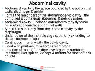
- 1. Abdominal cavity is the space bounded by the abdominal walls, diaphragm & pelvis Forms the major part of the abdominopelvic cavity—the combined & continuous abdominal & pelvic cavities Abdominal cavity - Enclosed anterolaterally by dynamic musculo-aponeurotic abdominal walls Separated superiorly from the thoracic cavity by the diaphragm Under cover of the thoracic cage superiorly extending to the 4th intercostal space Continuous inferiorly with the pelvic cavity Lined with peritoneum, a serous membrane Location of most of the digestive organs – stomach, intestines, liver, spleen, kidneys & ureters for most of their course Abdominal cavity
- 2. 2 types Abdominal cavity proper- Pelvic cavity – Abdominal cavity proper is divided into 9 regions Transtubercular line: from one iliac tubercle to the other iliac crest & at the upper border of L5 Transpyloric plane: lower border of L1 Midclavicular plane: midpoint of the clavicle to the mid- inguinal point Above the diaphragm – thoracic cavity Below the diaphragm – abdominal cavity Abdominal cavity..cont’d
- 3. 1. 2. 3. 4. 5. 6. 7. 8. 9. Epigastric region Umbilical region Hypogastric ‘’ Hypochondrial (right) ‘’ Hypochondrial (left) ‘’ Lumbar ‘’ (right) Lumbar ‘’ (left) Iliac ‘’ (right) Iliac ‘’ (left) Abdominal regions
- 4. Abdominal cavity…cont’d In the abdominal cavity there is viscera, peritoneum, vessels & lymphatics Viscera – contents (organs) of the abdominal cavity Peritoneum – serous membrane (peritoneal cavity) -Lines -Covers -Reflected Abdominal aorta, Inferior vena cava (IVC)
- 5. Peritoneum is a glistening, transparent serous membrane that consists of two continuous layers Parietal peritoneum, lining the internal surface of the abdominopelvic wall Lines - Parietal peritoneum Covers – Visceral peritoneum Reflected – Folds of peritoneum Parietal peritoneum devs from somatopleuric mesoderm, thus lines the parietis (muscles of the AAW & posterior abdominal wall, PAW) – pain sensitive Visceral peritoneum devs from splanchnopleuric layer of mesoderm - pain insensitive Peritoneal cavity – visceral & peritoneal layers Peritoneum & peritoneal cavity
- 6. Visceral peritoneum - invest viscera such as the spleen & stomach Peritoneum & viscera are in the abdominal cavity Relationship of the viscera to the peritoneum: Intraperitoneal organs are almost completely covered with visceral peritoneum (e.g., the spleen & stomach) Intraperitoneal organs have conceptually, if not literally, invaginated into a closed sac, like pressing your fist into an inflated balloon Extraperitoneal & retroperitoneal organs are outside the peritoneal cavity—external or posterior to the parietal peritoneum & are only partially covered with peritoneum Organs such as the kidneys are between the parietal peritoneum & PAW & have parietal peritoneum only on their anterior surfaces, often with amount of intervening fatty tissue Peritoneum & peritoneal cavity
- 8. Peritoneal cavity Parietal layer Visceral layer Peritoneal fluid - moist Connective tissue Squamous epithelium It is a potential space within the abdominal cavity & continues with the pelvic cavity – Male (closed) female (communication through UT,V) Dev from intra-embryonic caelom Squamous epithelium is mesodermal – mesothelium Property to secrete fluid – peritoneal fluid Visceral layer of peritoneum covered the uncovered part of the viscera No organs in the peritoneal cavity
- 9. Parietal peritoneum is: Served by the same blood & lymphatic vasculature & the same somatic nerve supply as the region of the abdominopelvic wall it lines - sensitive to pressure, pain, heat, and cold; pain from the parietal peritoneum is generally well localized Visceral peritoneum is: supplied by the same blood & lymphatic vessels & the same visceral nerve supply as the organs it covers -Insensitive to touch, heat, cold & laceration & is stimulated primarily by stretching & chemical irritation. Pain from the visceral peritoneum is poorly localized & is referred to the dermatomes of the spinal ganglia providing the sensory fibers Pain from the foregut derivatives is usually experienced in the epigastric region; that from the midgut derivatives, in the umbilical region & that from the hindgut derivatives, in the pubic region Peritoneal Vessels & Nerves
- 10. 1. Omentum is a double-layered of peritoneum passing from the stomach, liver & pancreas 2. Mesentery is a double layer of peritoneum - invagination of the peritoneum by an organ & constitutes a continuity of the visceral & parietal peritoneum (e.g. small intestine & transverse mesocolon) Mesenteries provide a means for neurovascular communication between the organ & the body wall. Thus, have a core of connective tissue containing blood & lymphatic vessels, nerves, fat & lymph nodes Viscera with a mesentery are mobile; the degree of mobility depends on the length of the mesentery 3. Ligament consists of a double layer of peritoneum that connects an organ with another organ or to the abdominal wall. For example, the liver is connected to the AAW by the falciform ligament Folds of peritoneum
- 11. Greater omentum extends superiorly, laterally to the left & inferiorly from the greater curvature of the stomach to the pancreas Gastrophrenic ligament - greater curvature of the stomach & the diaphragm Gastrosplenic ligament - greater curvature of the stomach & the spleen Gastrocolic ligament - inferior portion of the greater curvature of the stomach to the transverse colon -Gastrocolic ligament is the largest part, descending anteriorly & inferiorly beyond the transverse colon & then ascending again posteriorly, fusing with the visceral peritoneum of the transverse colon & the superior layer of its mesentery The descending & ascending portions of the gastrocolic part of the greater omentum usually fuse together, forming a four-layered fatty “omental apron” Other peritoneal formations
- 13. Abdominal Aorta – Artery of the gut
- 14. a. b. i. ii. iii. iv. i. ii. Origin: Continuation of descending, thoracic aorta, pierces the diaphragm at the aortic opening – T12 Above the aortic opening there is medial arcuate ligament Course: Descends downward in front of the vert column Relations: Anterior Posterior Anterior relations – Body of pancreas 3rd part of the duodenum Root of mesentery Splenic & left renal veins Posterior relations – Lumbar vert (body of T12-L4) Left lumbar veins passes post to the aorta Termination: at the lower border of L4 vert into common Iliac arteries Abdominal Aorta
- 15. 1. 2. Visceral Parietal Visceral Abdominal Aorta - Branches Visceral (Unpaired) Foregut – Celiac artery..T12 Midgut –Superior Mesenteric Artery.. L1 Hindgut – Inferior Mesenteric Artery.. L3 Visceral (Paired) Right & Left renal arteries – L1 Right & Left Middle Sacral Arteries – T12 Right & Left Testicular or Ovarian Arteries – L2 Parietal (Unpaired) Median Sacral Artery Parietal (Paired) Right & Left Phrenic Arteries 1st – 4th Lumbar Arteries
- 17. From foregut – stomach & proximal part of the duodenum, stomach & pancreas At the side of the foregut – spleen but not from foregut ONLY at the side At the junction of the foregut – liver & pancreas Thus, celiac artery gives off 3 branches to supply these structures 1. Left gastric artery 2. Splenic artery 3. Hepatic artery Foregut dev structures – 3 sides/places
- 18. Derivatives of the midgut – distal part of duodenum, jejunum, ileum, cecum, appendix, ascending colon & proximal 1/3 of transverse colon Thus, superior mesenteric artery gives off 6 branches to supply these structures 1. Inferior pacreatico-duodenal artery 2. Jejunal artery 3. Ileal artery 4. Ileocolic artery 5. Right colic artery 6. Middle colic artery Midgut
- 19. Derivatives of the hindgut – Proximal 2/3 of transverse colon, descending colon, sigmoid colon, rectum & the upper portion of the anal canal Therefore, inferior mesenteric artery (IMA) gives off 3 branches to supply these structures 1. Left colic artery 2. Sigmoid artery 3. Superior rectal artery The hindgut continues as the rectum & thus the IMA becomes the superior rectal artery Hindgut
- 20. Peritoneal cavity is divided into a greater sac & an omental bursa (lesser sac) Greater sac is the main & larger part of the peritoneal cavity. A surgical incision through the AAW enters the greater sac. The omental bursa (lesser sac), the smaller part of the peritoneal cavity, lies posterior to the stomach, lesser omentum & adjacent structures Omental bursa permits free movement of the stomach on adjacent structures because the anterior & posterior walls of the omental bursa slide smoothly over each other Subdivisions of Peritoneal Cavity
- 22. The omental bursa has 2 recesses: 1. Superior recess - limited superiorly by the diaphragm & the posterior layers of the coronary ligament of the liver 2. Inferior recess - superior part of the layers of the greater omentum. Most of the inferior recess of the omental bursa is a potential space sealed off from the main part of the omental bursa posterior to the stomach after adhesion of the anterior & posterior layers of the greater omentum Lesser sac communicates with the greater sac through the omental foramen (epiploic foramen of Winslow), an opening situated posterior to the free edge of the lesser omentum forming the hepatoduodenal ligament Boundaries of the omental foramen Anteriorly—the hepatoduodenal ligament (free edge of lesser omentum) containing the portal vein, hepatic artery & bile duct Posteriorly—IVC & right crus of diaphragm, covered with parietal peritoneum (retroperitoneal) Superiorly—the liver, covered with visceral peritoneum Inferiorly—superior or first part of the duodenum Omental bursa (lesser sac)
- 23. Peritoneum & Surgical Procedures Because the peritoneum is well innervated, patients undergoing abdominal surgery experience more pain with large, invasive, open incisions of the peritoneum (laparotomy) than they do with small laparoscopic incisions or vaginal operations Peritonitis & Ascites: Bacterial contamination occurs during laparotomy or when the gut is traumatically penetrated or ruptured due to infection & inflammation (e.g., appendicitis), allowing gas, faecal matter & bacteria to enter the peritoneal cavity, the result is infection & inflammation of the peritoneum—peritonitis Peritoneal Adhesions & Adhesiotomy: If the peritoneum is damaged, by a stab wound for example, or infected, the peritoneal surfaces become inflamed, making them sticky with fibrin. As healing occurs, the fibrin may be replaced with fibrous tissue, forming abnormal attachments between the visceral peritoneum of adjacent viscera, or between the visceral peritoneum of a viscus and the parietal peritoneum of the adjacent abdominal wall. Adhesions (scar tissue) may also form after an abdominal operation (e.g., owing to a ruptured appendix) & limit the normal movements of the viscera Abdominal Paracentesis: Treatment of generalized peritonitis includes removal of the ascitic fluid and, in the presence of infection, administration of large doses of antibiotics. Surgical puncture of the peritoneal cavity for the aspiration or drainage of fluid is called paracentesis. After injection of a local anesthetic agent, a needle or trocar and a cannula are inserted through the anterolateral abdominal wall into Applied Anatomy of the peritoneal cavity
- 25. Food blender & reservoir; its chief function is enzymatic digestion. The gastric juice gradually converts a mass of food into a semiliquid mixture, chyme (G. juice) PARTS: The shape of the stomach is dynamic (changing in shape as it functions) & highly variable from person to person. The stomach has 3 or 4 parts Cardiac part - surrounding the cardial orifice, the trumpet-shaped opening of the esophagus into the stomach Fundus - superior part of the stomach that is related to the left dome of the diaphragm The superior part of the fundus usually reaches the level of the left 5th intercostal space. The cardial notch is between the esophagus & the fundus The fundus may be dilated by gas, fluid, food, or any combination of these Body, the major part of the stomach, lies between the fundus & the pyloric antrum. The pyloric part of the stomach is the funnel-shaped region; its wide part, the pyloric antrum, leads into the pyloric canal, its narrow part Pylorus, the distal sphincteric region, is a thickening of the circular layer of smooth muscle, which controls discharge of the stomach contents through the pyloric orifice into the duodenum The stomach
- 27. Rules of 2’s • Cardiac & Pyloric 2 ENDS • Antero superior & Postero inferior 2 SURFACES • Cardiac & Pyloric 2 ORIFICES • Cardiac & Angularis 2 INCISURA • Greater & Lesser 2 CURVATURES • Greater & Lesser 2 OMENTUMS • Cardio-oesophageal & Pyloric 2 SPHINCTERS • Anterior & Posterior Vagal Nerves 2 NERVES • R & L Gastric & Gasto-epiploic A 2 SETS OF ARTERIES • Gastric & Pyloric 2 CANALS • Approximate volume of stomach 2 PINTS
- 28. i. ii. The lesser curvature forms the shorter concave border of the stomach; the angular incisure (notch) is the sharp indentation approximately 2/3 of the distance along the lesser curvature that approximates the junction of the body& pyloric part of the stomach The greater curvature forms the longer convex border of the stomach Pyloric part is divided into 2 Pyloric antrum Pyloric canal
- 29. 1. 2. 3. 4. 5. i. ii. iii. i. ii. iii. Mucus membrane Submucosal layer Muscular layer Serous layer Cavity of the stomach Mucus membrane is made up of 3 layers Inner side is epithelial layer – lines, cover & produce glands Lamina propria Muscularis mucosa – secretion of glands Muscular layer Oblique layer Inner circular Outer circular These are layers are the same in the whole alimentary canal – stomach & intestine Layers of the stomach (Inner to outside)
- 30. 1. 2. 3. 4. 5. The stomach is a derivative of the foregut. Thus, it supplied by the celiac artery directly or indirectly Anastomoses -Lesser curvature by the right & left gastric arteries -Greater curvature, by the right & left gastro-omental artery (gastro-epiploic artery) -Fundus & upper part of stomach receive blood from the short & posterior gastric arteries, branches of the splenic artery Left gastric artery - celiac artery (direct) Right gastric artery – branch of hepatic artery Short gastric artery – branch of splenic artery Left gastroepiploic artery – branch splenic artery Right gastroepiploic artery – branch of gastroduodenal & branch of hepatic artery Arterial supply
- 32. 1. 2. 3. 4. 5. Gastric veins - parallel the arteries in position & course Left gastric vein – portal vein directly Right gastric vein – portal vein directly Short gastric vein – splenic vein Left gastroepiploic vein – splenic vein Right gastroepiploic vein – superior mesenteric vein or portal vein Venous drainage
- 33. Gastric lymphatic vessels accompany arteries - greater & lesser curvatures & drain lymph from its anterior & posterior surfaces toward its curvatures, where the gastric & gastro-omental lymph nodes are located Efferent vessels from these nodes via the pancreaticosplenic, pyloric & pancreaticoduodenal lymph nodes accompany the large arteries to the celiac lymph nodes Sympathetic nerve supply is from the T1-L1 or L2, which passes to the celiac plexus via the greater splanchnic nerves & is distributed as plexuses around the gastric & gastro-omental arteries Parasympathetic nerve supply is from the anterior vagal trunk (left vagus nerve) & posterior vagal trunk (right vagus nerve) - enter the abdomen through the esophageal hiatus Lymphatic drainage & nerve supply
- 35. a. b. c. d. 1. Stomach is covered by peritoneum, except where blood vessels run along its curvatures & in a small area posterior to the cardial orifice 2. Folds of peritoneum attached to the stomach Lesser omentum Greater omentum Gastrosplenic ligament Gastrophrenic ligament2 layers of the lesser omentum separate to extend around the stomach & come together again to leave its greater curvature as the greater omentum 3. Lesser sac – behind the stomach (special relation) Relations Stomach
- 36. Anteriorly - diaphragm, the left lobe of the liver & the AAW Posteriorly - omental bursa & pancreas; the posterior surface of the stomach forms most of the anterior wall of the lesser sac Spleen separated by the greater sac Stomach bed on which the stomach rests when a person is in the supine position is formed by the structures forming the posterior wall of the omental bursa Relations Stomach – other relations
- 37. i. ii. i. ii. i. ii. iii. iv. v. Introduction: Is a fold of peritoneum connecting the stomach with the liver Description: made up of 2 layers Anterior layer Posterior layer It has 2 margins Free margin – on the right side Attached margin In the free margin the following structures are present Bile duct on the right Hepatic artery on the left Portal vein posterior to both Left & right gastric vessels (artery, veins & lymphatics) Plexus of nerves & lymphatic vessels Lesser Omentum
- 38. i. ii. iii. Attached margin Below to the lesser curvature of the stomach & first 2cm of the duodenum Also attached to the lips of portahepatis of the liver In the floor of fissure for ligamentum venosum Lesser Omentum…Cont’d
- 39. Is a fold of peritoneum – stomach & the pancreas Description: Has 4 layers, layers 1 & 2 are known as anterior layer & layers 3 & 4 are called posterior layer Layers 1 & 2 are attached to the greater curvature of the stomach & from their they descend downward Layers 3 & 4 are going upwards & posteriorly to be attached at the ant border of the pancreas while going upwards its loosely attached to the mesocolon Greater Omentum
- 40. Right & left gastroepiploic vessels (artery & vein) The vessels are supplying the stomach as well as greater omentum Greater Omentum…..Contents i. ii. Greater & Lesser Sacs The peritoneal cavity is made up of Greater Sac Lesser Sac Greater Sac = peritoneal cavity – Lesser Sac
- 41. i. ii. iii. i. ii. i. ii. iii. Introduction: Is a part of the peritoneal cavity which devs separately as the lesser sac. The Lesser Sa is connected to the Greater Sac through the Epiploic foramen (of Winslow) Description: It has ant & post layer or surfaces; has upper, right & left borders Boundaries: Anteriorly: 2nd layer of lesser omentum Peritoneum behind the stomach 2nd layer of greater omentum Posteriorly 3rd layer of greater omentum Its also formed by the peritoneum covering the PAW Lesser Sac is 2 things & in between thing Lesser omentum Stomach In between greater omentum Lesser Sac
- 42. i. ii. iii. i. ii. Right border Leino-Renal ligament Gastrosplenic ligament Gastrophrenic ligament Left border Greater omentum Above the stomach Lesser Sac…Cont’d
- 43. i. ii. i. ii. iii. iv. Introduction: Foramen connecting the lesser sac with the greater sac especially with Monsun’s pouch (Hepato-Renal pouch), part of the greater sac Boundaries: Anteriorly it is bounded by Free margin of the lesser omentum in which there is bile duct on the right, hepatic artery on the left & portal vein on posterior 2/3 Posteriorly Inferior vena cava Right crus of the diaphragm Upper border Caudate process of the liver & caudate lobe covered by visceral peritoneum Lower border 1st part of the duodenum Portal vein Hepatic artery Bile duct Foramen of Winslow
- 44. 1. 2. 3. 4. i. ii. iii. iv. v. vi. vii. There may be infection due to perforated peptic ulcer lesser sac pus – pus is drained b cutting the greater omentum Morrison’s pouch takes pus due to olicysticis (inflammation of the bladder) due to appendicitis, due to distention of the abdominal cavity Internal hernia Posterior lesion of the stomach on the posterior surface of the stomach bed (stomach bed is made up of structures on the post surface of the stomach separated by lesser sac) Stomach bed is formed by (superior to inferior) Splenic artery Left crus/dome of the diaphragm Left Kidney Left suprarenal gland Neck & body of pancreas Left colic flexure Transverse colon (transverse mesocolon) Applied Anatomy
- 45. Duodenum, jejunum & ileum, extends from the pylorus of the stomach to the ileocecal junction, where the ileum joins the cecum, the first part of the large intestine DUODENUM: 1st & shortest (25 cm) part of the small intestine, is also the widest & most fixed part Begins at the pylorus on the right side & ends at the duodenojejunal junction on the left side The small intestine
- 46. 1. 2. 3. 4. Superior (1st) part: short (5 cm), mostly horizontal & lies anterolateral to the body of L1 vertebra Descending (2nd) part: longer (7-10 cm) and runs inferiorly along the right sides of the L2 and L3 vertebrae, curving around the head of the pancreas; initially it lies to the right and parallel to the IVC. The bile duct and main pancreatic ducts via the hepatopancreatic ampulla enter its posteromedial wall Inferior (horizontal/3rd) part: 6 to 8 cm long and crosses anterior to the IVC & aorta & posterior to the superior mesenteric artery (SMA) & superior mesenteric vein (SMV) at the level of the L3 vertebra Ascending (4th) part: short (5 cm) & begins at the left of the L3 vertebra & rises superiorly as far as the superior border of the L2 vertebra, 2 to 3 cm to the left of the midline. It passes on the left side of the aorta to reach the inferior border of the body of the pancreas. Here it curves anteriorly to join the jejunum at the duodenojejunal junction, which takes the form of an acute angle, the duodenojejunal flexure . The flexure is supported by the attachment of the suspensory muscle of the duodenum (ligament of Treitz) 4 parts of the Duodenum
- 47. 4 parts of the duodenum
- 48. The suspensory muscle of the duodenum is commonly composed of a slip of skeletal muscle from the diaphragm & a fibromuscular band of smooth muscle from the 3rd & 4th parts of the duodenum Contraction of this suspensory muscle widens the angle of the duodenojejunal flexure, facilitating movement of the intestinal contents The suspensory muscle passes posterior to the pancreas and splenic vein and anterior to the left renal vein. The 1st 2 cm of the superior part of the duodenum has a mesentery & is mobile. This free part—relatively dilated and smooth-walled—is called the ampulla or duodenal cap The distal 3 cm of the superior part & the other three parts of the duodenum have no mesentery and are immobile because they are retroperitoneal
- 49. Arterial supply - 2 different vessels An important transition in the blood supply of the alimentary tract occurs over the course of the descending (2nd) part of the duodenum, approximately where the bile duct enters. The basis of this transition is embryological - junction of the foregut & midgut. Consequently, the duodenal arteries arise from 2 different sources Proximally - celiac trunk & the 1st & 2nd parts of the duodenum are supplied via the gastroduodenal artery & its branch, the superior pancreaticoduodenal artery Distally - extending as far as the left colic flexure) is supplied by the SMA & the 3rd & 4th parts of the duodenum are supplied by its branch, the inferior pancreaticoduodenal artery The superior & inferior pancreaticoduodenal arteries form an anastomotic loop between the celiac trunk & the SMA; consequently, there is potential for collateral circulation here Venous drainage Follow the arteries & drain into the hepatic portal vein - directly & others indirectly through the superior mesenteric & splenic veins Lymphatic vessels, which follow the arteries in a retrograde direction The anterior lymphatic vessels drain into the pancreaticoduodenal lymph nodes located along the superior and inferior pancreaticoduodenal arteries, and into the pyloric lymph nodes, which lie along the gastroduodenal artery The posterior lymphatic vessels pass posterior to the head of the pancreas and drain into the superior mesenteric lymph nodes. Efferent lymphatic vessels from the duodenal lymph nodes drain into the celiac lymph nodes Nerve supply Parasympathetic innervation from the vagus & sympathetic innervation from the greater & lesser splanchnic nerves by way of the celiac & superior mesenteric plexuses & then via periarterial plexuses extending to the pancreaticoduodenal arteries Vasculature, lymphatics & nerve supply
- 51. The jejunum begins at the duodenojejunal junction & the ileum ends at the ileocecal junction, the union of the terminal ileum and cecum Jejunum & ileum are 6 to 7 m long in cadavers; however, tonic contraction makes them substantially shorter in living persons Jejunum about 2/5 of the length & the ileum the remainder The terminal ileum usually lies in the pelvis, from which it ascends to end in the medial aspect of the cecum. Although no clear line of demarcation between the jejunum and ileum exists, they have distinctive characteristics for most of their lengths The mesentery, a fan-shaped fold of peritoneum, attaches the jejunum & ileum to the PAW. The root of the mesentery (15 cm long) is directed obliquely, inferiorly & to the right It extends from the duodenojejunal junction on the left side of the L2 vertebra to the ileocolic junction & the right sacro-iliac joint. The root of the mesentery crosses (successively) the ascending & horizontal parts of the duodenum, abdominal aorta, inferior vena cava, right ureter, right psoas major muscle & right testicular or ovarian vessels Arterial Supply Arterial supply from the SMA The SMA runs between the layers of the mesentery and sends many branches to the jejunum and ileum. The arteries unite to form loops or arches—arterial arcades—that give rise to straight arteries—the vasa recta Venous drainage The SMV lies anterior and to the right of the SMA in the root of the mesentery. The SMV ends posterior to the neck of the pancreas, where it unites with the splenic vein to form the hepatic portal vein. Specialized lymphatic vessels, called lacteals, in the intestinal villi that absorb fat and drain into the lymphatic plexuses in the walls of the jejunum and ileum. The lymphatic plexuses drain into lymphatic vessels between the layers of the mesentery and then sequentially through three groups of lymph nodes juxta -intestinal lymph nodes (close to the intestinal wall), mesenteric lymph nodes (scattered among the arterial arcades) & central superior nodes (along the proximal part of the SMA) Efferent lymphatic vessels from these nodes drain into the superior mesenteric lymph nodes. Lymphatic vessels from the terminal ileum follow the ileal branch of the ileocolic artery to the ileocolic lymph nodes Jejunum & Ileum
- 54. Sympathetic & parasympathetic innervation In general, sympathetic stimulation reduces secretion & motility of the intestine & acts as a vasoconstrictor, reducing or stopping digestion & making blood (& energy) available for “fleeing or fighting.” Parasympathetic stimulation increases secretion and motility of the intestine, restoring digestive activity after a sympathetic reaction. The SMA & its branches are surrounded by a dense perivascular nerve plexus through which the nerve fibers are conducted to the parts of the intestine supplied by the SMA. The sympathetic fibers originate in the T7-T12 segments of the spinal cord & reach the superior mesenteric nerve plexus through the sympathetic trunks & thoracic abdominopelvic (greater, lesser & least) splanchnic nerves The small intestine also has sensory (visceral afferent) fibers The intestine is insensitive to most pain stimuli, including cutting and burning; however, it is sensitive to sudden distention (“gas pains”) and transient ischemia from abnormally long contractions that are perceived as colic (spasmodic abdominal pains) Nerve supply
- 55. Jejunum Ileum Deeper red Paler pink Normally is in the upper left part of the abdominal cavity Normally is in the lower right part of the abdominal cavity, it may be in the pelvis Jejunum is practically empty i.e. there is no food Ileum is practically full due to presence of food Long few branches of duodenal artery (Vasa recta) Many short branches of Iliac arteries (Vasa recta) Windows between jejunal arteries No windows Between the branches of arteries there is no fat Presence of too much fats between the arterial branches A few large loops Arcades Many short loops Arcades Differences between Jejunum & Ileum
- 57. i. ii. iii. Caecum Appendix Ascending colon (right colon) Transverse colon Descending colon (left colon) Sigmoid (pelvic) colon Rectum & anal canal Development Duodenum, 1st part or above the opening of the bile duct devs from the foregut below the opening of the bile duct devs from midgut Jejunum, Ileum, caecum, appendix, A. colon, right 2/3 of transverse colon all dev from the midgut The left 1/3 of the transverse colon, d. colon, sigmoid colon, rectum & anal canal above the pectineal line devs from the hindgut. Below the pectineal line devs from the ectoderm Large intestine
- 58. Differences between large & small intestines Large Intestine Small Intestine Taenia coli (three thickened bands of longitudinal smooth muscle fibers) No taenia coli Sacculations (Haustra) - pouches of the colon between the taeniae No Sacculations Appendices epiploic - small, fatty appendices (projections) of colon No appendices epiploic No villi & microvilli Presence of villi & microvilii
- 59. Large intestine The three taeniae coli make up most of the longitudinal muscle of the large intestine, except in the rectum Because the taeniae are shorter than the large intestine, the colon has the typical sacculated shape formed by the haustra The taeniae begin at the base of the appendix and run the length of the large intestine, merging at the rectosigmoid junction into a continuous layer around the rectum
- 60. The caecum, the first part of the large intestine, which is continuous with the A. colon, is a blind intestinal pouch in the right lower quadrant, where it lies in the iliac fossa inferior to the junction of the terminal ileum & caecum The cecum is usually almost entirely enveloped by peritoneum & can be lifted freely; however, the caecum has no mesentery The ileum enters the caecum obliquely & partly invaginates into it, forming folds superior & inferior to the ileal orifice. These folds form the ileocecal valve Caecum
- 61. Caecum is supplied by the ileocolic artery, the terminal branch of the SMA. The appendix is supplied by the appendicular artery, a branch of the ileocolic artery A tributary of the SMV, the ileocolic vein, drains blood from the caecum & appendix The lymphatic vessels from the cecum & appendix pass to lymph nodes in the mesoappendix and to the ileocolic lymph nodes that lie along the ileocolic artery Efferent lymphatic vessels pass to the superior mesenteric lymph nodes. The nerve supply to the cecum and appendix derives from sympathetic and parasympathetic nerves from the superior mesenteric plexus The sympathetic nerve fibers originate in the lower thoracic part of the spinal cord (T10-T12) & the parasympathetic nerve fibers derive from the vagus nerves. Afferent nerve fibers from the appendix accompany the sympathetic nerves to the T10 segment of the spinal cord Vasculature & nerve supply of Caecum & Appendix
- 62. The vermiform (L. worm-like) appendix, a blind intestinal diverticulum, extends 2cm behind the ileocecal junction (inferior to the caecum) Length varies & has a short triangular mesentery (mesoappendix) - derives from the posterior side of the mesentery of the terminal ileum Mesoappendix attaches to the caecum & the proximal part of the appendix Position of the appendix is variable, but it is usually retrocecal (behind the caecum) Base of the appendix most often lies deep to a point that is 1/3 of the way along the oblique line joining the right ASIS to the umbilicus - McBurney point Appendix
- 64. i. ii. iii. iv. v. i. ii. Retrocaecal (behind the caecum)- – 65% Pelvic appendix (in pelvis) Pre-Ileal (in front of the ileum) Post-ileal (behind ileum) Subcaecal (below the caecum) Arterial supply: appendicular artery – ileocolic artery Venous drainage: appendicular veins – ileocolic veins – SMV Lymphatic drainage: ileocolic lymph nodes Nerve supply: Appendicular plexus of nerves which is sympathetic (T10) & parasympathetic nerve fibres Peritoneal Relations Covered by peritoneum Attached to the PAW by its mesentery (mesoappendix). This mesentery is triangular in shape & contain appendicular vessels & lymph nodes Positions
- 66. i. ii. iii. i. ii. Appendicitis Symptoms Pain Fever – high Vomiting The pain is of 2 types In appendicitis, the pain is also referred to the umbilicus because umbilicus is supplied by the same nerve that supplies the appendix Referred pain: if there is an infection of viscera, the pain is referred to the skin supplied by the same segment of the spinal cord Real pain (visceral pain): There is a point known as McBurney’s point. If this point is palpated by finger, it gives the maximum tenderness (pain on touching) McBurney’s point which is right 2/3 & left 1/3 on a line drawn between the umbilicus & the ASIS Applied Anatomy
- 67. Four parts—ascending, transverse, descending & sigmoid—that succeed one another in an arch The ascending colon passes superiorly on the right side of the abdominal cavity from the cecum to the right lobe of the liver, where it turns to the left as the right colic flexure (hepatic flexure) The A. colon, narrower than the cecum, lies retroperitoneally along the right side of the PAW. The A. colon is covered by peritoneum anteriorly & on its sides; in about 25% of people, it has a short mesentery. The A. colon is separated from the AAW by the greater omentum. A vertical groove lined with parietal peritoneum (the right paracolic gutter) lies lateral to the ascending colon The arterial supply to the ascending colon & right colic flexure is from branches of the SMA—the ileocolic and right colic arteries Tributaries of the SMV, the ileocolic & right colic veins, drain blood from the ascending colon The lymphatic vessels first pass to the epicolic & paracolic lymph nodes, next to the ileocolic and intermediate right colic lymph nodes & from them to the superior mesenteric nodes The nerves to the A. colon derive from the superior mesenteric plexus Colon
- 68. Largest & most mobile part of the large intestine, crosses the abdomen from the right colic flexure to the left colic flexure, where it bends inferiorly to become the descending colon The left colic flexure (splenic flexure)—usually more superior, more acute & less mobile than the right colic flexure—lies anterior to the inferior part of the left kidney & attaches to the diaphragm through the phrenicocolic ligament The mesentery of the transverse colon, the transverse mesocolon, loops down, often inferior to the level of the iliac crests, and is adherent to the posterior wall of the omental bursa The root of the transverse mesocolon lies along the inferior border of the pancreas & is continuous with the parietal peritoneum posteriorly Arterial supply of the transverse colon is mainly from the middle colic a branch of the SMA; may also be supplied to variable degrees by the right & left colic arteries via anastomoses Venous drainage of the transverse colon is through the SMV. Lymphatic drainage is to the middle colic lymph nodes, which in turn drain to the superior mesenteric lymph nodes Nerves of the transverse colon arise from the superior mesenteric plexus & follow the right & middle colic arteries nerves transmit sympathetic and parasympathetic (vagal) nerve fibers. Some nerves derived from the inferior mesenteric plexus may follow anastomoses from the left colic artery Transverse colon
- 69. Passes retroperitoneally from the left colic flexure into the left iliac fossa, where it is continuous with the sigmoid colon. Covered with peritoneum anteriorly & laterally & binds it to the PAW. Retroperitoneal, especially in the iliac fossa, has a short mesentery in about 33% of people. As it descends, the colon passes anterior to the lateral border of the left kidney. As with the A. colon, a left paracolic gutter lies on the lateral side of the descending colon The sigmoid colon, characterized by its S-shaped loop of variable length, links the descending colon & the rectum The sigmoid colon extends from the iliac fossa to the 3rd sacral segment, where it joins the rectum. The termination of the taeniae coli indicates the rectosigmoid junction. Has a long mesentery (sigmoid mesocolon). The root of the sigmoid mesocolon has an inverted V-shaped attachment, extending first medially & superiorly along the external iliac vessels & then medially & inferiorly from the bifurcation of the common iliac vessels to the anterior aspect of the sacrum The left ureter & the division of the left common iliac artery lie retroperitoneally posterior to the apex of the root of the sigmoid mesocolon nd Descending & the Sigmoid colon
- 70. Arterial supply of the descending & sigmoid colon - left colic & sigmoid arteries (IMA) The left colic & sigmoid arteries pass to the left, where they divide into ascending & descending branches. Usually all or most of the branches of the arteries supplying blood to the colon (ileocolic; right, middle, & left colic; & sigmoid arteries) anastomose with each other as they approach the colon, thus forming a continuous anastomotic channel, the marginal artery - important collateral circulation IMV returns blood from the descending & sigmoid colon, flowing usually into the splenic vein & then the hepatic portal vein The lymphatic vessels from the descending & sigmoid colon pass to the epicolic & paracolic lymph nodes & then through the intermediate colic lymph nodes along the left colic artery Lymph from these nodes passes to inferior mesenteric lymph nodes that lie around the IMA; Lymph from the left Vasculature of the descending & sigmoid colon
- 71. Sympathetic nerve supply of the descending & sigmoid colon - lumbar part of the sympathetic trunk via lumbar (abdominopelvic) splanchnic nerves, the inferior mesenteric ganglion & the periarterial plexuses on the IMA Parasympathetic nerve supply is from the pelvic splanchnic nerves via the inferior hypogastric (pelvic) plexus & nerves, which ascend retroperitoneally from the plexus, independent of the arterial supply Proximal to the middle of the sigmoid colon the visceral afferents conveying pain pass retrogradely with sympathetic fibers to thoracolumbar spinal sensory ganglia, whereas those carrying reflex information travel with the parasympathetic fibers to vagal sensory ganglia Nerve supply to the descending & sigmoid colon
- 72. The rectum, the fixed terminal part of the large intestine, is continuous with the sigmoid colon at the level of the S3 vertebra. The junction is at the lower end of the mesentery of the sigmoid colon The rectum is continuous inferiorly with the anal canal Rectum & Anal Canal
- 74. Hiatal Hernia Is a protrusion of part of the stomach into the mediastinum through the esophageal hiatus of the diaphragm Occur most often in people after middle age, due to the weakening of the muscular part of the diaphragm & widening of the esophageal hiatus Although clinically there are several types of hiatal hernias, the two main types are paraesophageal hiatal hernia & sliding hiatal hernia. Some regurgitation of stomach contents into the esophagus is possible because the clamping action of the right crus of the diaphragm on the inferior end of the esophagus is weak. Applied Anatomy
- 75. When the body or pyloric part of the stomach contains a malignant tumor, the mass may be palpable. Using gastroscopy, physicians can inspect the lining of the air-inflated stomach, enabling them to observe gastric lesions & take biopsies Partial gastrectomy may be performed to remove the region of the stomach involved by carcinoma Because of the anastomoses of the arteries supplying the stomach provide good collateral circulation, one or more arteries may be ligated during this procedure without seriously affecting the blood supply of the remaining part of the stomach Partial gastrectomy to remove a carcinoma usually also requires removal of all involved regional lymph nodes. Because cancer frequently occurs in the pyloric region, removal of the pyloric lymph nodes as well as the right gastro-omental lymph nodes also receiving lymph drainage from this region is especially important. As stomach cancer becomes more advanced, the lymphogenous dissemination of malignant cells involves the celiac lymph nodes, to which all gastric nodes drain. Carcinoma of Stomach & Gastrectomy
- 76. Gastric ulcers are open lesions of the mucosa of the stomach, whereas peptic ulcers are lesions of the mucosa of the pyloric canal or, more often, the duodenum. Most ulcers of the stomach & duodenum are associated with an infection of a specific bacterium Helicobacter pylori (H. pylori). People experiencing severe chronic anxiety are most prone to the development of peptic ulcers They often have gastric acid secretion rates that are markedly higher than normal between meals. It is thought that the high acid level in the stomach & duodenum overwhelms the bicarbonate normally produced by the duodenum & reduces the effectiveness of the mucous lining, leaving it vulnerable to H. pylori The bacteria erode the protective mucous lining of the stomach, inflaming the mucosa and making it vulnerable to the effects of the gastric acid & digestive enzymes (pepsin) produced by the stomach If the ulcer erodes into the gastric arteries, it can cause life-threatening bleeding. Because the secretion of acid by parietal cells of the stomach is largely controlled by the vagus nerves, vagotomy (surgical section of the vagus nerves) is performed in some people with chronic or recurring ulcers to reduce the production of acid A posterior gastric ulcer may erode through the stomach wall into the pancreas, resulting in referred pain to the back. In such cases, erosion of the splenic artery results in severe hemorrhage into the peritoneal cavity Gastric Ulcers, Peptic Ulcers, Helicobacter pylori & Vagotomy
- 77. Duodenal (Peptic) Ulcers: Inflammatory erosions of the duodenal wall, duodenal ulcers, are in the posterior wall of the superior part of the duodenum within 3 cm of the pylorus. Occasionally, an ulcer perforates the duodenal wall, permitting its contents to enter the peritoneal cavity & produce peritonitis Erosion of the gastroduodenal artery, a posterior relation of the superior part of the duodenum, by a duodenal ulcer results in severe hemorrhage into the peritoneal cavity Ileal Diverticulum: An ileal diverticulum (of Meckel) is a congenital anomaly that occurs in 1% to 2% of people A remnant of the proximal part of the embryonic omphalo-enteric duct (yolk stalk), the diverticulum usually appears as a finger-like pouch 3 to 6 cm long It is always on the border of the intestine opposite the mesenteric attachment An ileal diverticulum may become inflamed & produce pain mimicking appendicitis Diverticulosis: Is a disorder in which multiple false diverticula (external evaginations or outpocketings of the mucosa of the colon) develop along the intestine It primarily affects middle aged & elderly people. Diverticulosis is commonly found in the sigmoid colon. Diverticula are subject to infection & rupture, leading to diverticulitis Duodenal (Peptic) Ulcers, Ileal Diverticulum & Diverticulosis
- 78. Colitis, Colectomy & Ileostomy Chronic inflammation of the colon (ulcerative colitis, Crohn disease) is characterized by severe inflammation & ulceration of the colon & rectum In some cases, a colectomy is performed, during which the terminal ileum & colon as well as the rectum & anal canal are removed An ileostomy is then constructed to establish an artificial cutaneous opening between the ileum and the skin of the anterolateral abdominal wall Following a partial colectomy, a colostomy or sigmoidostomy is performed to create an artificial cutaneous opening for the terminal part of the colon Colonoscopy The interior surface of the colon can be observed & photographed in a procedure called colonoscopy or coloscopy, using a long fiberoptic endoscope (colonoscope) inserted into the colon through the anus & rectum Small instruments can be passed through the colonoscope to perform minor operative procedures, such as biopsies or removal of polyps Most tumors of the large intestine occur in the rectum; about 12% of them appear near the rectosigmoid junction The interior of the sigmoid colon is observed with a sigmoidoscope, a shorter endoscope, in a procedure called sigmoidoscopy Colitis, Colectomy & Ileostomy
- 79. Introduction: It is the largest, ovoid & mobile lymphatic organ Shape & length: usually about 12 cm long & 7 cm wide, roughly the size & shape of a clenched fist Location: Lies intraperitoneally in the left hypochondrial region (upper quadrant) Gross Anatomy – 2s 2 borders – superior (has a notch) & inferior 2 ends – anterior end (lateral end) & posterior border 2 surfaces – visceral surface (contains hilum) & diaphragmatic surface (lateral surface) Spleen
- 81. The spleen lies superficially in the left upper abdominal quadrant between the 9th -11th ribs Its convex, costal surface fits the inferior surface of the diaphragm & the curved bodies of the ribs In the supine position, the long axis of the spleen is roughly parallel to the long axis of the 10th rib The spleen is not often palpable through the AAW unless it is enlarged Surface Anatomy
- 82. Arterial supply: Splenic artery (caeliac artery) enters through the hilum & divide into 5-6 branches to supply the spleen Venous drainage: Splenic vein – portal vein Lymphatics: Pancreatico- splenic lymph nodes Nerve supply: Splenic plexuses of nerve which is a branch of caeliac plexuses which contains sympathetic & parasympathetic fibres Vasculature, lymphatic drainage & nerve supply
- 83. i. ii. a. i. ii. i. a. Peritoneal relations b. Others a. Peritoneal relations: Is entirely surrounded by peritoneum except at the hilum, where the splenic branches of the splenic artery & vein enter & leave It has 2 ligaments: Lienorenal ligament –extends from the left kidney to the hilum of the spleen From the kidney the ligament comes, the ligament then attaches to the tips of the hilum & then from the hilum it goes to the stomach Contents of the Lienorenal ligament Tail of pancreas Splenic vessels b. Gastrosplenic ligament – extends from the fundus of the stomach to the hilum of the spleen Contents of the gastrosplenic ligament Short gastric vessels Phrenico-Colic ligament – support the spleen but NOT attached to the spleen. Its from diaphragm to left Colic Flexure Relations
- 84. Relations on the visceral surface Other relations Related to Make impression Fundus of the stomach Gastric impression (above the hilum) Left Kidney Renal impression (below the hilum) Tail of pancreas Pancreatic impression (related to the hilum) Left colic Flexure (colon) Colic impression (at the end of visceral surface)
- 85. i. Its related to ribs: 9th – 11th 9th rib is near the upper border Longitudinal axis of the spleen is in the 10th rib Inferior border is in the 11th rib Longitudinal axis of the spleen is the axis coming from the anterior to the posterior ends ii. Its related to the diaphragm iii. Its related to the left lung above iv. Its related to the pleura below Relations on the diaphragmatic surface
- 86. Rupture of Spleen & Splenomegaly: Despite being well protected by the 9th -11th ribs, the spleen is the most commonly injured organ in the abdomen Severe blows on the left side may fracture one or more ribs, resulting in sharp bone fragments that can lacerate the spleen Blunt trauma to other regions of the abdomen that cause a sudden, marked increase in intra-abdominal pressure (e.g., by impalement on the steering wheel of a car) can also rupture the spleen because its capsule is thin & its parenchyma (essential substance) is soft & pulpy If ruptured, the spleen bleeds profusely. Rupture of the spleen causes severe intraperitoneal hemorrhage & shock Repair of a ruptured spleen is difficult; consequently, splenectomy (removal of the spleen) or subtotal (partial) splenectomy (removal of one or more segments of the spleen) is often performed to prevent the patient from bleeding to death Even total splenectomy usually does not produce serious side effects, especially in adults, because most of its functions are assumed by other reticuloendothelial organs (e.g., liver & bone marrow) - more susceptible to certain bacterial infections. When the spleen is diseased, resulting from, for example, granulocytic leukemia (high leukocyte & white blood cell count), it may enlarge to 10 or more times its normal size & weight (splenomegaly) Applied Anatomy
- 87. Introduction: Liver is the largest internal organ & largest most important gland of the digestive system Function: Stores glycogen & secretes bile Types of glands: The liver contains both types of glands i.e. endocrine & exocrine Weight: 1500 g Location: Below the diaphragm mainly in the right hypochondrial region. It runs through the whole length Liver
- 88. i. ii. iii. iv. v. i. ii. iii. iv. v. 5 letter word It has 5 surfaces Anterior surface Superior surface Right lateral surface Inferior surface Posterior surface The 1st three surfaces are related to the diaphragm & can be called Diaphragmatic (convex) surface The last two surfaces are related to the viscera & can be called Visceral surface (concave) It has 5 ligaments Falciform ligament – ant & sup surf to the diaphragm Superior coronary ligament – sup surf to diaphragm Inferior coronary ligament – inf surf to diaphragm Right triangular ligament – right surf to diaphragm Left triangular ligament – left surf to diaphragm Gross Anatomy
- 89. i. ii. i. ii. iii. i. ii. 2 Lobes: Anatomically, the liver has 2 lobes Right lobe – bigger Left lobe - smaller Anatomical lobes are divided by the following structures Falciform ligament present on the ant & sup surfaces Ligamentum teres present from the inferior surface Ligamentum venosum present on the posterior surface On the right lobe, there are 2 other lobes present Quadrate (4 sided) lobe – inferior surface Caudate (triangular) lobe – posterior surface In congenital error, there may be an additional lobe known as the Reidle's lobe Maximally, the liver can contain 5 lobes Gross Anatomy
- 90. Anterior & Superior Surfaces of the Liver
- 91. Visceral surface of the liver
- 92. Physiologically & functionally, the portion of the liver which is supplied by the right hepatic duct, right branch of the hepatic artery & the right branch of the portal vein is the right lobe Likewise, the portion of the liver supplied by the left branch of the hepatic artery, left branch of the hepatic duct & left branches of the portal vein is the left lobe of the liver In anatomical lobe - right lobe is bigger In physiological lobe - right & left lobes are equal Physiological lobes
- 93. i. ii. On the surface, the physiological lobe is marked by the following imaginary lines On the inferior surface, the imaginary line is passing through the gallbladder On the posterior surface, the imaginary line is passing through the inferior vena cava (IVC) Thus, on the right side of the imaginary side is the right lobe & on the left side is the left lobe This is to say that the imaginary line divide the liver into 2 equal physiological lobes Anatomically, Quadrate & Caudate lobes belongs to the right lobe Physiologically & functionally, the belong to the left lobe & both lobes are equal Physiological lobes
- 94. Physiological lobe marked by an Imaginary line passing through IVC & gallbladder
- 95. i. ii. Liver receives blood from two sources Hepatic portal vein (80%) Hepatic artery (20%) The hepatic portal vein carries poorly oxygenated blood & is bringing the product of digestion & absorption to the liver for metabolic activity (purified, detoxified, metabolized & changed) & sent to the heart The hepatic artery, a branch of the celiac trunk, carries well- oxygenated blood from the aorta & mainly supplies the connective tissues of the liver. Thus, the hepatocytes mainly receive blood from the hepatic artery Close to the porta hepatis, the hepatic artery & hepatic portal vein terminate by dividing into right & left branches, which supply the right & left livers, respectively. Within each lobe, the primary branches of the hepatic portal vein & hepatic artery are consistent enough to form vascular segments Between the segments are the right, intermediate (middle) & left hepatic veins, which drain parts of adjacent segments. The hepatic veins open into the IVC just inferior to the diaphragm & to the right atrium of the heart Arterial supply & Venous drainage
- 96. Nerve supply: Hepatic plexuses of nerve i.e. both sympathetic & parasympathetic The liver is a major lymph-producing organ; between one quarter & one half of the lymph received by the thoracic duct comes from the liver. The lymphatic vessels of the liver occur as superficial lymphatics in the subperitoneal fibrous capsule of the liver (Glisson capsule), which form its outer surface & as deep lymphatics in the connective tissue that accompany the ramifications of the portal triad & hepatic veins Thus, drains by hepatic lymph nodes but few goes to the thorax Nerve supply & Lymphatic drainage
- 97. a. b. i. ii. a. b. Peritoneal Others a. Peritoneal relations Covered by peritoneum except at the bare area on the posterior surface Folds of peritoneum Lesser omentum Ligament of the liver: 5 in number Falciform, Sup. Coronary, Inf. Coronary, Right triang. & left triang. ligaments Relations of the Liver
- 98. a. b. i. ii. iii. a. b. c. Visceral surface Inferior surface Posterior surface a. Inferior surface is related to Stomach giving gastric impression Ligamentum teres – left umbilical vein of embryonic life Porta hepatis – to the tips of the lesser omentum attaches & in side the porta hepatis there are Hepatic artery – right & left Hepatic duct – right & left Portal vein – right & left Other relations of the Liver
- 99. i. ii. iii. iv. v. iv. Fossa for gallbladder v. Quadrate lobe vi. Duodenal impression related to the duodenum vii. Renal impression related to the right kidney viii. Right colic flexure related to the right colon (A. colon) b. Posterior surface Its related to the oesophagus (oesophaeal impression) Fissure for ligamentum venosum – ductus venosum of the embryonic life Caudate lobe with caudate process Fossa for IVC Bare area of the liver not covered by peritoneum & related to the right suprarenal gland Other relations of the Liver
- 100. i. ii. iii. i. ii. i. ii. Right Lateral Surface is related to Ribs i.e. 5 ribs – from 7th – 11th Right lung Right pleura Anterior Surface is related to Diaphragm AAW Superior Surface is related to The heart on the midline & the pericardium Right & left luns
- 101. The hepatic portal vein is the main channel of the portal venous system It collects poorly oxygenated but nutrient-rich blood from the abdominal part of the alimentary tract, including the gallbladder, pancreas, and spleen, and carries it to the liver. Within the liver, its branches are distributed in a segmental pattern and end in expanded capillaries, the venous sinusoids of the liver Portal-systemic anastomoses, in which the portal venous system communicates with the systemic venous system, are in the following locations Between the esophageal veins draining into either the azygos vein (systemic system) or the left gastric vein (portal system); when dilated these are esophageal varices Between the rectal veins, the inferior & middle veins draining into the IVC (systemic system) & the superior rectal vein continuing as the IMV (portal system); when abnormally dilated these are hemorrhoids Paraumbilical veins of the anterior abdominal wall (portal system) anastomosing with superficial epigastric veins (systemic system); when dilated these veins produce caput medusae—varicose veins radiating from the umbilicus. These dilated veins were called caput medusae because of their resemblance to the serpents on the head of Medusa, a character in Greek mythology. Twigs of colic veins (portal Hepatic Portal Vein & Portal-Systemic Anastomoses
- 102. Liver Biopsy: Hepatic tissue may be obtained for diagnostic purposes by liver biopsy. The needle puncture is commonly made through the right 10th intercostal space in the MAL Rupture of Liver Although less so than the spleen, the liver is vulnerable to rupture because it is large, fixed in position & friable. Often the liver is torn by a fractured rib that perforates the diaphragm. Because of the liver's great vascularity & friability, liver lacerations often cause considerable hemorrhage & right upper quadrant pain Cirrhosis of Liver: In cirrhosis of the liver, hepatocytes are destroyed & replaced by fibrous tissue. This tissue surrounds the intrahepatic blood vessels & biliary ducts, making the liver firm and impeding circulation of blood through it. Cirrhosis, the most common of many causes of portal hypertension, frequently develops in chronic alcoholics Applied Anatomy
- 103. Hepatic Lobectomies & Segmentectomy: When it was discovered that the right & left hepatic arteries & ducts, as well as branches of the right & left hepatic portal veins, do not communicate significantly, it became possible to perform hepatic lobectomies - with minimal bleeding. If a severe injury or tumor involves one segment or adjacent segments, it may be possible to resect only the affected segment(s) segmentectomy Portal Hypertension: Obstruction of the hepatic portal vein due to cirrhosis, pressure rises in the hepatic portal vein & its tributaries, producing portal hypertension. At the sites of anastomoses between portal & systemic veins, portal hypertension produces enlarged varicose veins & blood flow from the portal to the systemic system of veins. A common method for reducing portal hypertension is to divert blood from the portal venous system to the systemic venous system by creating a communication between the portal vein & the IVC or by joining the splenic and left renal veins—a portacaval anastomosis or portosystemic shunt Applied Anatomy..Cont’d
- 104. Bile is produced continuously in the liver & stored in the gallbladder, In addition to storing bile, the gallbladder concentrates it by absorbing water & salts Bile emulsifies the fat so it can be absorbed in the distal intestine The hepatocytes secrete bile into the bile canaliculi formed between them. The canaliculi drain into the small interlobular biliary ducts & then into large collecting bile ducts of the intrahepatic portal triad, which merge to form the right & left hepatic ducts Right & left hepatic ducts drain the right & left livers (portal lobes), respectively. Shortly after leaving the porta hepatis, the right and left hepatic ducts unite to form the common hepatic duct, which is joined on the right side by the cystic duct to form the bile duct Biliary Ducts & Gallbladder
- 105. Formed in the free edge of the lesser omentum by the union of the cystic duct & common hepatic duct The bile duct descends posterior to the superior part of the duodenum & lies in a groove on the posterior surface of the head of the pancreas On the left side of the descending part of the duodenum, the bile duct comes into contact with the main pancreatic duct The two ducts run obliquely through the wall of this part of the duodenum, where they unite to form the hepatopancreatic ampulla (ampulla of Vater) The distal end of the ampulla opens into the duodenum through the major duodenal papilla The muscle around the distal end of the bile duct is thickened to form the sphincter of the bile duct. When this sphincter contracts, bile cannot enter the ampulla and/or the duodenum; hence bile backs up and passes along the cystic duct to the gallbladder for concentration & storage Bile Duct
- 107. The arteries supplying the bile duct include the Posterior superior pancreaticoduodenal artery & gastroduodenal artery, supplying the retroduodenal part of the duct Cystic artery, supplying the proximal part of the duct. Right hepatic artery, supplying the middle part of the duct Veins from the proximal part of the bile duct & the hepatic ducts generally enter the liver directly The posterior superior pancreaticoduodenal vein drains the distal part of the bile duct & empties into the hepatic portal vein or one of its tributaries Lymphatic vessels from the bile duct pass to the cystic lymph node near the neck of the gallbladder, the node of the omental foramen and the hepatic lymph nodes Efferent lymphatic vessels from the bile duct pass to the celiac lymph nodes Vasculature
- 108. Pear-shaped gallbladder (7-10 cm long) lies in the gallbladder fossa on the visceral surface of the liver Peritoneal relation: Peritoneum completely surrounds the fundus of the gallbladder & binds its body and neck to the liver Hepatic surface of the gallbladder attaches to the liver by connective tissue of the fibrous capsule of the liver Gallbladder has three parts The fundus, the wide end, projects from the inferior border of the liver & is usually located at the tip of the right ninth costal cartilage in the midclavicular line The body contacts the visceral surface of the liver, the transverse colon, and the superior part of the duodenum. The neck is narrow, tapered & directed toward the porta hepatis Neck makes an S-shaped bend & joins the cystic duct. Internally, the mucosa of the neck spirals into a spiral fold (spiral valve), which keeps the cystic duct open so that bile can easily divert into the gallbladder when the distal end of the bile duct is closed by the sphincter of the bile duct and/or the hepatopancreatic sphincter, or when bile passes to the duodenum as the gallbladder contracts The cystic duct (4 cm long) connects the neck of the gallbladder to the common hepatic duct. The cystic duct passes between the layers of the lesser omentum, usually parallel to the common hepatic duct, which it joins to form the bile duct Gallbladder
- 110. Cystic artery, which supplies the gallbladder & cystic duct, commonly - right hepatic artery in the angle between the common hepatic duct & the cystic duct Variations in the origin & course of the cystic artery are common Cystic veins draining the biliary ducts & the neck of the gallbladder may pass to the liver directly or drain through the hepatic portal vein to the liver The veins from the fundus & body pass directly into the visceral surface of the liver and drain into the hepatic sinusoids Lymphatic drainage of the gallbladder is to the hepatic lymph nodes often by way of the cystic lymph node located near the neck of the gallbladder Efferent lymphatic vessels from these nodes pass to the celiac lymph nodes Nerves to the gallbladder & cystic duct pass along the cystic artery - celiac nerve plexus (sympathetic & visceral [pain] afferents), the vagus nerve (parasympathetic) & the right phrenic nerve (somatic afferent fibers) Vasculature, lymphatic drainage & nerve supply
- 111. Blood supply to the gallbladder
- 112. Gallstones Gallstones: Concretions (L. calculi) in the gallbladder cystic duct, hepatic ducts, or bile duct. The distal end of the hepatopancreatic ampulla is the narrowest part of the biliary passages & is the common site for impaction of a gallstone. Gallstones may produce biliary colic (pain in the epigastric region). When the gallbladder relaxes, the stone in the cystic duct may pass back into the gallbladder. If a stone blocks the cystic duct, cholecystitis (inflammation of the gallbladder) occurs because of bile accumulation. Pain develops in the epigastric region & later shifts to the right hypochondriac region at the junction of the 9th costal cartilage & the lateral border of the rectus sheath. Inflammation of the gallbladder may cause pain in the posterior thoracic wall or right shoulder as a result of irritation of the diaphragm. If bile cannot leave the gallbladder, it enters the blood & causes obstructive jaundice Cholecystectomy: Removal of gallbladders. Laparoscopic cholecystectomy often replaces the openincision surgical method. The cystic artery most commonly arises from the right hepatic artery in the cystohepatic triangle (Calot triangle) Applied Anatomy