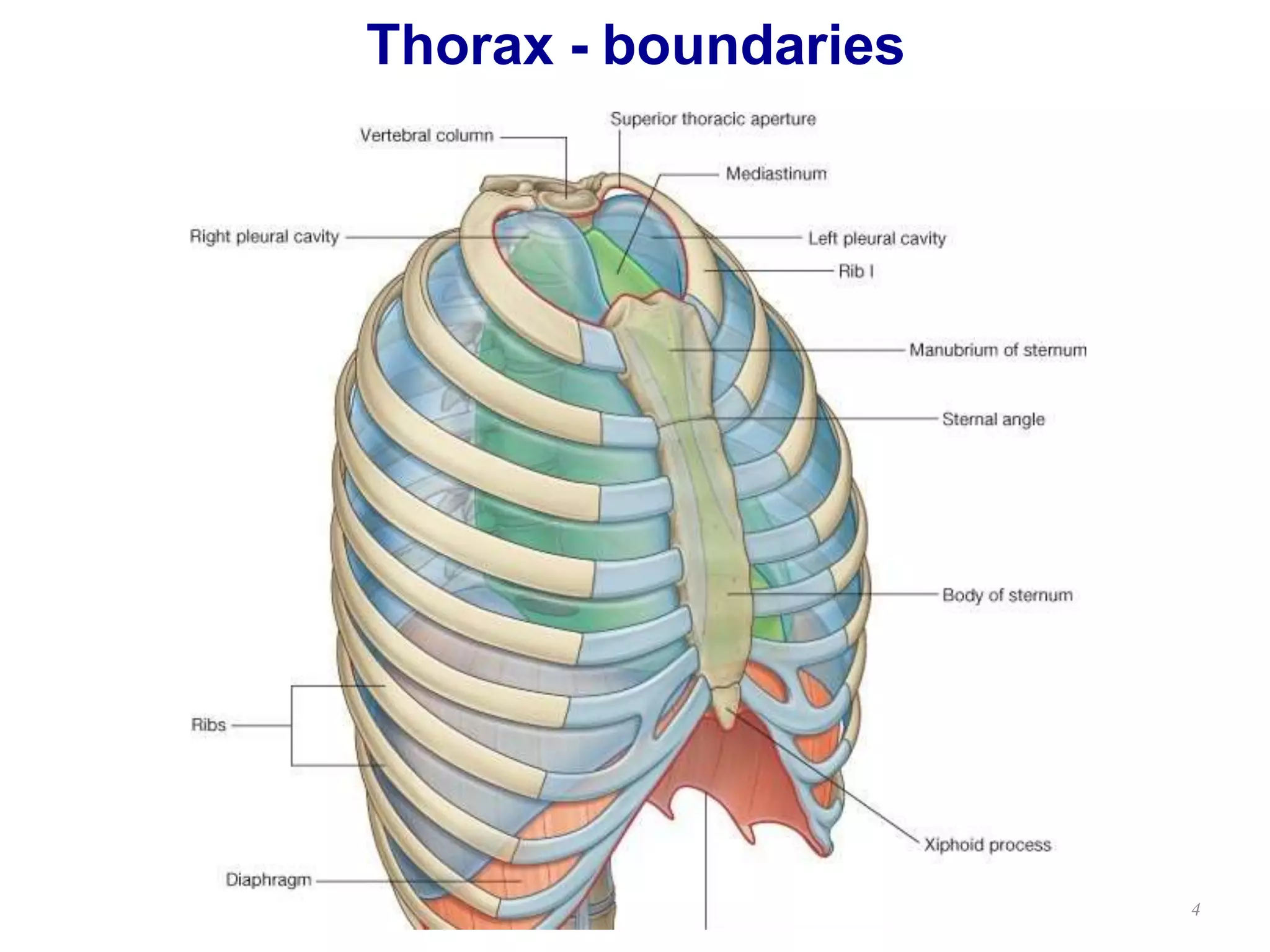
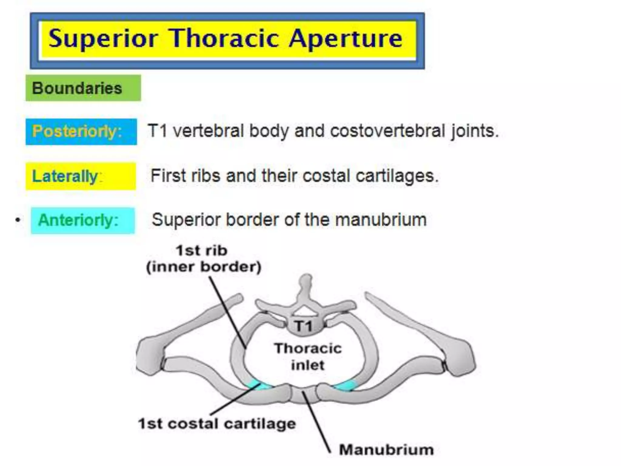
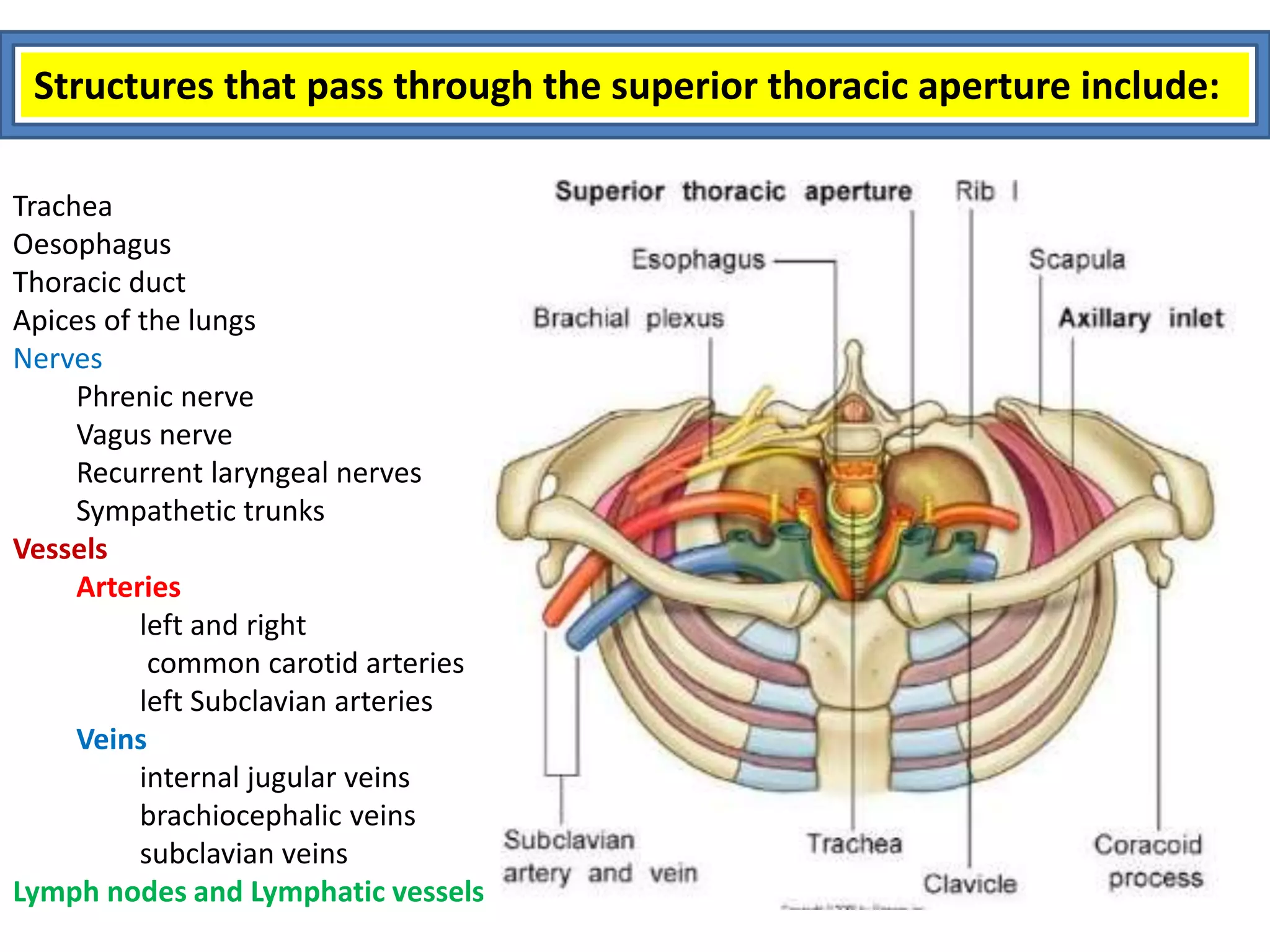
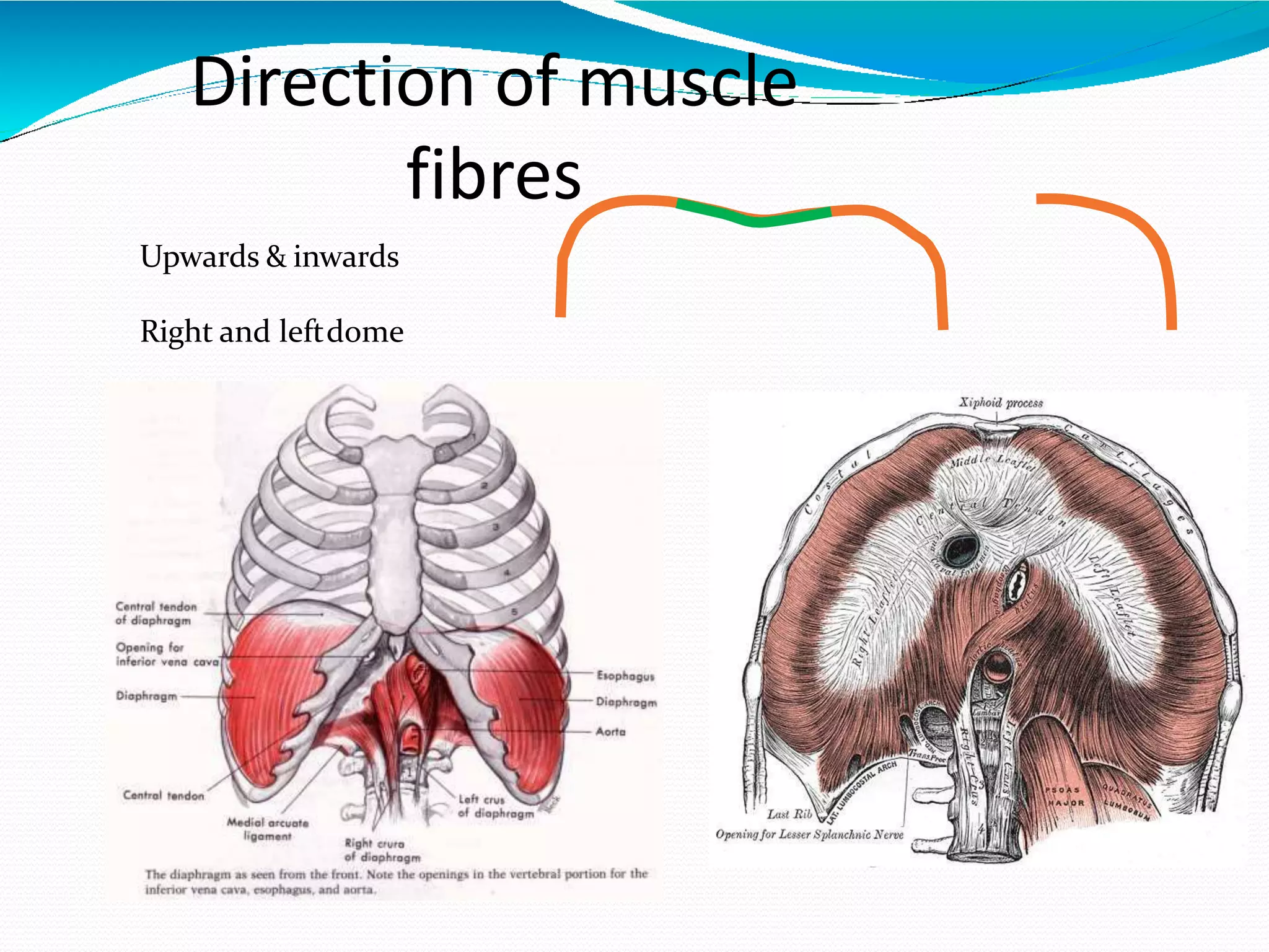
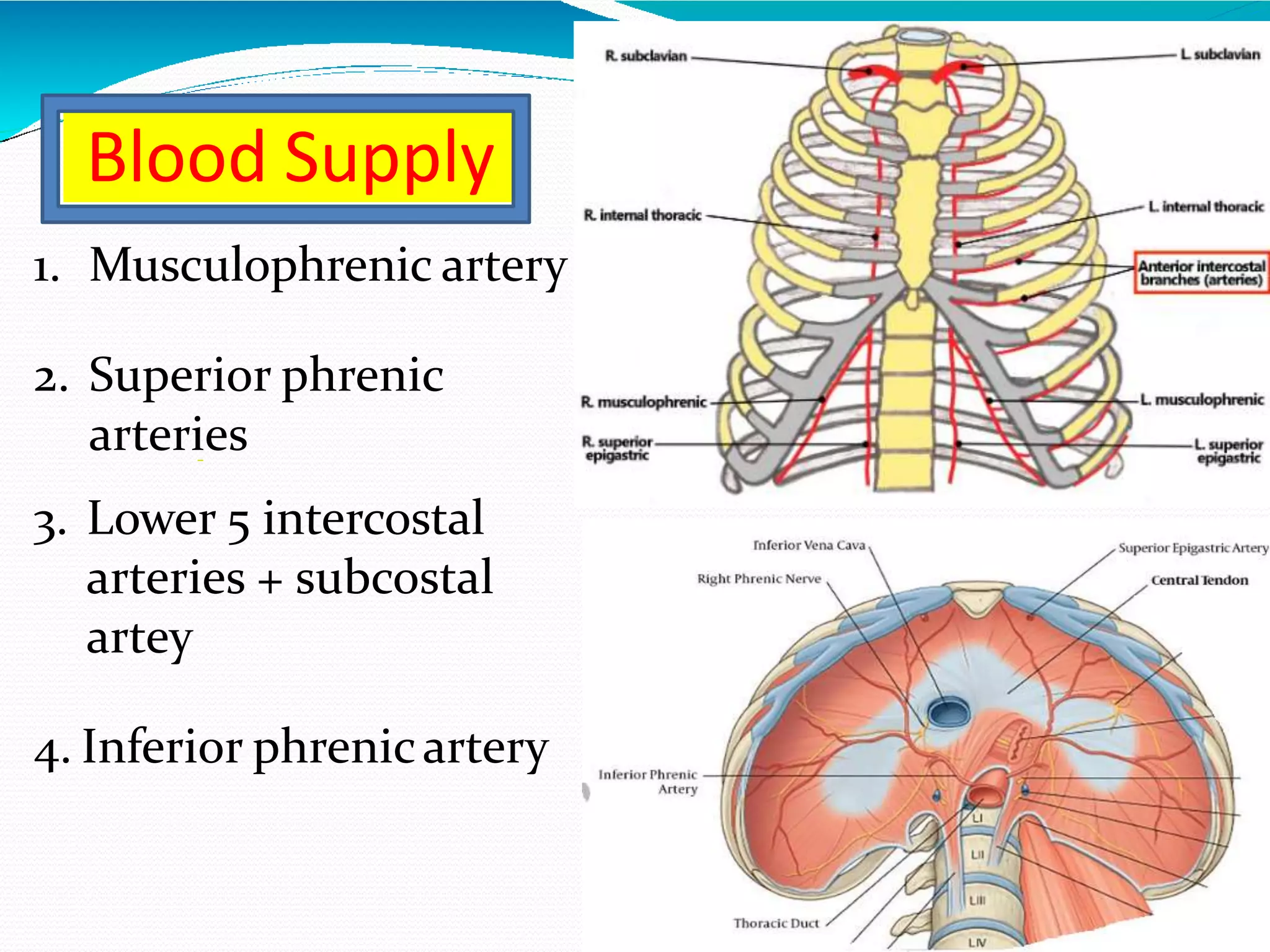
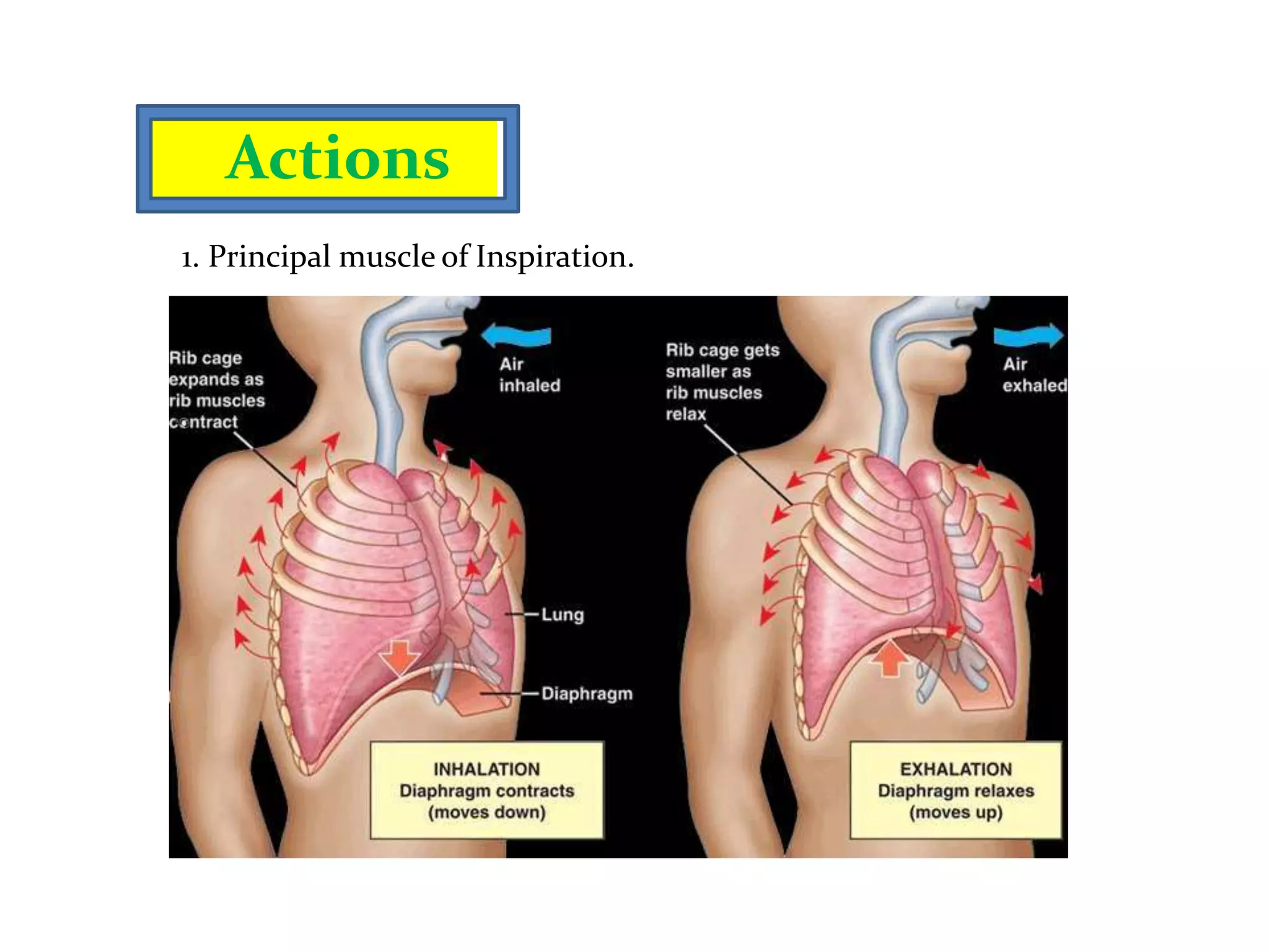
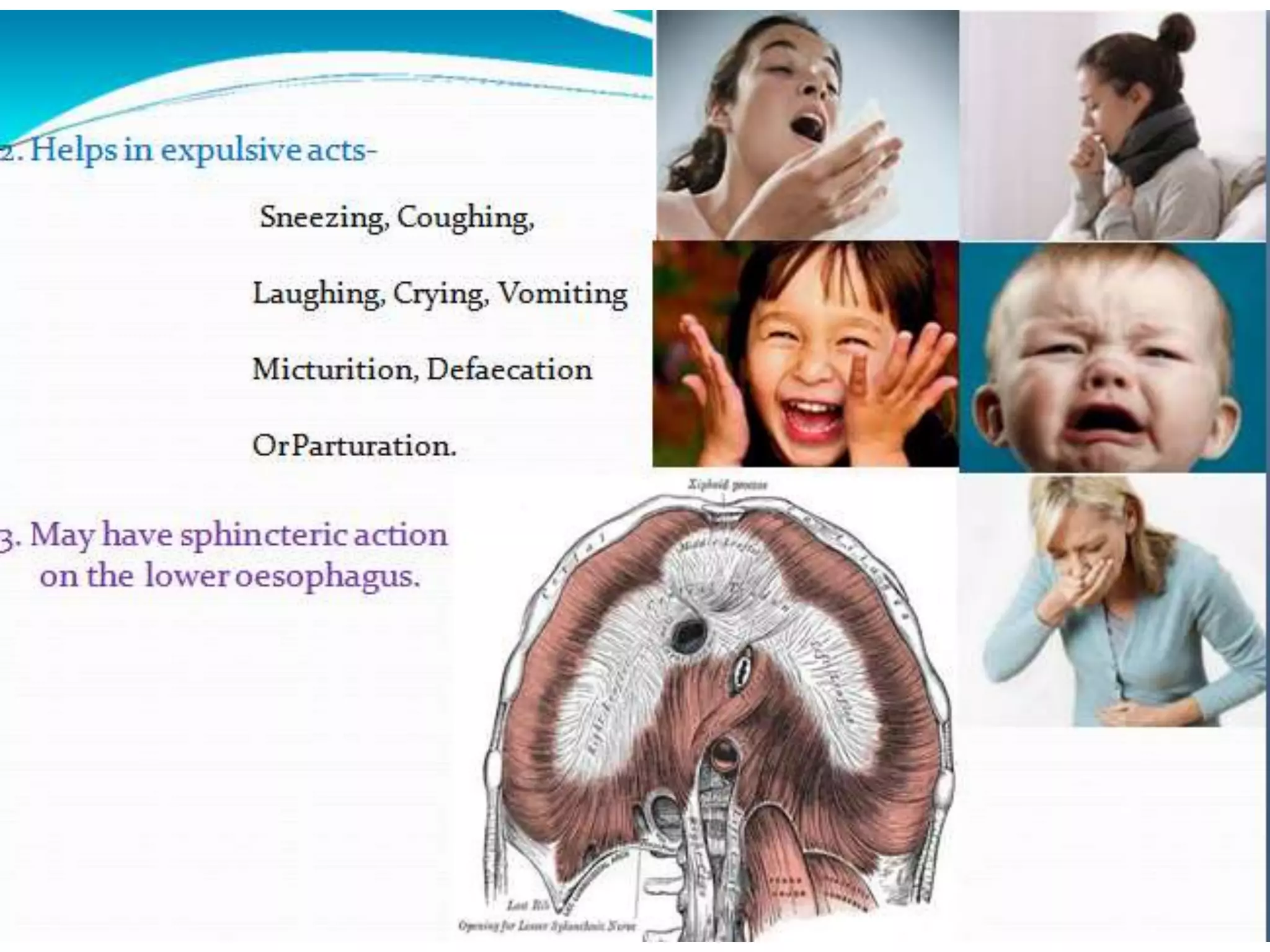
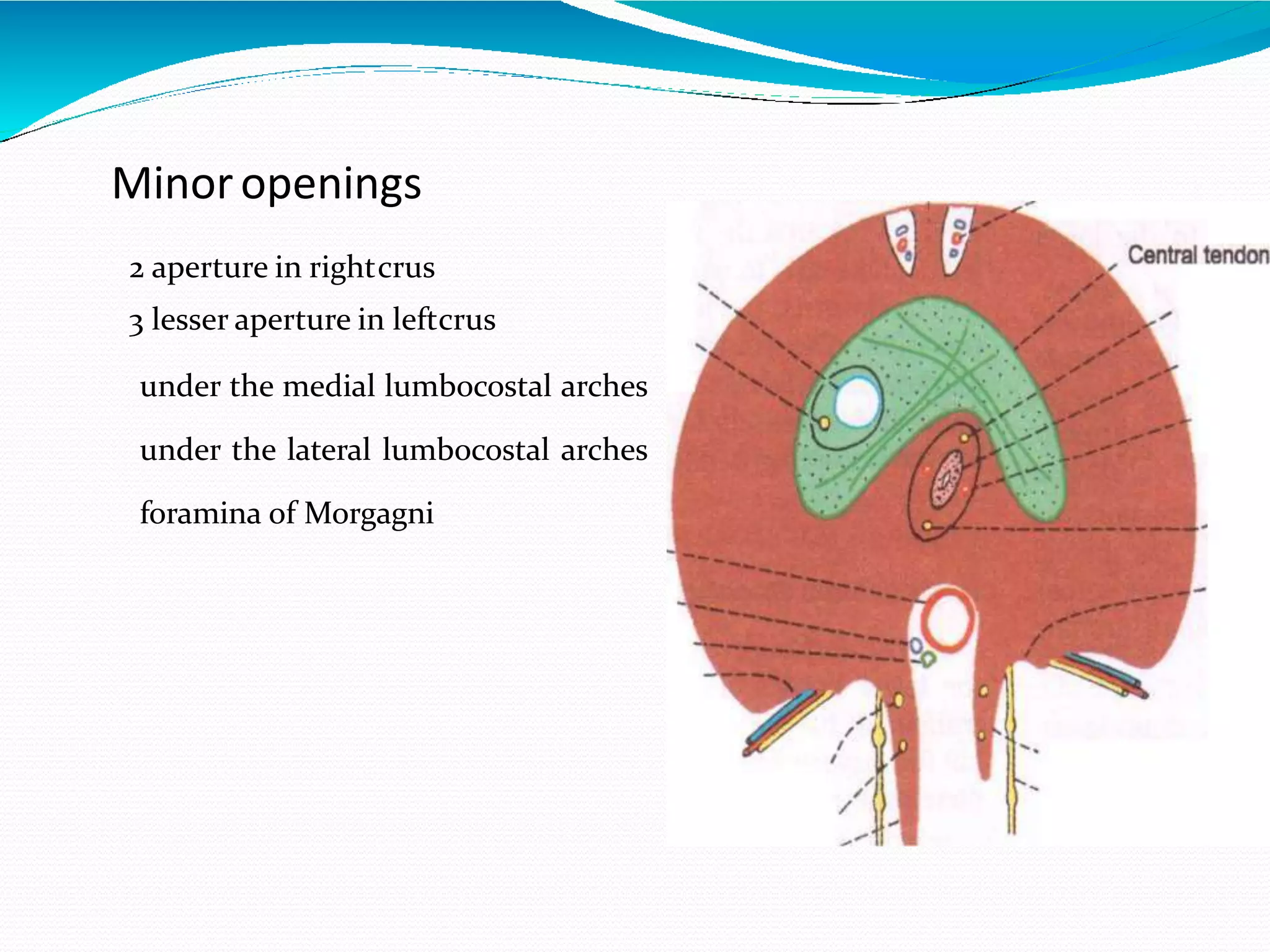
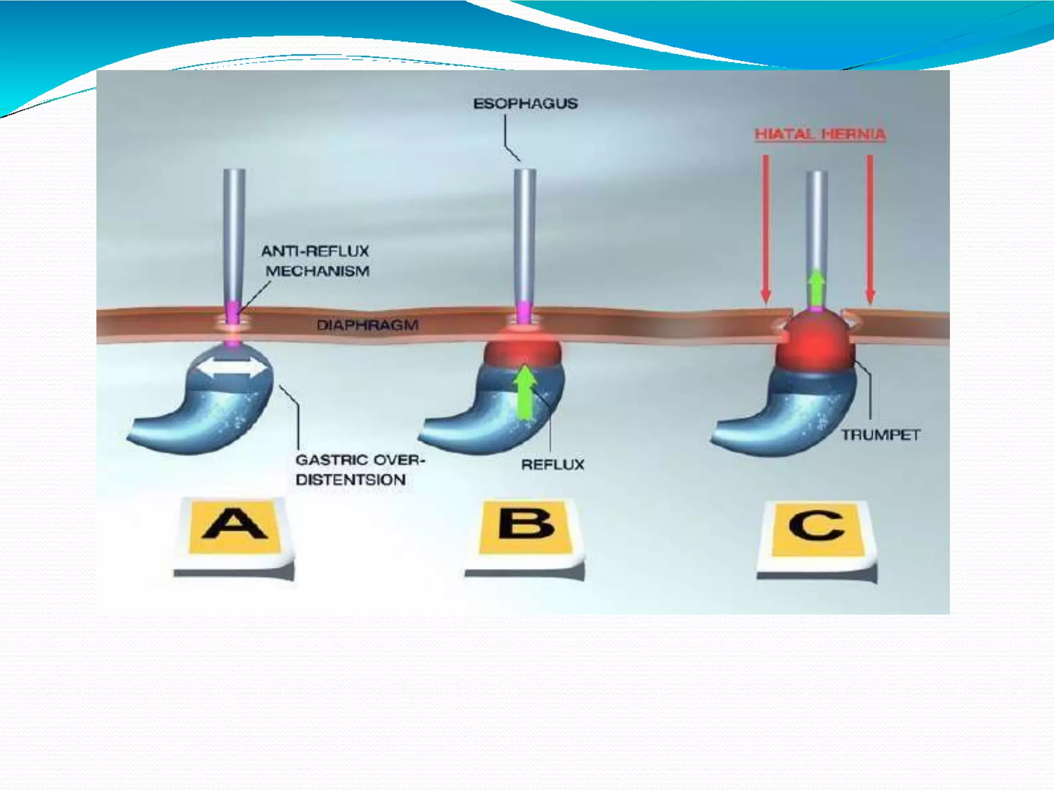
2
Learning objectives
At the end of session students should be able to
• Describe the thoracic cage and its boundaries, thoracic Inlet and outlet.
• Describe the position & component of muscular & tendinous part of
diaphragm
• Describe the attachments of diaphragm along with its blood supply and
nerve supply
• Describe the openings present in the diaphragm and their respective levels
• Enumerate the structures passing through the openings and piercing the
diaphragm.
• List the functions of diaphragm
• Enumerate the conditions related to damage to Phrenic Nerve
• Describe the main features of these condition
3
Thorax - boundaries
4
Superior Thoracic Aperture
6
Plane of Inlet
7
45°
Suprapleural membrane/ Sibson’s fascia
8
Suprapleural membrane: Relations
9
Trachea
Oesophagus
Thoracic duct
Apices of the lungs
Nerves
Phrenic nerve
Vagus nerve
Recurrent laryngeal nerves
Sympathetic trunks
Vessels
Arteries
left and right
common carotid arteries
left Subclavian arteries
Veins
internal jugular veins
brachiocephalic veins
subclavian veins
Lymph nodes and Lymphatic vessels
Structures that pass through the superior thoracic aperture include:
Inf thoracic aperture-Diaphragm- ant view
12
Thoracic skeleton ant view
13
12
Thoracic skeleton post view
Typical rib
15
Atypical ribs
16
Thoracic vertebrae
17
Sternum and Joints
18
19
Intercostal muscles
External
Internal
Innermost



Subcostalis
Intercostalis intima
Sternocostalis {transversus thoracis }
Intercostal muscles
20
Externalandinternalintercostalmuscles
21
Externalandinternalintercostalmuscles
22
Cervical Rib Syndrome
43
Diaphragm
•Greek, from dia 'through, apart'
+ phragma 'a fence'.
•Fibro-muscular sheet
•Separates the thoracic cavity
from the abdominal cavity
•Performs an important
function in respiration.
Origin
3 Parts:-
1. Sternal
2. Costal
3. lumbar
Direction of muscle
fibres
Upwards & inwards
Right and leftdome
Nerve Supply
Motor – Phrenic
nerve(C3,4,5)
Sensory –
1.central part- phrenic
nerve
2.peripheral part-
lower six thoracic
nerve
Blood Supply
1. Musculophrenic artery
2. Superior phrenic
arteries
3. Lower 5 intercostal
arteries + subcostal
artey
4. Inferior phrenicartery
Actions
1. Principal muscle of Inspiration.
2. Helps in expulsiveacts-
Sneezing, Coughing,
Laughing, Crying,Vomiting
Micturition, Defaecation
OrParturation.
3. May have sphincteric action
on the lower oesophagus.
Position of Diaphragm
3 Factors :-
1.The elastic recoil of Lung
2.Thepressureexerted byabdominal viscera
3.The muscles of abdominalwall
Highest- in supineposition
Lowest- in sitting
Intermediate- in standing
Relations
Superiorly-
pleurae
pericardium
Inferiorly -
peritoneum
liver
fundus of stomach
spleen
kidneys
suprarenal
Openin
gs
Majoropenings
Caval opening
Oesophagealopening
Aorticopening
Minoropenings
2 aperture in rightcrus
3 lesser aperture in leftcrus
under the medial lumbocostal arches
under the lateral lumbocostal arches
foramina of Morgagni
Developme
nt
The Diaphragm 'develops from the followingsources.
1. Septum transversum
2. Pleuroperitoneal membranes
3. Lateral thoracicwall
4. Dorsal mesentery of oesophagus
Applied
Anatomy
1. Hiccough- a)peripheral
b)central
2. Shoulder tip pain
3. Unilateral paralysis of Diaphragm
4. Eventration
5. Diaphragmatic hernia – a)Congenital
b) Acquired
Congenital
Hernia
a) Morgagni hernia or Retrosternalhernia
b) Bochdalek hernia orPosterolateral
c) Central hernia
e) Congenital hiatal hernia
d) Posteriorhernia
Bochdalek hernia
Acquired
Hernia
a) Traumatic hernia
b) Acquired hiatal hernia
Thoracic inlet & outlet

Thoracic inlet & outlet