
Recommended
More Related Content
What's hot
What's hot (20)
Similar to Anatomy of the stomach
Similar to Anatomy of the stomach (20)
Recently uploaded
Recently uploaded (20)
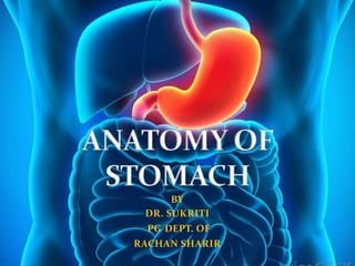
Anatomy of the stomach
- 1. BY DR. SUKRITI PG DEPT. OF RACHAN SHARIR
- 2. The stomach is a muscular bag forming the widest and most distensible part of digestive tube. It is connected above with the lower end of oesophagus, and below with the duodenum. It acts as a reservoir of food and helps in digestion of carbohydrates , protein and fats.
- 3. LOCATION- • Stomach is located at level of T10 - L3 vertebrae. • It is obliquely situated in the upper and left part of the abdomen, occupying the epigastric , umbilical and left hypochondriac regions. • most of it lies under cover of the left costal margins and the ribs. Shape: J-shaped in normal persons • Shape of stomach is depends upon the degree of tone of muscles. • Empty stomach – j shaped • When partially distended- piriform in shape • In obese persons – horizontal Size- Length – 25cm or 10 inch CAPACITY At birth- 30 ml At puberty- 1 liter Adult – 1.5 -2 liter or more
- 4. EXTERNAL FEATURES The stomach has: two openings cardiac orifice pyloric orifice; Two curvatures- greater curvature; lesser curvature Two surfaces- anterior posterior surfaces.
- 5. Curvatures of the stomach • The lesser curvature- Forms the right border of the stomach and extends from the cardiac orifice to the pylorus. • It is suspended from the liver by the lesser omentum. greatercurvature- Much longer than the lesser curvature and extends from the left of the cardiac orifice, over the dome of the fundus, and along the left border of the stomach to the pylorus. It provide attachments to the greater omentum , gastrosplenic ligament the gastrophrenic ligaments.
- 7. Openings of the stomach Gastroesophageal/Cardiac orifice- Jioned by lower end of oesophagus. It lies behind the 7th costal cartilage 2.5 cm from its junction with sternum, at the level of T11. Pyloric orifice- Opens into duodenum . In empty stomach and in supine position , it lies ½ inch right to the median plane , at the level of the L1.
- 8. the cardial notch, which is the superior angle created whenthe esophagus enters the stomach. the angular notch, which is a bend onthe lesser curvature.
- 9. PARTS OF STOMACH The stomach is divided into two Parts- (1) Cardiac part (2) Pyloric part The cardiac part is subdivided into two parts- (1)Fundus (2)Body The smaller pyloric part is subdivided into two parts- (1) Pyloric antrum (2) Pyloric canal
- 10. 1.Fundus: This is upper convex dome-shaped part situated above a horizontal line drawn at the level of cardiac orifice. It is usually full of gas. 2.Body: Lies b/w fundus and pyloric antrum. The gastric glands distributed in the fundus and body of stomach. contain all three types of secretory cells, namely a. The mucous cells b. The chief peptic . cells which secrete the digestive enzymes. c. The parietal or oxyntic cells which secrete HCL
- 11. Pyloric Part (1)The pyloric antrum- is separated from the pyloric canal by an inconstant sulcus, sulcus intermedius present on the greater curvature. It is about 7.5 cm long. The pyloric glands are richest in mucous cells. (2) The pyloric canal – is about 2.5 cm long. It is narrow and tubular. At its right end, it terminates at the pylorus.
- 12. Relations of stomach Peritoneal Relations stomach is lined by peritoneum on both its surfaces. • At the lesser curvature, the layers of peritoneum lining the anterior and posterior surfaces meet and become continuous with the lesser omentum. • Along the greater part of the greater curvature, the two lawyers meet to form the greater omentum. • Near the fundus, the two layers meet to form the gastrosplenic ligament. • Near the cardiac end, the peritoneum on the posterior surface is reflected on to the diaphragm as the gastrophrenic ligament. • Cranial to this ligament a small part of the posterior surface of the stomach is in direct contact with the diaphragm (left crus). This is the bare area of the stomach.
- 14. Visceral Relations The anterior surface of the stomach is related to the liver, the diaphragm, transverse colon and the anterior abdominal wall. The diaphragm separates the stomach from the left pleura, the pericardium, and the sixth to ninth ribs. The space between left . costal margin and lower edge of left lung on stomach is known as Traube's space Normally, on percussion, there is resonant note over this space; but in splenomegaly or pleural effusion, a dull note is felt at this site.
- 15. Posterior relations The posterior surface of the stomach is related to structures forming the stomach bed. These structures are: a. Diaphragm b. Left kidney c. Left suprarenal gland d. Pancreas . e. Transverse mesocolon f. Splenic flexure of the colon g. Splenic artery
- 17. Interior of the stomach The stomach wall composed four layers- (1)The mucosa – • innermost layer of stomach . Mucosa of an empty stomach is thrown into folds termed as gastric rugae. The rugae are longitudinal along the lesser curvature and may be irregular elsewhere. • The rugae are flattened in a distended stomach. On the mucosal surface there are numerous small depressions that can be seen with a hand lens. These are the gastric pits. The gastric glands open into these pits.
- 18. (2)Submucous coat - is made of connective tissue, arterioles and nerve plexus. (3)Muscle coat is arranged as under: a. Longitudinal fibres are most superficial, mainly along the curvature. b. Inner circular fibres encircle the body and are thickened at pylorus to form pyloric sphincter c. The deepest layer consists of oblique fibres which loop over the cardiac notch. Some fibres spread in the fundus and body of stomach. Rest from a well-developed ridge on each side of the lesser curvature. These fibres on contraction form gastric canal" for the passage of fluids. (4) Serous coat consists of the peritoneal covering.
- 19. Arterial blood supply: • Arteries of the stomach: All are branches of the celiac artery • 1. Left gastric artery: arises from the celiac artery. It supplies the lower third of the esophagus and the upper right part of the stomach. • 2. Right gastric artery: arises from the hepatic artery at the upper border of the pylorus and supplies the lower right part of the stomach.
- 20. • 3. Short gastric arteries: arise from the splenic artery and supply fundus of the stomach • 4. Left gastroepiploic artery: arises from splenic artery and supply the greater curvature. • 5. Right gastroepiploic artery: arises from the gastroduodenal branch of the hepatic artery, and supplies the stomach along the lower part of the greatercurvature.
- 24. the drains gastric superior nodes surrounding the Left Gastric Artery. Distal portion of lesser curvature through drains the suprapyloric nodes.
- 25. The stomach can be divided into four lymphatic territories. The drainage of these areas is as follows. (a)upper part of left 1/3rd drains into the pancreaticosplenic nodes lying along the splenic artery, i.s. on the back of the stomach. Lymph vessels from these nodes travel along the splenic artery to reach the coeliac nodes. (b), i.e. right 2/3rd drains into the left gastric nodes lying along the artery of the same name. These nodes also drain the abdominal part of the oesophagus. Lymph from these nodes drains into the coeliac nodes. Area (c), i.e. lower part of left 1/3rd drains into the right gastroepiploic nodes that lie along the artery of the same name. Lymph vessels arising in these nodes drain into the subpyloric nodes which lie in the angle between the first and second parts of
- 26. Proximal portion of the greater supplied lymphatic traverse curvature is by the vessels that the pancreaticosplenic nodes. Antral portion of the greater curvature drains into the subpyloric and omental nodal groups.
- 27. The main innervations are Left and Right Vagus Nerves. The sympathetic nerve supply- derived from thoracic six to ten segments of the spinal cord, via the greater splanchnic nerves, coeliac and hepatic plexus. They travel along the arteries supplying the stomach.
- 28. The parasympathetic nerves are derived from the vagi, through the esophageal plexus and gastric nerves. 1. The anterior gastric nerve- contains mainly the left vagal fibres. but it does contain fibers from the right vagus. The anterior gastric nerve divides into A number of gastric branches for the anterior surface of the fundus and body of the stomach. Two pyloric branches,one for the pyloric antrum and another for the pylorus.
- 29. posterior gastric nerve -contains mainly the right vagal fibres. but contains fibers from the left vagus nerve. The posterior gastric nerve divides into: Smaller, gastric branches for the posterior surface of the fundus, the body and the pyloric antrum. Larger, coeliac branches for the coeliac plexus.
- 30. Clinical anatomy of stomach Gastric pain - is felt in the epigastrium because the stomach is supplied from segments T6 to T9 of the spinal cord, which also supply the upper part of the abdominal wall. Pain is produced either by spasm of muscle, or by over-distension. Pyloric stenosis-Pyloric stenosis is a narrowing of the opening from the stomach to the first part of the small intestine (the pylorus). • Normally, a muscular valve (pylorus) between the stomach and small intestine holds food in the stomach until it is ready for the next stage in the digestive process. In pyloric stenosis, the pylorus muscles thicken and become abnormally large, blocking food from reaching the small intestine.
- 31. Gastric ulcer - occurs typically along the lesser curvature This is possibly due to the following peculiarities of lesser curvature. A. Mucosa is not freely movable over the muscular coat. B. The epithelium is comparatively thin. C. Blood supply is less abundant and there are fewer anastomoses. D. Nerve supply is more abundant, with large ganglia E. Because of the gastric canal, it receives most of the insult from irritating drinks. • Gastric carcinoma is common and occurs along the greater curvature. On this account, the lymphatic drainage of stomach assumes importance. Metastasis can occur through the thoracic duct to the left supraclavicular lymph node (Troisier's sign). These lymph nodes are called as "signal vomiting after meals (thin and long).