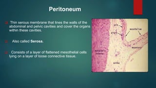
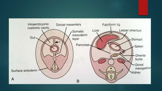
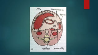
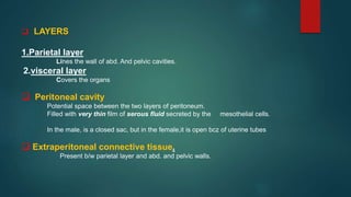
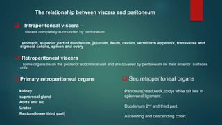
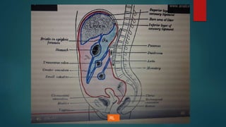
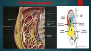
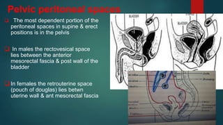
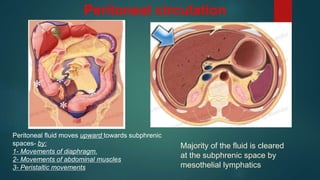
Peritoneum
DR SOUMITRA HALDER
JR-2
KOLKATA MEDICAL COLLEGE
PART -1
Peritoneum
 Thin serous membrane that lines the walls of the
abdominal and pelvic cavities and cover the organs
within these cavities.
 Also called Serosa.
 Consists of a layer of flattened mesothelial cells
lying on a layer of loose connective tissue.
 LAYERS
1.Parietal layer
Lines the wall of abd. And pelvic cavities.
2.visceral layer
Covers the organs
 Peritoneal cavity
Potential space between the two layers of peritoneum.
Filled with very thin film of serous fluid secreted by the mesothelial cells.
In the male, is a closed sac, but in the female,it is open bcz of uterine tubes
 Extraperitoneal connective tissue.
Present b/w parietal layer and abd. and pelvic walls.
The relationship between viscera and peritoneum
 Intraperitoneal viscera -
viscera completely surrounded by peritoneum
stomach, superior part of duodenum, jejunum, ileum, cecum, vermiform appendix, transverse and
sigmoid colons, spleen and ovary
 Retroperitoneal viscera
some organs lie on the posterior abdominal wall and are covered by peritoneum on their anterior surfaces
only.
 Primary retroperitoneal organs
kidney
suprarenal gland
Aorta and ivc
Ureter
Rectum(lower third part)
Pancreas(head,neck,body) while tail lies in
splenrenal ligament.
Duodenum 2nd and third part.
Ascending and descending colon.
 Sec.retroperitoneal organs
`
Definitions
LIGAMENT
 Two folds of peritoneum
 supporting structers
Mysentry
 Two folds of peritoneum
 Connecting to posterior abdominal wall
Omentum
 Connecting the stomach to other organs
Peritoneal reflections
RL
Falciform ligament
Connects liver to the posterior
aspect of the anterior abdominal
wall just right to the midline
Coronary ligament
 Peritoneum that lines the under
surface of diaphragm is reflected on
to superior surface of rt lobe of liver
forming the upper layer of
coronary ligament
 Then, it descends from sup surface of
liver to ant surface then inferior surface
of liver.
 From post part of inferior surface,
peritoneum reflected on to front of right
kidney & rt suprarenal gland forming the
lower layer of coronary ligament.
 The lower & upper layers of coronary
ligament bound a large area on the post
surface of the liver called bare area of
the liver which has no peritoneal
covering.
Rt triangular ligament.
 long v shaped fold.
 Formed by approximation of two
layers of coronary ligament on rt
lateral end.
Left triangular ligament.
 On upper border of lt lobe.
 Upper layer continues with left layer
of falciform ligament.
 Lower layer with lt layer of lesser
omentum.
 is short and does not
compartmentalize
Peritoneal Ligaments of the Stomach
Lesser omentum
Gastro hepatic ligament
 attaches liver to lesser
curvature,
 left gactric artery &coronary
vein
Hepatoduodenal ligament
 attaches liver to duodenum
 CBD,hepatic artery&PV
OVARIAN CA
Greater omentum
Anterior sheet
Anterior layer
From ant surface of stomach and duodenum.
Posterior layer
From post surface of stomach and pylorus.
Two layers of anterior sheet decend from greater curvature to a
variable distance in peritoneal cavity
Ascend back on itself to form posterior sheet.
Posterior sheet
Anterior layer forms posterior wall of lesser sac.
Post layer n mesocolon n attatched to t.mesocolon passes infront
of transvers colon at its root to form gastrocolic ligament.
.
The gastrosplenic ligament
 Connects the stomach to spleen
 contains the short gastric vessels,spenic
collaterals
 It’s a major route of escape for
pancreatitis arising in the peripheral body
&tail
Phrenicocolic lig
Spleno renal ligament
 Posterior aspect of spleen to anterior
pararenal space
 Forms post-lat border of lesser sac
 encloses tail of pancreas& distal
splenic artery
Ligaments of lower abdomen
 Transverse mesocolon
Small bowel mesentry
Sigmoid mesocolon
Peritoneal spaces
Potential space between the parietal & visceral peritoneum
 Contains fluid that lubricates the surface of the peritoneum
 Not depicted on conventional radiologic studies or by cross
secectional imaging unless they are distended by fluid or air.
In men, the peritoneal cavity is closed,
In women, it communicates with the extraperitoneal
pelvis exteriorly through the fallopian tubes.
Peritoneal spaces
Supramesocolic space
Right
 R subphrenic(subdiaphragmatic)
 R subhepatic( hepatorenal or
morrisons pouch)
 Lesser sac (omental bursa)
Left
 L perihepatic space
 L subdiaphragmatic space
Left sided peritoneal spaces
Perihepatic spaces-
The anterior perihepatic
The posterior perihepatic
Left Subphrenic space
 LAS- diaphragm ant& lat,
stomach post.
 communicates with the post
subphrenic(perisplenic) space
 The perisplenic space surrounds
most of the spleen except for a
portion of spleen lying within the
splenorenal ligament
Right sided supra mesocolic spaces
The right sub diaphragmatic
space is limited anteriorly by
the falciform ligament and
posteriorly by the upper layer
coronary ligament
Hepatorenal space –morrisons pouch
 Just beneath the bare area the right
peritoneal space courses between the
posterior surface of segment 6 and the
anterior renal fascia .
 This is a relatively small potential space.
 It is the most dependent portion of the
right supramesocolic spaces
Lesser sac
 Lies behind the stomach
 Ant to pancreas
 Left margin-gastro splenic
ligament
 Right margin–medial surface of
coronary ligament
 Post boundary –gastrocolic
reflection&mesocolon
Lesser sac boundaries
Lesser sac division
 Superior recess-seen
surrounding the caudate
lobe.
 Inf recess-lies behind the
stomach,extending into the
layers of GO
The sup & inf recess are
separated by a peritoneal fold
(ghl)that accompanies the left
gastric artery
Foramen of winslow
 Communication betn the greater & lesser
sac.
 Ant margin –hepatoduodenal ligament ( L
omentum ) contain P.Triad
 Post margin-peritoneum covering IVC
 Roof-peritoneum covering caudate lobe
 Floor-peritoneum covering 1st part of
duodenum
Foramen of winslow
Inframesocolic spaces
below the transverse mesocolon and
transverse colon as far as the true pelvis.
 divided in two unequal spaces
RIC/LIC by the root of the mesentery
of the small intestine.
 It contains the right and left paracolic
gutters lateral to the ascending and
descending colon.
Pelvic peritoneal spaces
 The most dependent portion of the
peritoneal spaces in supine & erect
positions is in the pelvis
 In males the rectovesical space
lies between the anterior
mesorectal fascia & post wall of the
bladder
 In females the retrouterine space
(pouch of douglas) lies betwn
uterine wall & ant mesorectal fascia
Peritoneal fluid
 Peritoneal fluid is pale yellow fluid rich in
leukocytes
 50-70 ml
 Mobile viscera glide easily on one another.
Peritoneal circulation
Majority of the fluid is cleared
at the subphrenic space by
mesothelial lymphatics
Peritoneal fluid moves upward towards subphrenic
spaces- by:
1- Movements of diaphragm.
2- Movements of abdominal muscles
3- Peristaltic movements
Functions of peritoneal fluid
 lubrication.
 Absorbption
 Support viscera
 MOVEMENT
……………….to be continue (PART -2 )
Thank you…….

Peritoneum Anatomy and pathology part 1

  • 1.
  • 2.
  • 3.
    Peritoneum  Thin serousmembrane that lines the walls of the abdominal and pelvic cavities and cover the organs within these cavities.  Also called Serosa.  Consists of a layer of flattened mesothelial cells lying on a layer of loose connective tissue.
  • 7.
     LAYERS 1.Parietal layer Linesthe wall of abd. And pelvic cavities. 2.visceral layer Covers the organs  Peritoneal cavity Potential space between the two layers of peritoneum. Filled with very thin film of serous fluid secreted by the mesothelial cells. In the male, is a closed sac, but in the female,it is open bcz of uterine tubes  Extraperitoneal connective tissue. Present b/w parietal layer and abd. and pelvic walls.
  • 8.
    The relationship betweenviscera and peritoneum  Intraperitoneal viscera - viscera completely surrounded by peritoneum stomach, superior part of duodenum, jejunum, ileum, cecum, vermiform appendix, transverse and sigmoid colons, spleen and ovary  Retroperitoneal viscera some organs lie on the posterior abdominal wall and are covered by peritoneum on their anterior surfaces only.  Primary retroperitoneal organs kidney suprarenal gland Aorta and ivc Ureter Rectum(lower third part) Pancreas(head,neck,body) while tail lies in splenrenal ligament. Duodenum 2nd and third part. Ascending and descending colon.  Sec.retroperitoneal organs
  • 9.
  • 10.
    Definitions LIGAMENT  Two foldsof peritoneum  supporting structers Mysentry  Two folds of peritoneum  Connecting to posterior abdominal wall Omentum  Connecting the stomach to other organs
  • 11.
  • 12.
    Falciform ligament Connects liverto the posterior aspect of the anterior abdominal wall just right to the midline
  • 13.
    Coronary ligament  Peritoneumthat lines the under surface of diaphragm is reflected on to superior surface of rt lobe of liver forming the upper layer of coronary ligament
  • 14.
     Then, itdescends from sup surface of liver to ant surface then inferior surface of liver.  From post part of inferior surface, peritoneum reflected on to front of right kidney & rt suprarenal gland forming the lower layer of coronary ligament.  The lower & upper layers of coronary ligament bound a large area on the post surface of the liver called bare area of the liver which has no peritoneal covering.
  • 15.
    Rt triangular ligament. long v shaped fold.  Formed by approximation of two layers of coronary ligament on rt lateral end. Left triangular ligament.  On upper border of lt lobe.  Upper layer continues with left layer of falciform ligament.  Lower layer with lt layer of lesser omentum.  is short and does not compartmentalize
  • 17.
  • 18.
    Lesser omentum Gastro hepaticligament  attaches liver to lesser curvature,  left gactric artery &coronary vein Hepatoduodenal ligament  attaches liver to duodenum  CBD,hepatic artery&PV
  • 19.
  • 20.
    Greater omentum Anterior sheet Anteriorlayer From ant surface of stomach and duodenum. Posterior layer From post surface of stomach and pylorus. Two layers of anterior sheet decend from greater curvature to a variable distance in peritoneal cavity Ascend back on itself to form posterior sheet. Posterior sheet Anterior layer forms posterior wall of lesser sac. Post layer n mesocolon n attatched to t.mesocolon passes infront of transvers colon at its root to form gastrocolic ligament. .
  • 22.
    The gastrosplenic ligament Connects the stomach to spleen  contains the short gastric vessels,spenic collaterals  It’s a major route of escape for pancreatitis arising in the peripheral body &tail
  • 25.
  • 26.
    Spleno renal ligament Posterior aspect of spleen to anterior pararenal space  Forms post-lat border of lesser sac  encloses tail of pancreas& distal splenic artery
  • 27.
    Ligaments of lowerabdomen  Transverse mesocolon Small bowel mesentry Sigmoid mesocolon
  • 29.
    Peritoneal spaces Potential spacebetween the parietal & visceral peritoneum  Contains fluid that lubricates the surface of the peritoneum  Not depicted on conventional radiologic studies or by cross secectional imaging unless they are distended by fluid or air. In men, the peritoneal cavity is closed, In women, it communicates with the extraperitoneal pelvis exteriorly through the fallopian tubes.
  • 30.
  • 31.
    Supramesocolic space Right  Rsubphrenic(subdiaphragmatic)  R subhepatic( hepatorenal or morrisons pouch)  Lesser sac (omental bursa) Left  L perihepatic space  L subdiaphragmatic space
  • 32.
    Left sided peritonealspaces Perihepatic spaces- The anterior perihepatic The posterior perihepatic
  • 33.
    Left Subphrenic space LAS- diaphragm ant& lat, stomach post.  communicates with the post subphrenic(perisplenic) space  The perisplenic space surrounds most of the spleen except for a portion of spleen lying within the splenorenal ligament
  • 35.
    Right sided supramesocolic spaces The right sub diaphragmatic space is limited anteriorly by the falciform ligament and posteriorly by the upper layer coronary ligament
  • 36.
    Hepatorenal space –morrisonspouch  Just beneath the bare area the right peritoneal space courses between the posterior surface of segment 6 and the anterior renal fascia .  This is a relatively small potential space.  It is the most dependent portion of the right supramesocolic spaces
  • 38.
    Lesser sac  Liesbehind the stomach  Ant to pancreas  Left margin-gastro splenic ligament  Right margin–medial surface of coronary ligament  Post boundary –gastrocolic reflection&mesocolon
  • 39.
  • 40.
    Lesser sac division Superior recess-seen surrounding the caudate lobe.  Inf recess-lies behind the stomach,extending into the layers of GO The sup & inf recess are separated by a peritoneal fold (ghl)that accompanies the left gastric artery
  • 42.
    Foramen of winslow Communication betn the greater & lesser sac.  Ant margin –hepatoduodenal ligament ( L omentum ) contain P.Triad  Post margin-peritoneum covering IVC  Roof-peritoneum covering caudate lobe  Floor-peritoneum covering 1st part of duodenum
  • 43.
  • 44.
    Inframesocolic spaces below thetransverse mesocolon and transverse colon as far as the true pelvis.  divided in two unequal spaces RIC/LIC by the root of the mesentery of the small intestine.  It contains the right and left paracolic gutters lateral to the ascending and descending colon.
  • 46.
    Pelvic peritoneal spaces The most dependent portion of the peritoneal spaces in supine & erect positions is in the pelvis  In males the rectovesical space lies between the anterior mesorectal fascia & post wall of the bladder  In females the retrouterine space (pouch of douglas) lies betwn uterine wall & ant mesorectal fascia
  • 48.
    Peritoneal fluid  Peritonealfluid is pale yellow fluid rich in leukocytes  50-70 ml  Mobile viscera glide easily on one another.
  • 49.
    Peritoneal circulation Majority ofthe fluid is cleared at the subphrenic space by mesothelial lymphatics Peritoneal fluid moves upward towards subphrenic spaces- by: 1- Movements of diaphragm. 2- Movements of abdominal muscles 3- Peristaltic movements
  • 50.
    Functions of peritonealfluid  lubrication.  Absorbption  Support viscera  MOVEMENT
  • 51.
  • 52.