Downloaded 153 times


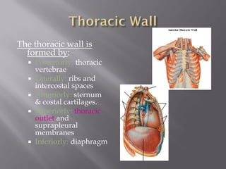
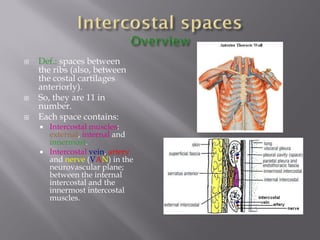
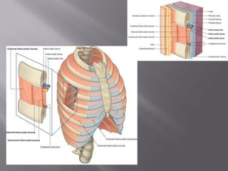

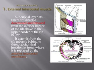


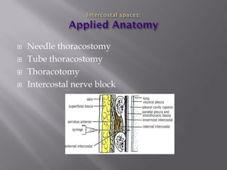
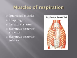


The document summarizes the anatomy of the thoracic wall. It is formed posteriorly by thoracic vertebrae, laterally by ribs and intercostal spaces, and anteriorly by the sternum and costal cartilages. The intercostal spaces contain intercostal muscles and neurovascular bundles. The document further describes the layers of intercostal muscles, blood supply, innervation, and clinical procedures relevant to the thoracic wall.


