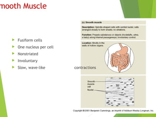
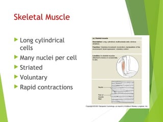
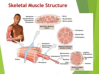
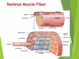
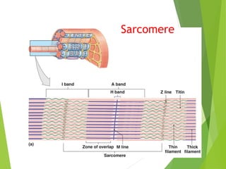
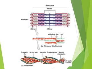
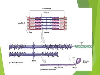
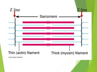
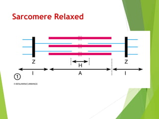
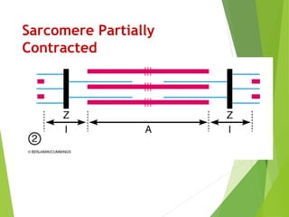
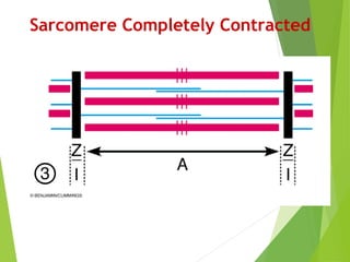
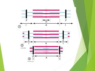
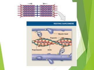
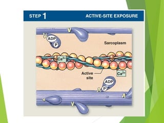
Muscle Physiology
Muscle Tissue
 Skeletal Muscle
 Cardiac Muscle
 Smooth Muscle
mooth Muscle
 Fusiform cells
 One nucleus per cell
 Nonstriated
 Involuntary
 Slow, wave-like contractions
Skeletal Muscle
 Long cylindrical
cells
 Many nuclei per cell
 Striated
 Voluntary
 Rapid contractions
Skeletal Muscle
 Produce movement
 Maintain posture & body position
 Support Soft Tissues
 Guard entrance / exits
 Maintain body temperature
 Store nutrient reserves
Skeletal Muscle Structure
Skeletal Muscle Fiber
Sarcomere
Z line Z line
Sarcomere Relaxed
Sarcomere Partially
Contracted
Sarcomere Completely Contracted
Neuromuscular Junction
Single Fiber Tension
The all–or–none principle
As a whole, a muscle fiber is
either contracted or relaxed
Tension of a Single Muscle
Fiber
Depends on
The number of pivoting cross-
bridges
The fiber’s resting length at the
time of stimulation
The frequency of stimulation
Length–tension relationship
-Number of pivoting cross-
bridges depends on:
amount of overlap between
thick and thin fibers
-Optimum overlap produces
greatest amount of tension:
too much or too little reduces
efficiency
-Normal resting sarcomere length:
is 75% to 130% of optimal length
Muscle Contraction Types
Isotonic contraction
Isometric contraction
Muscle Contraction Types
Isotonic contraction
Isometric contraction
Muscle Contraction Types
Isotonic contraction
Isometric contraction
ATP as Energy Source
Creatine
Molecule capable of storing ATP energy
Creatine + ATP Creatine phosphate + ADP
ADP + Creatine phosphate ATP + Creatine
Metabolism
 Aerobic metabolism
 95% of cell demand
 Kreb’s cycle
 1 pyruvic acid molecule  17 ATP
 Anaerobic metabolism
 Glycolysis  2 pyruvic acids + 2 ATP
 Provides substrates for aerobic metabolism
 As pyruvic acid builds converted to lactic acid
Muscle Fatigue
 Muscle Fatigue
 When muscles can no longer perform a required
activity, they are fatigued
 Results of Muscle Fatigue
 Depletion of metabolic reserves
 Damage to sarcolemma and sarcoplasmic reticulum
 Low pH (lactic acid)
 Muscle exhaustion and pain
Muscle Hypertrophy
• Muscle growth from
heavy training
 Increases diameter of
muscle fibers
 Increases number of
myofibrils
 Increases
mitochondria,
glycogen reserves
Muscle Atrophy
– Lack of muscle
activity
• Reduces muscle
size, tone, and
power
Steroid Hormones
 Stimulate muscle growth and hypertrophy
 Growth hormone
 Testosterone
 Thyroid hormones
 Epinephrine
Muscle Tonus
 Tightness of a muscle
 Some fibers always contracted
Tetany
 Sustained contraction of a muscle
 Result of a rapid succession of nerve impulses
Tetanus
Refractory Period
 Brief period of time in which muscle cells will not
respond to a stimulus
Refractory
Skeletal Muscle Cardiac Muscle
Refractory Periods

Muscular physiology

Editor's Notes

  • #3 There are three types of muscle tissue in the body. Skeletal muscle is the type that attaches to our bones and is used for movement and maintaining posture. Cardiac muscle is only found in the heart. It pumps blood. Smooth muscle is found in organs of the body such as the GI tract. Smooth muscle in the GI tract moves food and its digested products.
  • #4 Smooth muscle is found in the walls of hollow organs. *Their muscle cells are fusiform in shape. *Smooth muscle cells have just on nucleus per cell. *Smooth muscle is nonstriated. *Smooth muscle is involuntary. *The contractions of smooth muscle are slow and wave-like.
  • #5 Skeletal muscle attaches to our skeleton. *The muscle cells a long and cylindrical. *Each muscle cell has many nuclei. *Skeletal muscle tissue is striated. It has tiny bands that run across the muscle cells. *Skeletal muscle is voluntary. We can move them when we want to. *Skeletal muscle is capable of rapid contractions. It is the most rapid of the muscle types.
  • #7 Organization of Connective Tissues Muscle attachments Endomysium, perimysium, and epimysium come together at ends of muscles to form connective tissue attachment to bone matrix i.e., tendon (bundle) or aponeurosis (sheet) Nerves Skeletal muscles are voluntary muscles, controlled by nerves of the central nervous system (brain and spinal cord) Blood Vessels Muscles have extensive vascular systems that 1) Supply large amounts of oxygen 2) Supply nutrients 3) Carry away wastes Skeletal Muscle fibers are: Are very long Develop through fusion of mesodermal cells (myoblasts) Become very large Contain hundreds of nuclei
  • #8 Internal Organization of Muscle Fibers The sarcolemma The cell membrane of a muscle fiber (cell) Surrounds the sarcoplasm (cytoplasm of muscle fiber) A change in transmembrane potential begins contractions Transverse tubules (T tubules) Transmit action potential through cell Allow entire muscle fiber to contract simultaneously Have same properties as sarcolemma Myofibrils Lengthwise subdivisions within muscle fiber Made up of bundles of protein filaments (myofilaments) Myofilaments are responsible for muscle contraction Types of myofilaments: thin filaments: made of the protein actin thick filaments: made of the protein myosin Sarcoplasmic reticulum (SR) A membranous structure surrounding each myofibril Helps transmit action potential to myofibril Similar in structure to smooth endoplasmic reticulum Forms chambers (terminal cisternae) attached to T tubules Triad Is formed by one T tubule and two terminal cisternae Cisternae: concentrate Ca2+ (via ion pumps) release Ca2+ into sarcomeres to begin muscle contraction
  • #9 Internal Organization of Muscle Fibers Sarcomeres The contractile units of muscle Structural units of myofibrils Form visible patterns within myofibrils Muscle striations A striped or striated pattern within myofibrils: alternating dark, thick filaments (A bands) and light, thin filaments (I bands) M Lines and Z Lines: M line: the center of the A band at midline of sarcomere Z lines: the centers of the I bands at two ends of sarcomere Zone of overlap: the densest, darkest area on a light micrograph where thick and thin filaments overlap The H Band: the area around the M line has thick filaments but no thin filaments Titin: Strands of protein reach from tips of thick filaments to the Z line stabilize the filaments
  • #10 Sarcomere Function Transverse tubules encircle the sarcomere near zones of overlap Ca2+ released by SR causes thin and thick filaments to interact Muscle Contraction Is caused by interactions of thick and thin filaments Structures of protein molecules determine interactions Four Thin Filament Proteins F-actin (Filamentous actin) Is two twisted rows of globular G-actin The active sites on G-actin strands bind to myosin Nebulin Holds F-actin strands together Tropomyosin Is a double strand Prevents actin–myosin interaction Troponin A globular protein Binds tropomyosin to G-actin Controlled by Ca2+
  • #11 Initiating Contraction Ca2+ binds to receptor on troponin molecule Troponin–tropomyosin complex changes Exposes active site of F-actinThick Filaments Contain twisted myosin subunits Contain titin strands that recoil after stretching The mysosin molecule Tail: binds to other myosin molecules Head: made of two globular protein subunits reaches the nearest thin filament Myosin Action During contraction, myosin heads Interact with actin filaments, forming cross-bridges Pivot, producing motion Skeletal Muscle Contraction Sliding filament theory Thin filaments of sarcomere slide toward M line, alongside thick filaments The width of A zone stays the same Z lines move closer together
  • #12 A small section of a myofibril is illustrated here. Note the thick myosin filaments are arranged between overlapping actin filaments. *The two Z lines mark the boundary of a sarcomere. The sarcomere is the functional unit of a muscle cell .We will examine how sarcomeres function to help us better understand how muscles work. A striped or striated pattern within myofibrils: alternating dark, thick filaments (A bands) and light, thin filaments (I bands) M Lines and Z Lines: M line: the center of the A band at midline of sarcomere Z lines: the centers of the I bands at two ends of sarcomere Zone of overlap: the densest, darkest area on a light micrograph where thick and thin filaments overlap The H Band: the area around the M line has thick filaments but no thin filaments
  • #13 Here is another diagram of a sarcomere. Note the A band. It is formed by both myosin and actin filaments. The part of the sarcomere with only actin filaments is called the I band. This is a sarcomere that is relaxed.
  • #14 This sarcomere is partially contracted. Notice than the I bands are getting shorter.
  • #15 The sarcomere is completely contracted in this slide. The I and H bands have almost disappeared.
  • #16 Which filament has moved as the sarcomere contracted? Note the thick myosin filaments have not changed, but the thin actin filaments have moved closer together.
  • #18 The Ca ions bind to the troponin This binding weakens troponin-tropomoysin complex and actin Troponin moiecule changes position, rolling the tropomyosin away from the active sites on actin Thus allowing them to interact with energized myosin heads
  • #19 With the active sites on the actin exposed, the myosin heads bind to the, forming cross-bridges
  • #20 After cross-bridge formation, the ATP present in the myosin is used to “cock” (the opposite direction from its resting state). As the ATP is used and the ADP + P is released, the “power stroke” occurs as the myosin pivots toward the M line.
  • #21 When another ATP molecule attaches to the myosin head, the cross-bridge between the active site of the actin molecule and myosin head is broken. Thus freeing up the head to make another bridge and complete the contraction.
  • #22 Myosin splits the ATP into ADP + P and uses the released energy to re-cock the myosin head (reaching forward). Cycle can be repeated endlessly as along as calcium ion concentration remain high and sufficient ATP is present. ATP produced in cells – aerobic vs. anaerobic Ca controlled by what?
  • #23 Summary of Muscle Contraction: Ca 2+ ion is released from the SR CA 2+ bind to troponin Myosin cross-bridges bind to the actin The myosin head pivots towards the center of the sarcomere The myosin head binds an ATP molecule and detaches from the actin The free myosin head splits the ATP
  • #24 Is the location of neural stimulation Action potential (electrical signal) Travels along nerve axon Ends at synaptic terminal Synaptic terminal: releases neurotransmitter (acetylcholine or ACh) into the synaptic cleft (gap between synaptic terminal and motor end plate)
  • #26 The Neurotransmitter Acetylcholine or ACh Travels across the synaptic cleft Binds to membrane receptors on sarcolemma (motor end plate) Causes sodium–ion rush into sarcoplasm Is quickly broken down by enzyme (acetylcholinesterase or AChE)
  • #27 The influx of sodium will create an action potential in the sarcolemma. Note: This is the same mechanism for generating action potentials for the nerve impulse. The action potential travels down a T tubule. As the action potential passes through the sarcoplamic reticulum it stimulates the release of calcium ions. Calcium binds with troponin to move tropomyosin and expose the binding sites. Myosin heads attach to the binding sites of the actin filament and create a power stroke. ATP detaches the myosin heads and energizes them for another contraction. The process will continue until the action potentials cease. Without action potentials the calcium ions will return to the sarcoplasmic reticulum. Five Steps of the Contraction Cycle Exposure of active sites Formation of cross-bridges Pivoting of myosin heads Detachment of cross-bridges Reactivation of myosin  
  • #28 Relaxation Ca2+ concentrations fall Ca2+ detaches from troponin Active sites are re-covered by tropomyosin Sarcomeres remain contracted Skeletal muscle fibers shorten as thin filaments slide between thick filaments Free Ca2+ in the sarcoplasm triggers contraction SR releases Ca2+ when a motor neuron stimulates the muscle fiber Contraction is an active process Relaxation and return to resting length are passive
  • #30 Tension Produced by Whole Skeletal Muscles Depends on Internal tension produced by muscle fibers External tension exerted by muscle fibers on elastic extracellular fibers Total number of muscle fibers stimulated Tension Produced by Whole Skeletal Muscles Motor units in a skeletal muscle Contain hundreds of muscle fibers That contract at the same time Controlled by a single motor neuron Tension Produced by Whole Skeletal Muscles Recruitment (multiple motor unit summation) In a whole muscle or group of muscles, smooth motion and increasing tension are produced by slowly increasing the size or number of motor units stimulated Maximum tension Achieved when all motor units reach tetanus Can be sustained only a very short time
  • #31 Tension Produced by Whole Skeletal Muscles Sustained tension Less than maximum tension Allows motor units rest in rotation Muscle tone The normal tension and firmness of a muscle at rest Muscle units actively maintain body position, without motion Increasing muscle tone increases metabolic energy used, even at rest Back muscles 1:100 Finger muscles 1:10 Eye muscles 1:1
  • #33 Two Types of Skeletal Muscle Tension Isotonic Contraction Skeletal muscle changes length: resulting in motion If muscle tension > load (resistance): muscle shortens (concentric contraction) If muscle tension < load (resistance): muscle lengthens (eccentric contraction)
  • #34 Two Types of Skeletal Muscle Tension Isometric contraction Skeletal muscle develops tension, but is prevented from changing length Note: iso- = same, metric = measure Produces no movement Used in Standing Sitting Posture
  • #35 ATP or adenosine triphosphate is the form of energy that muscles and all cells of the body use. *The chemical bond between the last two phosphates has just the right amount of energy to unhook myosin heads and energize them for another contraction. Pulling of the end phosphate from ATP will release the energy. ADP and a single phosphate will be left over. New ATP can be regenerated by reconnecting the phosphate with the ADP with energy from our food.
  • #36 Creatine is a molecule capable of storing ATP energy. It can combine with ATP to produce creatine phosphate and ADP. The third phosphate and the energy from ATP attaches to creatine to form creatine phosphate. When needed again, the reaction is reversed and facilitated by enzyme creatine phosphokinase (CPK or CK). When cell muscles are damaged, this leaks across capillary beds and into the blood stream. Thus a high blood concentration of CPK is indicative of muscle tissue injury
  • #37 Muscle fatigue is often due to a lack of oxygen that causes ATP deficit. Lactic acid builds up from anaerobic respiration in the absence of oxygen. Lactic acid fatigues the muscle.
  • #41 The Recovery Period The time required after exertion for muscles to return to normal Oxygen becomes available Mitochondrial activity resumes The Cori Cycle The removal and recycling of lactic acid by the liver Liver converts lactic acid to pyruvic acid Glucose is released to recharge muscle glycogen reserves Oxygen Debt After exercise or other exertion The body needs more oxygen than usual to normalize metabolic activities Resulting in heavy breathing
  • #42 Hypertrophy is the enlargement of a muscle. Hypertrophied muscles have more capillaries and more mitochondria to help them generate more energy. Strenuous exercise and steroid hormones can induce muscle hypertrophy. Since men produce more steroid hormones than women, they usually have more hypertrophied muscles.
  • #44 Growth hormone & testosterone – stimulate synthesis of contractile proteins & enlargement of skeletal muscles Thyroid hormones – elevate rate of energy consumption in resting & active skeletal muscles Epinephrine – stimulate muscle metabolism and increase the duration of stimulation and force of contraction
  • #45 Muscle tonus or muscle tone refers to the tightness of a muscle. In a muscle some fibers are always contracted to add tension or tone to the muscle.
  • #46 Tetany is a sustained contraction of a muscle. It results from a rapid succession of nerve impulses delivered to the muscle.
  • #47 This slide illustrates how a muscle can go into a sustained contraction by rapid neural stimulation. In number four the muscle is in a complete sustained contraction or tetanus.
  • #48 The refractory period is a brief time in which muscle cells will not respond to stimulus.
  • #49 The area to the left of the red line is the refractory period for the muscle contraction. If the muscle is stimulated at any time to the left of the line, it will not respond. However, stimulating the muscle to the right of the red line will produce a second contraction on top of the first contraction. Repeated stimulations can result in tetany.
  • #50 Cardiac muscle tissue has a longer refractory period than skeletal muscle. This prevents the heart from going into tetany.