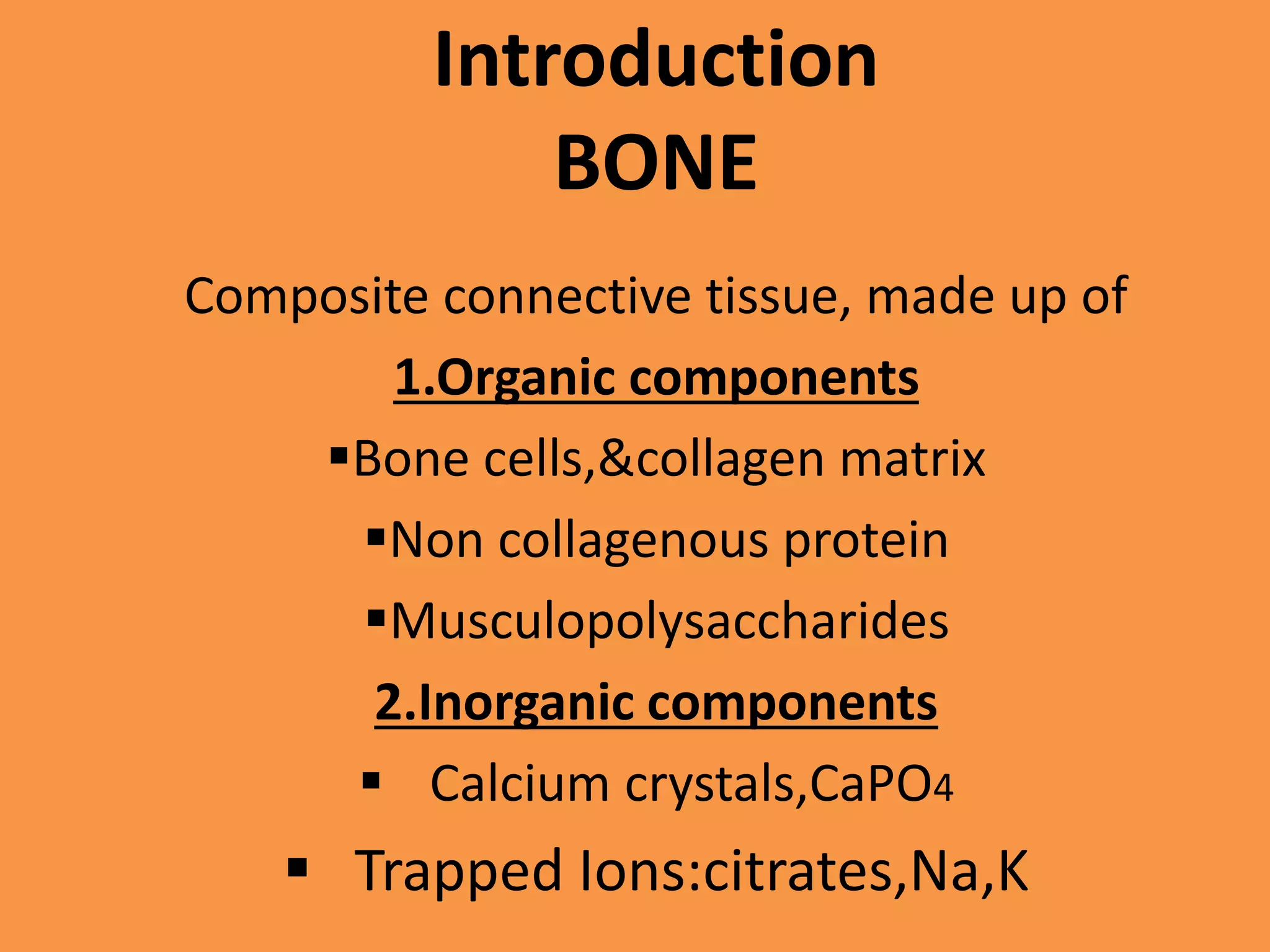
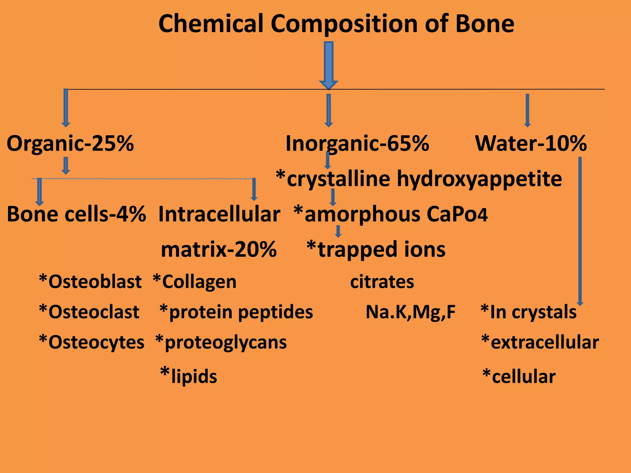
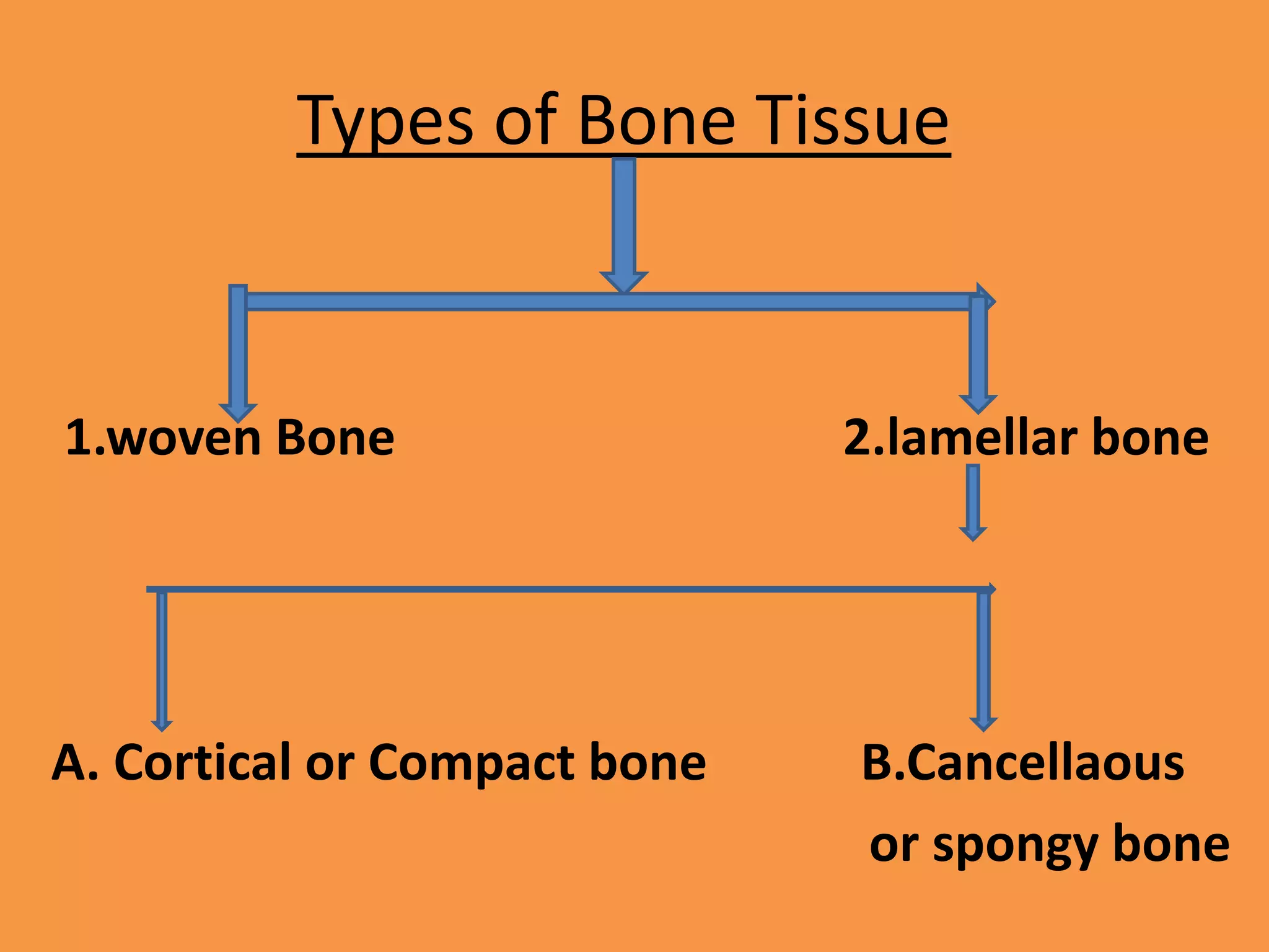
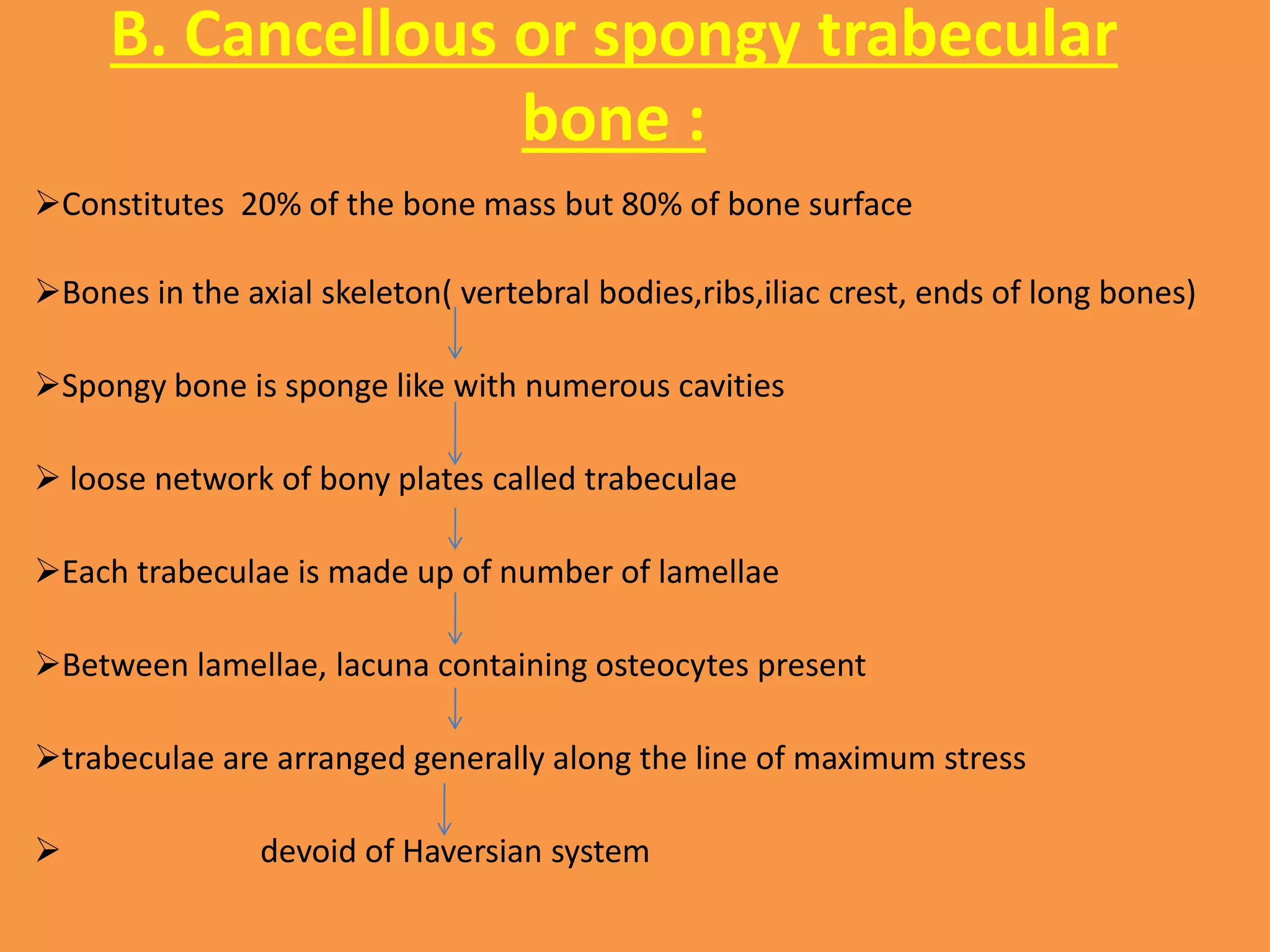
ANATOMY OF BONE
STRUCTURE,FUNCTION,CLASSIFICATION,
DEVELOPMENT AND BLOOD SUPPLY OF BONE.
Presenter:Dr.Siddique H.Ranna
PG Student
Moderator:Dr.Pranjal Tahbildar
Prof&HOD
Introduction
BONE
Composite connective tissue, made up of
1.Organic components
Bone cells,&collagen matrix
Non collagenous protein
Musculopolysaccharides
2.Inorganic components
 Calcium crystals,CaPO4
 Trapped Ions:citrates,Na,K
Chemical Composition of Bone
Organic-25% Inorganic-65% Water-10%
*crystalline hydroxyappetite
Bone cells-4% Intracellular *amorphous CaPo4
matrix-20% *trapped ions
*Osteoblast *Collagen citrates
*Osteoclast *protein peptides Na.K,Mg,F *In crystals
*Osteocytes *proteoglycans *extracellular
*lipids *cellular
FUNCTIONS OF BONE
1.MECHANICAL
*Protection
*Gives structure
*Facilitates movement
*Facilitates hearing
2.HEAMOPOITIC- *Produce Blood Cells
3.METABOLIC
*Mineral Storage
*Growth factor storage(IGF,TGF,BMP,etc)
*Fat storage
*Endocrine (FGF-23,Osteocalcin
Classification of Bones on the
Basis of Shape
Copyright © 2003 Pearson Education, Inc. publishing as Benjamin Cummings
Figure 5.1
Types of Bone Tissue
1.woven Bone 2.lamellar bone
A. Cortical or Compact bone B.Cancellaous
or spongy bone
1. WOVEN BONE OR IMMATURE
BONE
• Collagen fibers run in various direction, less
organized than lamellar bone
• Found in early phase of bone development,
bone remodeling or fracture healing
• Mineralized but less biomechanical strength
than lamellar bone
• This type of bone replaced by lamellar bone
latter.
2. LAMELLAR BONE OR MATURE
BONE
Collagen fibers are oriented in layers(lamellae)
Within each layer collagen fibers are parallel.
Fiber direction differ from layer to layer.
It provides tensile strength to the bone
Osteocytes are located at the borders between
these lamellae
A. Cortical or compact bone :
Constitute 80% of bone mass.
But only 20% of bone surface.
 consists of numerous cylindrical units known as
Haversian systems.
B. Cancellous or spongy trabecular
bone :
Constitutes 20% of the bone mass but 80% of bone surface
Bones in the axial skeleton( vertebral bodies,ribs,iliac crest, ends of long bones)
Spongy bone is sponge like with numerous cavities
 loose network of bony plates called trabeculae
Each trabeculae is made up of number of lamellae
Between lamellae, lacuna containing osteocytes present
trabeculae are arranged generally along the line of maximum stress
 devoid of Haversian system
Copyright 2009 John Wiley & Sons, Inc.
Histology of spongy bone
Ossification of Bone
Intramembranous Enchondral
form flat bones form long bones
Begins at 8wk begins at 2 months
Develops from fibrous develops from carti-
membrane -lage model
Intramembranous Ossification
(prenatal)
Mesenchymal cells create
fibrous CT framework for
ossification
Some mesenchymal cells
differentiate into osteoblasts
in an ossification center
Osteoblasts secrete bone matrix, osteoid
Intramembranous Ossification
Mineralization and
calcification of osteoid
Trapped osteoblasts become
osteocytes
Intramembranous Ossification (prenatal)
Osteoid accumulates in
between embryonic
blood vessels, creating
trabeculae of woven
bone.
Mesenchyme on bone
face condense and
differentiate into
periosteum
Intramembranous Ossification (prenatal)
A bone collar of thickly woven
osteoid forms around
trabeculae and ossifies into
compact bone
Spongy bone (diploë)
cavities made up of
trabeculae fill with red
marrow created from
vessels (vascular tissue)
overview
Endochondral Ossification
Bone collar formed around
diaphysis by osteoblasts
located on inner side of
periosteum
Endochondral Ossification
Cartilage in primary
ossification center calcifies,
then the cells die and
cavities form (cavitates)
Bone collar provides stability
during cavitation
Cartilage elsewhere continues to
elongate
Endochondral Ossification
Periosteal bud (lymph, blood
vessels, nerves, red marrow,
osteoblasts and osteoclasts)
enters cavity and builds
spongy bone
Endochondral Ossification
Osteoclasts dissolve spongy
bone to create medullary
cavity
Secondary Ossification Center
forms in epiphysis
Endochondral Ossification
Secondary Ossification Center
does NOT calcify. Spongy bone
retained.
Hyaline only remains on
epiphyseal surface (articular
cartilage) and at diaphysis and
epiphysis junction, to form the
epiphyseal plates.
Growing taller
throughout childhood!
When does lengthening stop?
• End of adolescence - lengthening stops
– Chondrocytes stop mitosis.
– Plate thins out and replaced by bone
– Diaphysis and epiphysis fuse to be one bone
• Epiphyseal plate closure (18 yr old females, 21 yr old
males)
• Thickening of bone continuous throughout life
Bone is Dynamic!
Bone is constantly remodeling and recycling
• Coupled process between:
1. Bone deposition (by osteoblasts)
2. Bone destruction/resorption (by osteoclasts)
• 5-7% of bone mass recycled weekly
• All spongy bone replaced every 3-4 years.
• All compact bone replaced every 10 years.
Prevents mineral salts from crystallizing; protecting against brittle
bones and fractures
Copyright 2009 John Wiley & Sons, Inc.
Bone tissue cell types
• Osteoprogenic cells – unspecialized stem
cells
• Osteoblasts – bone builders
• Osteocytes – mature bone cells derived
from osteoblasts
• Osteoclasts – bone ‘breakers’ are
multinucleate
Osteoblast Differentiation
Multinucleated cell the originates from hematopoietic stem cells
Characterized by possessing tartrate-resistant acid phosphatase (TRAP) within its
cytoplasmic vesicles and vacuoles
Found against bone surface in hollowed depressions called Howship’s lacunae
RANK: receptor-activated nuclear factor κB and RANKL: RANK ligand
Copyright 2009 John Wiley & Sons, Inc.
Blood supply of a long bone
THANK YOU
ALL

Anatomy of bone..latest

  • 1.
    ANATOMY OF BONE STRUCTURE,FUNCTION,CLASSIFICATION, DEVELOPMENTAND BLOOD SUPPLY OF BONE. Presenter:Dr.Siddique H.Ranna PG Student Moderator:Dr.Pranjal Tahbildar Prof&HOD
  • 2.
    Introduction BONE Composite connective tissue,made up of 1.Organic components Bone cells,&collagen matrix Non collagenous protein Musculopolysaccharides 2.Inorganic components  Calcium crystals,CaPO4  Trapped Ions:citrates,Na,K
  • 3.
    Chemical Composition ofBone Organic-25% Inorganic-65% Water-10% *crystalline hydroxyappetite Bone cells-4% Intracellular *amorphous CaPo4 matrix-20% *trapped ions *Osteoblast *Collagen citrates *Osteoclast *protein peptides Na.K,Mg,F *In crystals *Osteocytes *proteoglycans *extracellular *lipids *cellular
  • 4.
    FUNCTIONS OF BONE 1.MECHANICAL *Protection *Givesstructure *Facilitates movement *Facilitates hearing 2.HEAMOPOITIC- *Produce Blood Cells 3.METABOLIC *Mineral Storage *Growth factor storage(IGF,TGF,BMP,etc) *Fat storage *Endocrine (FGF-23,Osteocalcin
  • 5.
    Classification of Boneson the Basis of Shape Copyright © 2003 Pearson Education, Inc. publishing as Benjamin Cummings Figure 5.1
  • 9.
    Types of BoneTissue 1.woven Bone 2.lamellar bone A. Cortical or Compact bone B.Cancellaous or spongy bone
  • 10.
    1. WOVEN BONEOR IMMATURE BONE • Collagen fibers run in various direction, less organized than lamellar bone • Found in early phase of bone development, bone remodeling or fracture healing • Mineralized but less biomechanical strength than lamellar bone • This type of bone replaced by lamellar bone latter.
  • 11.
    2. LAMELLAR BONEOR MATURE BONE Collagen fibers are oriented in layers(lamellae) Within each layer collagen fibers are parallel. Fiber direction differ from layer to layer. It provides tensile strength to the bone Osteocytes are located at the borders between these lamellae
  • 12.
    A. Cortical orcompact bone : Constitute 80% of bone mass. But only 20% of bone surface.  consists of numerous cylindrical units known as Haversian systems.
  • 13.
    B. Cancellous orspongy trabecular bone : Constitutes 20% of the bone mass but 80% of bone surface Bones in the axial skeleton( vertebral bodies,ribs,iliac crest, ends of long bones) Spongy bone is sponge like with numerous cavities  loose network of bony plates called trabeculae Each trabeculae is made up of number of lamellae Between lamellae, lacuna containing osteocytes present trabeculae are arranged generally along the line of maximum stress  devoid of Haversian system
  • 14.
    Copyright 2009 JohnWiley & Sons, Inc. Histology of spongy bone
  • 15.
    Ossification of Bone IntramembranousEnchondral form flat bones form long bones Begins at 8wk begins at 2 months Develops from fibrous develops from carti- membrane -lage model
  • 16.
    Intramembranous Ossification (prenatal) Mesenchymal cellscreate fibrous CT framework for ossification Some mesenchymal cells differentiate into osteoblasts in an ossification center Osteoblasts secrete bone matrix, osteoid
  • 17.
    Intramembranous Ossification Mineralization and calcificationof osteoid Trapped osteoblasts become osteocytes
  • 18.
    Intramembranous Ossification (prenatal) Osteoidaccumulates in between embryonic blood vessels, creating trabeculae of woven bone. Mesenchyme on bone face condense and differentiate into periosteum
  • 19.
    Intramembranous Ossification (prenatal) Abone collar of thickly woven osteoid forms around trabeculae and ossifies into compact bone Spongy bone (diploë) cavities made up of trabeculae fill with red marrow created from vessels (vascular tissue) overview
  • 20.
    Endochondral Ossification Bone collarformed around diaphysis by osteoblasts located on inner side of periosteum
  • 21.
    Endochondral Ossification Cartilage inprimary ossification center calcifies, then the cells die and cavities form (cavitates) Bone collar provides stability during cavitation Cartilage elsewhere continues to elongate
  • 22.
    Endochondral Ossification Periosteal bud(lymph, blood vessels, nerves, red marrow, osteoblasts and osteoclasts) enters cavity and builds spongy bone
  • 23.
    Endochondral Ossification Osteoclasts dissolvespongy bone to create medullary cavity Secondary Ossification Center forms in epiphysis
  • 24.
    Endochondral Ossification Secondary OssificationCenter does NOT calcify. Spongy bone retained. Hyaline only remains on epiphyseal surface (articular cartilage) and at diaphysis and epiphysis junction, to form the epiphyseal plates.
  • 25.
  • 27.
    When does lengtheningstop? • End of adolescence - lengthening stops – Chondrocytes stop mitosis. – Plate thins out and replaced by bone – Diaphysis and epiphysis fuse to be one bone • Epiphyseal plate closure (18 yr old females, 21 yr old males) • Thickening of bone continuous throughout life
  • 28.
    Bone is Dynamic! Boneis constantly remodeling and recycling • Coupled process between: 1. Bone deposition (by osteoblasts) 2. Bone destruction/resorption (by osteoclasts) • 5-7% of bone mass recycled weekly • All spongy bone replaced every 3-4 years. • All compact bone replaced every 10 years. Prevents mineral salts from crystallizing; protecting against brittle bones and fractures
  • 29.
    Copyright 2009 JohnWiley & Sons, Inc. Bone tissue cell types • Osteoprogenic cells – unspecialized stem cells • Osteoblasts – bone builders • Osteocytes – mature bone cells derived from osteoblasts • Osteoclasts – bone ‘breakers’ are multinucleate
  • 30.
  • 31.
    Multinucleated cell theoriginates from hematopoietic stem cells Characterized by possessing tartrate-resistant acid phosphatase (TRAP) within its cytoplasmic vesicles and vacuoles Found against bone surface in hollowed depressions called Howship’s lacunae RANK: receptor-activated nuclear factor κB and RANKL: RANK ligand
  • 32.
    Copyright 2009 JohnWiley & Sons, Inc. Blood supply of a long bone
  • 33.