The document discusses the skeletal system and connective tissues. It covers the definitions of osteology and arthrology, the study of bones and joints. The skeletal system is composed of bones, cartilage, ligaments and other connective tissues. Cartilage is weaker but more flexible than bone. There are three types of cartilage - hyaline, fibrocartilage, and elastic cartilage. Bones provide structure, protection, movement, mineral storage and blood cell formation. The two types of ossification that form bones are intramembranous and endochondral ossification.
Bone tissue also called (osseous tissue) is a type of specialized dense connective tissue.
Histology
Junqueira’s Basic Histology Text and Atlas, 15th Ed
The presentation include general definition of bone and it's functions. Also, describe the chemical composition of bone and then specifically describe alveolar process.
Ossification (Intracartilaginous and Intramembranous)Mohiuddin Masum
This presentation includes:
* Ossification definition
* Types of ossification
* Center of ossification
* Intramembranous ossification process
* Intracartilaginous ossification process
a brief ppt description about cartilage which may be usefull for teaching for first year mbbs, bds and paramedical students, hope it is helpfull to everyone
Bone tissue is the major structural and supportive connective tissue of the body. Osseous tissue forms the rigid part of the bones that make up the skeletal system.
TEST BANK for Operations Management, 14th Edition by William J. Stevenson, Ve...kevinkariuki227
TEST BANK for Operations Management, 14th Edition by William J. Stevenson, Verified Chapters 1 - 19, Complete Newest Version.pdf
TEST BANK for Operations Management, 14th Edition by William J. Stevenson, Verified Chapters 1 - 19, Complete Newest Version.pdf
Prix Galien International 2024 Forum ProgramLevi Shapiro
June 20, 2024, Prix Galien International and Jerusalem Ethics Forum in ROME. Detailed agenda including panels:
- ADVANCES IN CARDIOLOGY: A NEW PARADIGM IS COMING
- WOMEN’S HEALTH: FERTILITY PRESERVATION
- WHAT’S NEW IN THE TREATMENT OF INFECTIOUS,
ONCOLOGICAL AND INFLAMMATORY SKIN DISEASES?
- ARTIFICIAL INTELLIGENCE AND ETHICS
- GENE THERAPY
- BEYOND BORDERS: GLOBAL INITIATIVES FOR DEMOCRATIZING LIFE SCIENCE TECHNOLOGIES AND PROMOTING ACCESS TO HEALTHCARE
- ETHICAL CHALLENGES IN LIFE SCIENCES
- Prix Galien International Awards Ceremony
New Directions in Targeted Therapeutic Approaches for Older Adults With Mantl...i3 Health
i3 Health is pleased to make the speaker slides from this activity available for use as a non-accredited self-study or teaching resource.
This slide deck presented by Dr. Kami Maddocks, Professor-Clinical in the Division of Hematology and
Associate Division Director for Ambulatory Operations
The Ohio State University Comprehensive Cancer Center, will provide insight into new directions in targeted therapeutic approaches for older adults with mantle cell lymphoma.
STATEMENT OF NEED
Mantle cell lymphoma (MCL) is a rare, aggressive B-cell non-Hodgkin lymphoma (NHL) accounting for 5% to 7% of all lymphomas. Its prognosis ranges from indolent disease that does not require treatment for years to very aggressive disease, which is associated with poor survival (Silkenstedt et al, 2021). Typically, MCL is diagnosed at advanced stage and in older patients who cannot tolerate intensive therapy (NCCN, 2022). Although recent advances have slightly increased remission rates, recurrence and relapse remain very common, leading to a median overall survival between 3 and 6 years (LLS, 2021). Though there are several effective options, progress is still needed towards establishing an accepted frontline approach for MCL (Castellino et al, 2022). Treatment selection and management of MCL are complicated by the heterogeneity of prognosis, advanced age and comorbidities of patients, and lack of an established standard approach for treatment, making it vital that clinicians be familiar with the latest research and advances in this area. In this activity chaired by Michael Wang, MD, Professor in the Department of Lymphoma & Myeloma at MD Anderson Cancer Center, expert faculty will discuss prognostic factors informing treatment, the promising results of recent trials in new therapeutic approaches, and the implications of treatment resistance in therapeutic selection for MCL.
Target Audience
Hematology/oncology fellows, attending faculty, and other health care professionals involved in the treatment of patients with mantle cell lymphoma (MCL).
Learning Objectives
1.) Identify clinical and biological prognostic factors that can guide treatment decision making for older adults with MCL
2.) Evaluate emerging data on targeted therapeutic approaches for treatment-naive and relapsed/refractory MCL and their applicability to older adults
3.) Assess mechanisms of resistance to targeted therapies for MCL and their implications for treatment selection
micro teaching on communication m.sc nursing.pdfAnurag Sharma
Microteaching is a unique model of practice teaching. It is a viable instrument for the. desired change in the teaching behavior or the behavior potential which, in specified types of real. classroom situations, tends to facilitate the achievement of specified types of objectives.
Pulmonary Thromboembolism - etilogy, types, medical- Surgical and nursing man...VarunMahajani
Disruption of blood supply to lung alveoli due to blockage of one or more pulmonary blood vessels is called as Pulmonary thromboembolism. In this presentation we will discuss its causes, types and its management in depth.
Tom Selleck Health: A Comprehensive Look at the Iconic Actor’s Wellness Journeygreendigital
Tom Selleck, an enduring figure in Hollywood. has captivated audiences for decades with his rugged charm, iconic moustache. and memorable roles in television and film. From his breakout role as Thomas Magnum in Magnum P.I. to his current portrayal of Frank Reagan in Blue Bloods. Selleck's career has spanned over 50 years. But beyond his professional achievements. fans have often been curious about Tom Selleck Health. especially as he has aged in the public eye.
Follow us on: Pinterest
Introduction
Many have been interested in Tom Selleck health. not only because of his enduring presence on screen but also because of the challenges. and lifestyle choices he has faced and made over the years. This article delves into the various aspects of Tom Selleck health. exploring his fitness regimen, diet, mental health. and the challenges he has encountered as he ages. We'll look at how he maintains his well-being. the health issues he has faced, and his approach to ageing .
Early Life and Career
Childhood and Athletic Beginnings
Tom Selleck was born on January 29, 1945, in Detroit, Michigan, and grew up in Sherman Oaks, California. From an early age, he was involved in sports, particularly basketball. which played a significant role in his physical development. His athletic pursuits continued into college. where he attended the University of Southern California (USC) on a basketball scholarship. This early involvement in sports laid a strong foundation for his physical health and disciplined lifestyle.
Transition to Acting
Selleck's transition from an athlete to an actor came with its physical demands. His first significant role in "Magnum P.I." required him to perform various stunts and maintain a fit appearance. This role, which he played from 1980 to 1988. necessitated a rigorous fitness routine to meet the show's demands. setting the stage for his long-term commitment to health and wellness.
Fitness Regimen
Workout Routine
Tom Selleck health and fitness regimen has evolved. adapting to his changing roles and age. During his "Magnum, P.I." days. Selleck's workouts were intense and focused on building and maintaining muscle mass. His routine included weightlifting, cardiovascular exercises. and specific training for the stunts he performed on the show.
Selleck adjusted his fitness routine as he aged to suit his body's needs. Today, his workouts focus on maintaining flexibility, strength, and cardiovascular health. He incorporates low-impact exercises such as swimming, walking, and light weightlifting. This balanced approach helps him stay fit without putting undue strain on his joints and muscles.
Importance of Flexibility and Mobility
In recent years, Selleck has emphasized the importance of flexibility and mobility in his fitness regimen. Understanding the natural decline in muscle mass and joint flexibility with age. he includes stretching and yoga in his routine. These practices help prevent injuries, improve posture, and maintain mobilit
These lecture slides, by Dr Sidra Arshad, offer a quick overview of physiological basis of a normal electrocardiogram.
Learning objectives:
1. Define an electrocardiogram (ECG) and electrocardiography
2. Describe how dipoles generated by the heart produce the waveforms of the ECG
3. Describe the components of a normal electrocardiogram of a typical bipolar leads (limb II)
4. Differentiate between intervals and segments
5. Enlist some common indications for obtaining an ECG
Study Resources:
1. Chapter 11, Guyton and Hall Textbook of Medical Physiology, 14th edition
2. Chapter 9, Human Physiology - From Cells to Systems, Lauralee Sherwood, 9th edition
3. Chapter 29, Ganong’s Review of Medical Physiology, 26th edition
4. Electrocardiogram, StatPearls - https://www.ncbi.nlm.nih.gov/books/NBK549803/
5. ECG in Medical Practice by ABM Abdullah, 4th edition
6. ECG Basics, http://www.nataliescasebook.com/tag/e-c-g-basics
Lung Cancer: Artificial Intelligence, Synergetics, Complex System Analysis, S...Oleg Kshivets
RESULTS: Overall life span (LS) was 2252.1±1742.5 days and cumulative 5-year survival (5YS) reached 73.2%, 10 years – 64.8%, 20 years – 42.5%. 513 LCP lived more than 5 years (LS=3124.6±1525.6 days), 148 LCP – more than 10 years (LS=5054.4±1504.1 days).199 LCP died because of LC (LS=562.7±374.5 days). 5YS of LCP after bi/lobectomies was significantly superior in comparison with LCP after pneumonectomies (78.1% vs.63.7%, P=0.00001 by log-rank test). AT significantly improved 5YS (66.3% vs. 34.8%) (P=0.00000 by log-rank test) only for LCP with N1-2. Cox modeling displayed that 5YS of LCP significantly depended on: phase transition (PT) early-invasive LC in terms of synergetics, PT N0—N12, cell ratio factors (ratio between cancer cells- CC and blood cells subpopulations), G1-3, histology, glucose, AT, blood cell circuit, prothrombin index, heparin tolerance, recalcification time (P=0.000-0.038). Neural networks, genetic algorithm selection and bootstrap simulation revealed relationships between 5YS and PT early-invasive LC (rank=1), PT N0—N12 (rank=2), thrombocytes/CC (3), erythrocytes/CC (4), eosinophils/CC (5), healthy cells/CC (6), lymphocytes/CC (7), segmented neutrophils/CC (8), stick neutrophils/CC (9), monocytes/CC (10); leucocytes/CC (11). Correct prediction of 5YS was 100% by neural networks computing (area under ROC curve=1.0; error=0.0).
CONCLUSIONS: 5YS of LCP after radical procedures significantly depended on: 1) PT early-invasive cancer; 2) PT N0--N12; 3) cell ratio factors; 4) blood cell circuit; 5) biochemical factors; 6) hemostasis system; 7) AT; 8) LC characteristics; 9) LC cell dynamics; 10) surgery type: lobectomy/pneumonectomy; 11) anthropometric data. Optimal diagnosis and treatment strategies for LC are: 1) screening and early detection of LC; 2) availability of experienced thoracic surgeons because of complexity of radical procedures; 3) aggressive en block surgery and adequate lymph node dissection for completeness; 4) precise prediction; 5) adjuvant chemoimmunoradiotherapy for LCP with unfavorable prognosis.
Ozempic: Preoperative Management of Patients on GLP-1 Receptor Agonists Saeid Safari
Preoperative Management of Patients on GLP-1 Receptor Agonists like Ozempic and Semiglutide
ASA GUIDELINE
NYSORA Guideline
2 Case Reports of Gastric Ultrasound
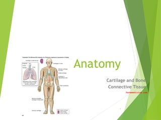
2. General Osteology
/Arthrology
Definitions:
Osteology: the study of bones
Bones: organs of the skeletal system
Skeletal System: bones and associated cartilages
Arthrology: the study of joints
Point of movement (fulcrum)
Endoskeleton: internal skeleton
endo- = inside
Versus exoskeleton
6-2
3. Skeletal System
Composed of dynamic living tissues
Osseous tissue, cartilage, fibrous CT, blood, nervous
tissue.
Continually rebuilds and remodels itself
Changes over a lifetime
Interacts with all of the other organ systems.
Includes:
bones of the skeleton
Cartilage
Ligaments
other connective tissues that stabilize or connect the
bones. 6-3
4. Skeletal System
Functions:
Supports our weight.
Interacts with muscles to produce
movements.
Protection
Blood cell formation
Red bone marrow
Mineral storage
Calcium
phosphate
6-4
5. Cartilage Connective Tissue
Characteristics:
Weaker than bone
More flexible than bone
Cells in an abundant matrix.
Cell Types
Chondroblasts
Chondrocytes in lacunae
Avascular
6-5
6. 3 Major Functions of
Cartilage
Supporting soft tissues.
Providing a gliding surface at articulations (joints)
Providing a model for the formation of most of the
bones in the body.
6-6
7. Types of Cartilage
Three types of cartilage:
Hyaline cartilage
Most abundant kind
Has a perichondrium (membrane)
Associated with synovial joints
Most bones first modeled in hyaline cartilage
Fibrocartilage
Has collagen fibers
Intervertebral discs, pubic symphysis
Elastic cartilage
Has elastic fibers
Ear, respiratory tubing
6-7
9. Growth Patterns of Cartilage
Two main types:
Interstitial Growth
Appositional Growth.
Interstitial Growth.
Chondrocytes in lacuna undergoes mitosis.
Two chondrocytes in one lacuna
Will push apart, form separate lacuna
6-9
11. Growth Patterns of Cartilage
Appositional Growth.
Undifferentiated cells divide (mitosis)
One daughter cell remains a stem cell, one
differentiates into a committed cell.
Committed cell further differentiates into
chondroblast
Located at edge of cartilage
Both types common during growth
Later, mostly appositional
In adult, usually no growth unless for repair
6-11
12. Bone
Bones are organs
Bones are composed of all tissue types.
Their primary component is osseous connective tissue.
The matrix is sturdy and rigid due to calcification (also called
mineralization).
6-12
13. Functions of Bone
Support.
Protection.
Movement
Hemopoiesis
Storage of minerals.
Energy Reserves (marrow)
6-13
14. Support and Protection
Bones provide structural support and serve as a
framework for the entire body.
Bones protect many delicate tissues and organs from
injury and trauma.
6-14
15. Movement
Muscles attach to the bones of the
skeleton
contract and pull on bone
functions as a series of levers.
6-15
16. Hemopoiesis
Blood cell production in red bone marrow
located in some spongy bone.
Red bone marrow contains stem cells
form all of the blood cell types.
6-16
17. Storage of Mineral and Energy
Reserves
More than 90% of the body’s reserves
of the minerals calcium and phosphate
are stored and released by bone.
Calcium: needed for
muscle contraction
blood clotting
nerve impulse transmission.
Phosphate: needed for
ATP utilization
structure of nucleic acids (DNA, RNA)
6-17
23. Structure of Long Bone
Endostium: lines marrow cavity, incomplete
Osteoprogenitor cells
Osteoblasts
Osteoclasts
Periostium: covers bone everywhere but articular
surfaces
Two layers
Fibrous layer: outermost, dense irregular CT
Site of tendon attachment
Inner layer: next to compact bone
Osteoblasts present in young bone
Anchored to bone by perforating fibers (collagen)
6-23
25. Flat Bones of the Skull
Two layers of compact bone
Inner table
Outer table
Region of spongy bone sandwiched between them
Called the diploe
Both layers of compact bone are covered by periosteum
6-25
27. Four Types of Bone Cells
Osteoprogenitor cells
stem cells derived from mesenchyme which produce
other stem cells and osteoblasts
Osteoblasts
produce new bone, and once osteoblasts become
entrapped in the matrix they produce and secrete, they
differentiate into osteocytes
Osteocytes
mature bone cells
Osteoclasts: not derived form osteoprogenitors
Related to macrophages
Formed from multiple cells; are multinucleated
are involved in bone resorption 6-27
33. Compact Bone Microanatomy
Osteon (Haversian) system: basic unit
Central (Haversian) canal
Concentric lamellae
Contain collagen fibers
Osteocytes
Lacunae
Canaliculi: permit intercellular communication
Cylinder that runs with long axis of long bone
6-33
34. Compact Bone Microanatomy
Perforating canals (Volkmann canals)
Contain blood vessels, nerve
Run perpendicular to central canals, connect them
Circumferential lamellae
Internal to periostium
External circumferential lamellae
Internal to endosteum
Internal circumferential lamellae
Run the entire circumference
Interstitial lamellae
Remains of osteons 6-34
41. Ossification
Osteogenesis: bone formation and
development
Begins in the embryo: By the eighth through
twelfth weeks:
the skeleton begins forming:
from mesenchyme
or from a hyaline cartilage model of bone.
These models are replaced by hard bone
Continues during childhood and adolescence.
In the adult, ossification contin6u-41es.
42. Intramembranous Ossification
Also called dermal ossification
Produces:
the flat bones of the skull (cranial vault)
some of the facial bones (zygomatic bone,
maxilla), the mandible (lower jaw)
the central part of the clavicle
(collarbone).
It begins when mesenchyme becomes
thickened and condensed with a dense
supply of blood capillaries.
6-42
43. Intramembranous Ossification
1. Ossification centers
form in thickened
mesenchyme
Osteoprogenitors
develop, become
osteoblasts
2. Osteoid (bone matrix)
calcifies
Trapped osteoblasts
become osteocytes
6-43
44. Intramembranous Ossification
3. Woven bone
(primary bone)
forms, periostium
forms (from
mesenchyme)
4. Lamellar bone
(secondary bone)
replaces woven
bone; compact and
spongy bone form
6-44
45. Endochondral Ossification
Begins with a hyaline cartilage model
Produces most of the other bones of the skeleton
Long bone will be used as an example.
6-45
46. Endochondral Ossification
Steps:
1. Cartilage model develops:
Chondroblasts become
chondrocytes
Perichondrium develops
2. Cartilage calcification, bone collar
develops in shaft
Chondrocytes hypertrophy, then
die
Blood vessels grow toward cartilage
Osteoblasts under perichondrium
form bone
3. Primary Ossification center forms:
Periosteal bud: osteoblasts and
blood vessels
12th week: most have formed 6-46
47. Endochondral Ossification
Steps:
3. Secondary Ossification centers:
In epiphysis
Some form post-natally
4. Cartilage replaced by bone
Except articular cartilage,
epiphyseal plate
5. Epiphyseal plate ossifies:
Forms epiphyseal line
Between 10 and 25
Last… clavicle 6-47
48. Epiphyseal Plate Morphology
Hyaline cartilage
5 zones: from epiphysis to diaphysis
Zone of resting cartilage
Small chondrocytes in cartilage matrix
Looks like healthy cartilage
Secures epiphyseal plate to epiphysis
Zone of proliferating cartilage
Chondrocytes here are undergoing rapid
mitosis
Stack up in columns
6-48
49. Epiphyseal Plate Morphology
Zone of hypertrophic cartilage
Chondrocytes stop dividing
Start hypertrophy
Absorb matrix
Zone of calcified cartilage
Few cells thick
Calcification of matrix
Kills the chondrocytes
Zone of ossification
Invasion by capillaries and osteoprogenitor
cells
6-49
51. Bone Growth
Interstitial growth occurs in the epiphyseal plate as
chondrocytes undergo mitosis
Growth in length
Appositional growth occurs within the periosteum.
Growth in diameter, thickness
6-51
52. Bone Remodeling
The continual deposition of new bone tissue and the
removal (resorption) of old bone tissue.
helps maintain calcium and phosphate levels in body
fluids, and can be stimulated by stress on a bone
occurs at both the periosteal and endosteal surfaces of a
bone
Relative rates differ with age, bone
6-52
53. Blood Supply and Innervation
Bone is highly vascularized, especially in regions containing red bone
marrow.
Kinds of blood vessels
Nutrient artery and the nutrient vein
supply the diaphysis of a long bone
Metaphyseal blood vessels
Diaphyseal face of epiphyseal plate
Periosteal blood vessels
Supply superficial osteons on diaphysis.
6-53
56. Effects of Hormones
Control and regulate growth patterns
in bone by altering the rates of both
osteoblast and osteoclast activity.
Growth hormone (Pituitary gland):
affects bone growth by stimulating the
formation of another hormone,
somatomedin which is produced by the
liver.
Somatomedin: directly stimulates
growth of cartilage in the epiphyseal
plate.
6-56
57. Effects of Hormones
Thyroid hormone (Thyroid gland): stimulates
bone growth.
Growth hormone and thyroid hormone
regulate and maintain normal activity at the
epiphyseal plates until puberty.
Calcitonin (Thyroid gland): inhibits osteoclast
activity.
Parathyroid Hormone (Parathyroid gland):
increases blood calcium levels, stimulates
osteoclast activity
Sex Hormones: gonads
Increase rate of bone formation
Production associated with puberty
6-57
58. Effects of Vitamins
Vitamin A: activates osteoblasts
Vitamin C: normal synthesis of collagen
Vitamin D: absorption and transport of calcium and
phosphate
6-58