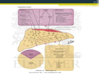
BONE
DR. IMAN ZUBAIR KHAN – JR 1
DEPT. OF ORAL MEDICINE &
RADIOLOGY
1
CONTENTS
• INTRODUCTION
• DEFINATION
• FUNCTIONS OF BONE
• CLASSIFICATION OF BONE
• COMPOSITION OF BONE
• STRUCTURE OF BONE
• BLOOD SUPPLY
• NERVE SUPPLY
• HISTOLOGY OF BONE
• OSSIFICATION OF BONE
• GROWTH OF BONE
• REMODELLING OF BONE
• EFFECT OF AGE ON BONE
• INVESTIGATIONS FOR BONE DISORDERS
• APPLIED ASPECTS
• REFERNCES
2
INTRODUCTION
• Skeleton is the main framework of our body that
supports and protects our organs. It is designed
for effective movements of the body
• Bone tissue and cartilage form the skeleton.
• Strong but light weight, bone is a dynamic, ever
changing tissue. Throughout life, it is continually
being broken down and formed.
3
DEFINATION
• According to Dorland’s medical dictionary:
Any distinct piece of the osseous framework, or skeleton,
of the body, called osseous tissue.
• According to GPT 8:
The hard portion of the connective tissue which
constitutes the majority of the skeleton; it consists of an
inorganic or mineral component and an organic
component (the matrix and cells); the matrix is
composed of collagenous fibers and is impregnated with
minerals, chiefly calcium phosphate (approx. 85%) and
calcium carbonate (approx. 10%), thus imparting the
quality of rigidity—called also osseous tissue .
4
FUNCTIONS OF BONE
• Shape and support to the body, and resist any
forms of stress.
• Provide surface for the attachment of muscles,
tendons, ligaments, etc.
• Levers for muscular actions.
• Protective in function.
• Bone marrow manufactures blood cells.
• Stores 97% of the body calcium and
phosphorus.
• Bone marrow contains reticuloendothelial cells
which are phagocytic in nature and take part in
immune responses of the body.
5
6
CLASSIFICATION OF BONE
• BY POSITION
• Axial skeleton :
Bones that are form the axis of body.
• Appendicular skeleton :
Bones that form the appendages of body.
7
8
• BY SHAPE
• Long bones: Each long bone has an elongated shaft and
two expanded ends which are smooth and articular.
(a) typical long bones like humerus, radius, ulna, femur,
tibia and fibula
(b) miniature long bones have only one epiphysis like
metacarpals, metatarsals and phalanges
(c) modified long bones have no medullary cavity like
clavicle
• Short bones: Their shape is usually cuboid, cuneiform,
trapezoid, or scaphoid. Eg- Tarsal and carpal bones
• Flat bones resemble shallow plates and form
boundaries of certain body cavities. Eg- Bones in the
vault of the skull, ribs, sternum and scapula.
9
• Irregular bones Eg- Vertebra, hip bone, and
bones in the base of the skull.
• Pneumatic bones: Certain irregular bones
contain large air spaces lined by epithelium Eg-
Maxilla, sphenoid, ethmoid
• Sesamoid bones: These are bony nodules found
embedded in the tendons or joint capsules. They
have no periosteum and ossify after birth. They
are related to an articular or nonarticular bony
surface, and the surfaces of contact are covered
with hyaline cartilage and lubricated by a
synovial membrane. Eg- Patella
10
11
• BY DEVELOPMENT
• Bones that ossify in membrane and are derived from
mesenchymal condensations.
Eg: Bones of the vault of skull and facial bones.
• Bones that ossify in cartilage and are derived from
pre-formed cartilaginous models.
Eg: Bones of limbs, vertebral column and thoracic
cage.
• Membrano-cartilaginous bones ossify partly in
membrane and partly in cartilage.
Eg: Clavicle, mandible, occipital,temporal, sphenoid.
12
• BY STRUCTURE
MACROSCOPICALLY
• Compact bone or cortical bone is dense in
texture . It is best developed in the cortex of the
long bones. This is an adaptation to bending and
twisting forces.
• Cancellous or spongy, or trabecular bone is open
in texture, and is made up of a meshwork of
trabeculae between which are marrow
containing spaces. This is an adaptation to
compressive forces.
13
14
• MICROSCOPICALLY
• Lamellar bone: Most of the mature human
bones,whether compact or cancellous, are
composed of thin plates of bony tissue called
lamellae. These are arranged in piles in a
cancellous bone, but in concentric cylinders
(Haversian system or secondary osteon) in a
compact bone.
• Woven Bone: seen in fetal bone, fracture repair
and in cancer of bone.
• Fibrous bone is found in young foetal bones, but
are common in reptiles and amphibia.
15
COMPOSITION OF BONE
16
17
ANATOMY OF BONE – Young bone
• Epiphysis - The ends and tips of a bone are
called epiphyses.
• Diaphysis - It is the elongated shaft of a long
bone.
• Metaphysis - The epiphysial ends of a diaphysis
are called metaphysis. Each metaphysis is the
zone of active growth. Before epiphysial fusion,
the metaphysis is richly supplied with blood
through end arteries forming 'hair-pin' bends.
18
• Epiphysial Plate of Cartilage - It separates
epiphysis from metaphysis.
Proliferation of cells in this cartilaginous plate is
responsible for lengthwise growth of a long bone.
After the epiphysial fusion, the bone can no longer
grow in length.
The growth cartilage is nourished by both the
epiphysial and metaphysial arteries.
19
20
ANATOMY OF BONE – Adult bone
• A typical long bone ossifies in three parts, the two ends
from secondary centres, and the intervening shaft from
a primary centre.
• Shaft: It is composed of periosteum, endosteum, cortex
and medullary cavity .
(a) Periosteum is a thick fibrous membrane covering the
surface of the bone. It is made up of an outer fibrous
layer, and an inner cellular layer .
Periosteum is united to the underlying bone by Sharpey's
fibres. At the articular margin the periosteum is
continuous with the capsule of the joint.
21
(b) Cortex is made up of a compact bone which gives
it the desired strength to withstand all possible
mechanical strains.
(c) Medullary cavity is filled with red or yellow bone
marrow.
At birth the marrow is red everywhere with
widespread active haemopoiesis. As the age
advances, the red marrow at many places atrophies
and is replaced by yellow, fatty marrow, with no
power of haemopoiesis. Red marrow persists in the
cancellous ends of long bones.
22
• The two ends of a long bone are made up of
cancellous bone covered with hyaline/articular
cartilage
23
24
25
BLOOD SUPPLY OF BONE
• Nutrient artery - It enters the shaft through the
nutrient foramen, runs through the cortex, and
divides into ascending and descending branches in
the medullary cavity.
Each branch divides into a number of small parallel
channels which terminate in the adult metaphysis by
anastomosing with the epiphysial, metaphysial and
periosteal arteries.
The nutrient artery supplies medullary cavity, inner
2/3 of cortex and metaphysis.
26
• Periosteal arteries - These are especially
numerous beneath the muscular and
ligamentous attachments.
They ramify beneath the periosteum and enter the
Volkmann's canals to supply the outer 1/3 of the
cortex.
• Epiphysial arteries - These are derived from
periarticular vascular arcades found on the
nonarticular bony surface.
• Metaphysial arteries - These are derived from
the neighbouring systemic vessels.
They pass directly into the metaphysis and
reinforce the metaphysial branches from the
primary nutrient artery.
27
28
NERVE SUPPLY OF BONE
• Nerves accompany the blood vessels. Most of them
are sympathetic and vasomotor in function
• A few of them are sensory which are distributed to
the articular ends and periosteum of the long
bones, to the vertebra, and to large flat bones.
29
HISTOLOGY OF BONE
• There are 4 types of cells in bone tissue.
• Osteoprogenitor cells - are unspecialized cells
derived from mesenchyme, the tissue from
which all connective tissues are derived. They
undergo mitosis and develop into osteoblasts.
They are found in the periosteum, endosteum
and in canals that contains blood vessels.
30
31
32
• Osteoblasts are the cells that form bone, but they
have lost the ability to divide by mitosis. They
secrete collagen and other organic components
needed to build bone tissue.
The differentiation of osteoproginitor cells into
osteoblast is acclererated by hormones and bone
proteins called Skeletal growth factors.
Their Functions are -
Role in formation of bone matrix
Role in calcification
Synthesis of proteins.
33
• Osteoclast - Concerned with bone resorption.
Giant phagocytic mutinucleated cells found in the
lacunae of bone matrix that are derived from
hemopoietic stem cells via monocytes.
• Their functions are -
Responsible for bone resorption during bone
remodelling.
Synthesis and release of lysosomal enzymes
necessary for bone resorption in to bone resorbing
compartment.
34
35
• Osteocytes- these cells are concerned with
maintenance of bone.
They are small flattened and rounded cells
embedded in bone lacunae and are derived from
mature Osteoblast.
• Their functions are -
Helping to maintain the bone as living tissue
because of their metabolic activity.
Maintain the exchange of calcium between the
bone and ECF.
36
37
38
• Bone lining cells
These cells form a continous epithelium like layer
on bony surfaces where active bone deposition or
removal is not taking place.
They are present on the periosteal as well as
endosteal surfaces.
39
• When we examine the structure of any adult
bone, we find that it is made up of layers or
LAMELLAE. This kind of bone is called
LAMELLAR BONE.
• Each lacuna contains 1 osteocyte. Spreading out
from each lacuna there are fine canals or
canaliculi that communicate with those from
other lacunae.
• The lamellar appearance of bone depends
mainly on the arrangement of collagen fibres.
The fibres of one lamellae run parallel to each
other
40
41
• In contrast to mature bone, newly formed bone
does not have lamellar structure. The bundles of
collagen fibers run randomly and interlace with
each other. Because of this, it is called WOVEN
BONE .
• All newly formed bones are woven bone. It is
later replaced by lamellar bone.
42
• COMPACT /CORTICAL BONE HISTOLOGY
• This type of bone is made up of lamellae, which
are arranged in the form of concentric rings that
surround a narrow HAVERSIAN CANAL present
at the center of each ring. The blood vessels,
lymphatic vessels and nerves from the
periosteum penetrate the compact bone through
PERFORATING or VOLKMANN’S CANALS. The
blood vessels and nerves of these canals connect
with blood vessels and nerves of medullary
cavity and periosteum and those of CENTRAL or
HAVERSIAN CANAL.
43
• One haversian canal and the lamellae around it
constitute a HAVERSIAN SYSTEM or OSTEON.
• The central canals run longitudinally through
bone. Around the canals are CONCENTRIC
LAMELLAE, rings of hard, calcified matrix.
• The area between osteons contains
INTERSTITIAL LAMELLAE. They also have
lacunae with osteocyte and canaliculi but their
lamellae are usually not connected to the
osteons.
44
Interstitial lamellae
Concentric lamellae
45
• SPONGY /TRABECULAR/CANCELLOUS BONE
HISTOLOGY
• In contrast to compact bone, spongy bone does
not contain true osteons. It consists of lamellae
arranged in an irregular latticework of thin
plates of bone called trabeculae.
• The spaces between the trabeculae of some
bones are filled with red marrow, which produce
blood cells.
46
47
48
OSSIFICATION OF BONE
• The process by which bone forms is called
OSSIFICATION.
• The skeleton of a human embryo is composed of
fibrous connective tissue membrane formed by
embryonic connective tissue (mesenchyme) and
hyaline cartilage that are loosely shaped like bones.
They provide supporting structure for ossification.
• Ossification begins around the 6th or 7th week of
embryonic life and continues throughout adulthood.
49
INTRAMEMBRANEOUS OSSIFICATION
• At the site where bone will develop,
mesenchymal cells become vascularized, cluster
and differentiate, first into osteoprogenitor cells
and then into osteoblasts.
• The site of such a cluster is called a CENTRE OF
OSSIFICATION.
• Osteoblasts secrete the organic matrix of bone
and gets surrounded to become osteocytes. Later
calcium & other minerals are deposited and
tissue calcifies
50
• As the bone matrix forms, it develops into
trabaculae. As trabaculae develop in various
ossification centres, they fuse with one another
to create the open latticework appearance of
spongy bone. Connective tissue in trabacular
spaces differentiates into red bone marrow.
• On the outside of bone, vascularized
mesenchyme develops into periosteum.
Eventually, Some of the spongy bone is replaced
by the cortical bone. This will remodeled to
reach its adult size & shape.
51
52
53
54
ENDOCHONDRAL OSSIFICATION
• It begins in the second month of development
and uses hyaline cartilage “bones” as models for
bone construction. It requires breakdown of
hyaline cartilage prior to ossification
• Development of the cartilage model -
Mesenchymal cells differentiate into
chondroblasts which form the hyaline cartilage
model and a membrane called
PERICHONDRIUM develops around the cartilage
.
55
• Growth of the cartilage model -
Cartilage model grows by interstitial & appositional
growth
The chondrocytes in mid-region calcify the matrix and
vacated lacunae form small cavities. The osteoblasts in
perichondrium produce periosteal bone collar.
• Development of primary ossification center -
Near the middle of the model, capillaries of the
periosteum grow into the disintegrating calcified
cartilage. These vessels and the osteoblasts, osteoclasts
& red marrow cells, are known as the PERIOSTEAL
BUD.
With the development of periosteal bud, the primary
ossification center and medullary cavity forms.
56
57
• Development of the diaphysis and epiphysis -
The diaphysis, which was once a solid mass of
hyaline cartilage, is replaced by compact bone.
When blood vessels enter the epiphysis, secondary
ossification centers develop.
58
59
60
BONE GROWTH
• Bones grow in length at the epiphyseal plate by a
process that is similar to endochondral ossification.
• The cartilage in the region of the epiphyseal plate
next to the epiphysis continues to grow by mitosis.
• The chondrocytes, in the region next to the
diaphysis, age and degenerate. Osteoblasts move in
and ossify the matrix to form bone. This process
continues throughout childhood and the adolescent
years until the cartilage growth slows and finally
stops.
• The epiphyseal plate completely ossifies so that only
a thin epiphyseal line remains and the bones can no
longer grow in length.
61
• Even though bones stop growing in length in
early adulthood, they can continue to increase in
thickness or diameter throughout life in
response to stress from increased muscle activity
or to weight. The increase in diameter is called
APPOSITIONAL GROWTH.
• Osteoblasts in the periosteum form compact
bone around the external bone surface. At the
same time, osteoclasts in the endosteum break
down bone on the internal bone surface, around
the medullary cavity. These two processes
together increase the diameter of the bone.
62
• ZONES IN BONE GROWTH
Growth zone – cartilage cells undergo mitosis,
pushing the epiphysis away from the diaphysis
Transformation zone – older cells enlarge, the
matrix becomes calcified, cartilage cells die, and
the matrix begins to deteriorate
Osteogenic zone – new bone formation occurs
63
REMODELLING OF BONE
• The ongoing replacement of old bone tissue by
new bone tissue is called REMODELLING OF
BONE.
• It takes place at different rates in various parts
of body.
• Osteoclasts are responsible for bone resorption
or destruction of matrix. A delicate homeostasis
exists between the actions of the osteoclasts in
removing minerals and collagen and of
osteoblasts in depositing them.
64
• ARF CYCLE -
ACTIVATION- osteoclasts are activated & begin
secreting acids to resorb bone.
RESORPTION- osteoclastic resoprtion occurs.
REVERSAL- resorption stops & osteoblast take
over.
FORMATION- osteoblast form bone on the
opposing surface to complete the bone reforming
process.
• This cycle takes about 100 days in Compact bone
& 200 days in Spongy bone.
65
66
67
68
ROLE OF VITAMINS MINERALS AND
HORMONES
• Sufficient amount of calcium and phosphorus
(component of hydroxy apatite) must be
included in the diet.
Magnesium deficiency - Inhibits the activity of
osteoblasts
Manganese deficiency - Inhibits laying down of
new bone and tissue
69
• Several vitamins like vitamins D, C, A, and B12, play
a role in bone remodeling.
• The most active form of vitamin D is calcitriol.
Acting as a hormone, it promotes removal of
calcium from bone. On the other hand,it retards
calcium loss in urine, which makes it available for
deposit in bone matrix.
• Vit C deficiency causes decrease collagen
production, which retards bone growth and delays
fracture healing .
• Vit A helps to control the activity , distribution, and
co-ordination of osteoblasts and osteoclasts during
development. Its deficiency results in a decreased
rate of growth in the skeleton.
• Vit B12 may play a role in osteoblast activity
70
71
72
73
AGEING EFFECT ON BONE
• There are 2 principal effects of aging on bone tissue.
The first is the loss of calcium and other minerals
from bone matrix (demineralization). This loss usually
begins after age 30 in females, accelerates greatly
around age 40 to 45 as levels of estrogen decrease,
and continues until as much as 30% of calcium is lost
by age 70.
In males calcium loss does not begin until after age
60.
The second principal effect of aging on the skeletal
system is a decrease in the rate of protein synthesis.
The bones become brittle and susceptible to fractures.
74
• Along with a complete medical history and physical exam,
other tests to diagnose bone disorders include:
• Lab tests on blood, urine, and other body fluids
• X-ray. An X-ray can show injuries, such as fractures,
infections, arthritis, and other changes.
• Computed tomography scan. A CT scan shows detailed
images of any part of the body, including the bones,
muscles, fat, and organs.
• Magnetic resonance imaging. An MRI scan provides
detailed images of soft tissue, the bone marrow cavity,
and bone tumors.
75
• Radionuclide bone scan. The bone scan is used to pinpoint
the location of bone tumors, as well as to detect spread to
other bones. It's also used to diagnose stress fractures or
tiny cracks in the bones
• Biopsy. Tissue samples are removed and examined under a
microscope. It's done to determine if cancer or other
abnormal cells are present. Two types of biopsy, including:
Needle biopsy. A needle is inserted into the bone to obtain
tissue.
Open biopsy. A surgical procedure in which an incision is
made through the skin to and allow a sample of tissue to be
cut or scraped away.
• Bone densitometry is often used to detect osteoporosis. The
test measures bone mass in the spine, hips, and arms. These
are the areas most likely to fracture when bone mass is low.
76
• Imaging
Radiographs are valuable in the diagnosis and
assessment of bone structure, suspected fractures and
bone deformity. They are of limited value, however, in
the detection of early osteolytic lesions and
osteoporosis as a large amount of bone mineral (30%)
must be lost from the skeleton before it can be
detected by radiography.
Bone mineral density (BMD) measurements are
invaluable for the assessment of patients with
suspected osteoporosis. Although there are several
ways of measuring BMD, dual energy X-ray
absorptiometry (DXA) is currently the method of
choice because of its sensitivity, precision and low
radiation dose. DXA scanning is based on the fact that
mineralized bone impedes the passage of X-rays
through bone tissue.
77
Quantitative ultrasound examination provides an
alternative to bone densitometry in the assessment of
patients with osteoporosis and fracture risk, but at
present this is mainly being used as a research tool.
Radionuclide bone scanning is of value in the
diagnosis of metastatic bone disease and Paget’s
disease. The technique is based on the incorporation
of radio-labelled bisphosphonate within newly formed
bone at sites of active remodeling, with imaging of
tracer uptake (‘hot spots’) by a gamma camera.
Although the incorporation of isotope is not specific to
a particular disease, the patterns of uptake in different
diseases usually allow a diagnosis to be made. Bone
scans are generally more sensitive than radiographs in
detecting metastatic disease but negative results can
occur in multiple myeloma where the osteoblastic
response is often suppressed.
78
• Bone biopsy
Bone biopsy is helpful in establishing the
diagnosis in selected patients with metabolic bone
disease when other tests have proved
inconclusive. The biopsy is taken using a large-
diameter (8 mm) trephine from the iliac crest
under local anaesthetic and the sample is
processed for histology, preferably without
decalcification. The biopsy sample can then be
analyzed for the presence of mineralization
defects (osteomalacia) or marrow infiltrates (e.g
secondary tumor), and to determine the extent of
osteoblast and osteoclast activity.
79
• Blood tests
Tests to determine the blood levels of calcium ,
phosphorus, Parathormone, Rhuematoid factor.
Osteomalacia, osteoporosis,osteopenia (low
calcium)
Hyperthyroidism ( increased blood calcium levels)
Pagets disease ( increased alkaline phosphatase)
Tmj disorders (increased RF)
HLA typing is done to assess genetic causes of
bone diseases.
• Synovial fluid analysis
80
81
APPLIED ASPECTS
• Estimation of Sex
Sex can be determined after the age of puberty.
Sexual differences are best marked in the pelvis
and skull, and accurate determination of sex can
be done in over 90% cases with either pelvis or
skull alone.
However, sexual dimorphism has been worked out
in a number of other bones, like sternum and
most of the limb bones
82
• Estimation of height
It is a common experience that trunk and limbs show
characteristic ratios among themselves and in
comparison with total height.
A number of regression formulae have been worked
out to determine height from the length of the
individual limb .
Height can also be determined from parts of certain
long bones from head length and from foot
measurements.
• Estimation of Race
It is of interest to anthropologists. A number of
metrical (like cranial
and facial indices) and non metrical features of the
skull, pelvis,
and certain other bones are of racial significance
83
REFERENCES
• B.D.C Human antomy,5th edition.
• Gray’s Anatomy ,40th edition
• Sembulingam K. essentials of medical physiology,4th edition
• Guyton & Hall textbook of medical physiology,10th edition
• Orban’s oral histology and embryology,10th edition
• I.B.Singh human histology , 6th edition
• Bloom and Fawcett concise histology ,2nd edition
• Khosla S. Rigg L. Pathophysiology of Age-Related Bone Loss
and osteoporosis Endocrinol Metab Clin N Am 34 (2005)
1015–1030
• http://www.score95.com/blog/blog/usmle-investigations-of-
bone-diseases/
• http://www.thehealthsite.com/diseases-conditions/tests-for-
diagnosis-of-diseases-and-conditions-related-to-bones/
84

Bone

  • 1.
    BONE DR. IMAN ZUBAIRKHAN – JR 1 DEPT. OF ORAL MEDICINE & RADIOLOGY 1
  • 2.
    CONTENTS • INTRODUCTION • DEFINATION •FUNCTIONS OF BONE • CLASSIFICATION OF BONE • COMPOSITION OF BONE • STRUCTURE OF BONE • BLOOD SUPPLY • NERVE SUPPLY • HISTOLOGY OF BONE • OSSIFICATION OF BONE • GROWTH OF BONE • REMODELLING OF BONE • EFFECT OF AGE ON BONE • INVESTIGATIONS FOR BONE DISORDERS • APPLIED ASPECTS • REFERNCES 2
  • 3.
    INTRODUCTION • Skeleton isthe main framework of our body that supports and protects our organs. It is designed for effective movements of the body • Bone tissue and cartilage form the skeleton. • Strong but light weight, bone is a dynamic, ever changing tissue. Throughout life, it is continually being broken down and formed. 3
  • 4.
    DEFINATION • According toDorland’s medical dictionary: Any distinct piece of the osseous framework, or skeleton, of the body, called osseous tissue. • According to GPT 8: The hard portion of the connective tissue which constitutes the majority of the skeleton; it consists of an inorganic or mineral component and an organic component (the matrix and cells); the matrix is composed of collagenous fibers and is impregnated with minerals, chiefly calcium phosphate (approx. 85%) and calcium carbonate (approx. 10%), thus imparting the quality of rigidity—called also osseous tissue . 4
  • 5.
    FUNCTIONS OF BONE •Shape and support to the body, and resist any forms of stress. • Provide surface for the attachment of muscles, tendons, ligaments, etc. • Levers for muscular actions. • Protective in function. • Bone marrow manufactures blood cells. • Stores 97% of the body calcium and phosphorus. • Bone marrow contains reticuloendothelial cells which are phagocytic in nature and take part in immune responses of the body. 5
  • 6.
  • 7.
    CLASSIFICATION OF BONE •BY POSITION • Axial skeleton : Bones that are form the axis of body. • Appendicular skeleton : Bones that form the appendages of body. 7
  • 8.
  • 9.
    • BY SHAPE •Long bones: Each long bone has an elongated shaft and two expanded ends which are smooth and articular. (a) typical long bones like humerus, radius, ulna, femur, tibia and fibula (b) miniature long bones have only one epiphysis like metacarpals, metatarsals and phalanges (c) modified long bones have no medullary cavity like clavicle • Short bones: Their shape is usually cuboid, cuneiform, trapezoid, or scaphoid. Eg- Tarsal and carpal bones • Flat bones resemble shallow plates and form boundaries of certain body cavities. Eg- Bones in the vault of the skull, ribs, sternum and scapula. 9
  • 10.
    • Irregular bonesEg- Vertebra, hip bone, and bones in the base of the skull. • Pneumatic bones: Certain irregular bones contain large air spaces lined by epithelium Eg- Maxilla, sphenoid, ethmoid • Sesamoid bones: These are bony nodules found embedded in the tendons or joint capsules. They have no periosteum and ossify after birth. They are related to an articular or nonarticular bony surface, and the surfaces of contact are covered with hyaline cartilage and lubricated by a synovial membrane. Eg- Patella 10
  • 11.
  • 12.
    • BY DEVELOPMENT •Bones that ossify in membrane and are derived from mesenchymal condensations. Eg: Bones of the vault of skull and facial bones. • Bones that ossify in cartilage and are derived from pre-formed cartilaginous models. Eg: Bones of limbs, vertebral column and thoracic cage. • Membrano-cartilaginous bones ossify partly in membrane and partly in cartilage. Eg: Clavicle, mandible, occipital,temporal, sphenoid. 12
  • 13.
    • BY STRUCTURE MACROSCOPICALLY •Compact bone or cortical bone is dense in texture . It is best developed in the cortex of the long bones. This is an adaptation to bending and twisting forces. • Cancellous or spongy, or trabecular bone is open in texture, and is made up of a meshwork of trabeculae between which are marrow containing spaces. This is an adaptation to compressive forces. 13
  • 14.
  • 15.
    • MICROSCOPICALLY • Lamellarbone: Most of the mature human bones,whether compact or cancellous, are composed of thin plates of bony tissue called lamellae. These are arranged in piles in a cancellous bone, but in concentric cylinders (Haversian system or secondary osteon) in a compact bone. • Woven Bone: seen in fetal bone, fracture repair and in cancer of bone. • Fibrous bone is found in young foetal bones, but are common in reptiles and amphibia. 15
  • 16.
  • 17.
  • 18.
    ANATOMY OF BONE– Young bone • Epiphysis - The ends and tips of a bone are called epiphyses. • Diaphysis - It is the elongated shaft of a long bone. • Metaphysis - The epiphysial ends of a diaphysis are called metaphysis. Each metaphysis is the zone of active growth. Before epiphysial fusion, the metaphysis is richly supplied with blood through end arteries forming 'hair-pin' bends. 18
  • 19.
    • Epiphysial Plateof Cartilage - It separates epiphysis from metaphysis. Proliferation of cells in this cartilaginous plate is responsible for lengthwise growth of a long bone. After the epiphysial fusion, the bone can no longer grow in length. The growth cartilage is nourished by both the epiphysial and metaphysial arteries. 19
  • 20.
  • 21.
    ANATOMY OF BONE– Adult bone • A typical long bone ossifies in three parts, the two ends from secondary centres, and the intervening shaft from a primary centre. • Shaft: It is composed of periosteum, endosteum, cortex and medullary cavity . (a) Periosteum is a thick fibrous membrane covering the surface of the bone. It is made up of an outer fibrous layer, and an inner cellular layer . Periosteum is united to the underlying bone by Sharpey's fibres. At the articular margin the periosteum is continuous with the capsule of the joint. 21
  • 22.
    (b) Cortex ismade up of a compact bone which gives it the desired strength to withstand all possible mechanical strains. (c) Medullary cavity is filled with red or yellow bone marrow. At birth the marrow is red everywhere with widespread active haemopoiesis. As the age advances, the red marrow at many places atrophies and is replaced by yellow, fatty marrow, with no power of haemopoiesis. Red marrow persists in the cancellous ends of long bones. 22
  • 23.
    • The twoends of a long bone are made up of cancellous bone covered with hyaline/articular cartilage 23
  • 24.
  • 25.
  • 26.
    BLOOD SUPPLY OFBONE • Nutrient artery - It enters the shaft through the nutrient foramen, runs through the cortex, and divides into ascending and descending branches in the medullary cavity. Each branch divides into a number of small parallel channels which terminate in the adult metaphysis by anastomosing with the epiphysial, metaphysial and periosteal arteries. The nutrient artery supplies medullary cavity, inner 2/3 of cortex and metaphysis. 26
  • 27.
    • Periosteal arteries- These are especially numerous beneath the muscular and ligamentous attachments. They ramify beneath the periosteum and enter the Volkmann's canals to supply the outer 1/3 of the cortex. • Epiphysial arteries - These are derived from periarticular vascular arcades found on the nonarticular bony surface. • Metaphysial arteries - These are derived from the neighbouring systemic vessels. They pass directly into the metaphysis and reinforce the metaphysial branches from the primary nutrient artery. 27
  • 28.
  • 29.
    NERVE SUPPLY OFBONE • Nerves accompany the blood vessels. Most of them are sympathetic and vasomotor in function • A few of them are sensory which are distributed to the articular ends and periosteum of the long bones, to the vertebra, and to large flat bones. 29
  • 30.
    HISTOLOGY OF BONE •There are 4 types of cells in bone tissue. • Osteoprogenitor cells - are unspecialized cells derived from mesenchyme, the tissue from which all connective tissues are derived. They undergo mitosis and develop into osteoblasts. They are found in the periosteum, endosteum and in canals that contains blood vessels. 30
  • 31.
  • 32.
  • 33.
    • Osteoblasts arethe cells that form bone, but they have lost the ability to divide by mitosis. They secrete collagen and other organic components needed to build bone tissue. The differentiation of osteoproginitor cells into osteoblast is acclererated by hormones and bone proteins called Skeletal growth factors. Their Functions are - Role in formation of bone matrix Role in calcification Synthesis of proteins. 33
  • 34.
    • Osteoclast -Concerned with bone resorption. Giant phagocytic mutinucleated cells found in the lacunae of bone matrix that are derived from hemopoietic stem cells via monocytes. • Their functions are - Responsible for bone resorption during bone remodelling. Synthesis and release of lysosomal enzymes necessary for bone resorption in to bone resorbing compartment. 34
  • 35.
  • 36.
    • Osteocytes- thesecells are concerned with maintenance of bone. They are small flattened and rounded cells embedded in bone lacunae and are derived from mature Osteoblast. • Their functions are - Helping to maintain the bone as living tissue because of their metabolic activity. Maintain the exchange of calcium between the bone and ECF. 36
  • 37.
  • 38.
  • 39.
    • Bone liningcells These cells form a continous epithelium like layer on bony surfaces where active bone deposition or removal is not taking place. They are present on the periosteal as well as endosteal surfaces. 39
  • 40.
    • When weexamine the structure of any adult bone, we find that it is made up of layers or LAMELLAE. This kind of bone is called LAMELLAR BONE. • Each lacuna contains 1 osteocyte. Spreading out from each lacuna there are fine canals or canaliculi that communicate with those from other lacunae. • The lamellar appearance of bone depends mainly on the arrangement of collagen fibres. The fibres of one lamellae run parallel to each other 40
  • 41.
  • 42.
    • In contrastto mature bone, newly formed bone does not have lamellar structure. The bundles of collagen fibers run randomly and interlace with each other. Because of this, it is called WOVEN BONE . • All newly formed bones are woven bone. It is later replaced by lamellar bone. 42
  • 43.
    • COMPACT /CORTICALBONE HISTOLOGY • This type of bone is made up of lamellae, which are arranged in the form of concentric rings that surround a narrow HAVERSIAN CANAL present at the center of each ring. The blood vessels, lymphatic vessels and nerves from the periosteum penetrate the compact bone through PERFORATING or VOLKMANN’S CANALS. The blood vessels and nerves of these canals connect with blood vessels and nerves of medullary cavity and periosteum and those of CENTRAL or HAVERSIAN CANAL. 43
  • 44.
    • One haversiancanal and the lamellae around it constitute a HAVERSIAN SYSTEM or OSTEON. • The central canals run longitudinally through bone. Around the canals are CONCENTRIC LAMELLAE, rings of hard, calcified matrix. • The area between osteons contains INTERSTITIAL LAMELLAE. They also have lacunae with osteocyte and canaliculi but their lamellae are usually not connected to the osteons. 44
  • 45.
  • 46.
    • SPONGY /TRABECULAR/CANCELLOUSBONE HISTOLOGY • In contrast to compact bone, spongy bone does not contain true osteons. It consists of lamellae arranged in an irregular latticework of thin plates of bone called trabeculae. • The spaces between the trabeculae of some bones are filled with red marrow, which produce blood cells. 46
  • 47.
  • 48.
  • 49.
    OSSIFICATION OF BONE •The process by which bone forms is called OSSIFICATION. • The skeleton of a human embryo is composed of fibrous connective tissue membrane formed by embryonic connective tissue (mesenchyme) and hyaline cartilage that are loosely shaped like bones. They provide supporting structure for ossification. • Ossification begins around the 6th or 7th week of embryonic life and continues throughout adulthood. 49
  • 50.
    INTRAMEMBRANEOUS OSSIFICATION • Atthe site where bone will develop, mesenchymal cells become vascularized, cluster and differentiate, first into osteoprogenitor cells and then into osteoblasts. • The site of such a cluster is called a CENTRE OF OSSIFICATION. • Osteoblasts secrete the organic matrix of bone and gets surrounded to become osteocytes. Later calcium & other minerals are deposited and tissue calcifies 50
  • 51.
    • As thebone matrix forms, it develops into trabaculae. As trabaculae develop in various ossification centres, they fuse with one another to create the open latticework appearance of spongy bone. Connective tissue in trabacular spaces differentiates into red bone marrow. • On the outside of bone, vascularized mesenchyme develops into periosteum. Eventually, Some of the spongy bone is replaced by the cortical bone. This will remodeled to reach its adult size & shape. 51
  • 52.
  • 53.
  • 54.
  • 55.
    ENDOCHONDRAL OSSIFICATION • Itbegins in the second month of development and uses hyaline cartilage “bones” as models for bone construction. It requires breakdown of hyaline cartilage prior to ossification • Development of the cartilage model - Mesenchymal cells differentiate into chondroblasts which form the hyaline cartilage model and a membrane called PERICHONDRIUM develops around the cartilage . 55
  • 56.
    • Growth ofthe cartilage model - Cartilage model grows by interstitial & appositional growth The chondrocytes in mid-region calcify the matrix and vacated lacunae form small cavities. The osteoblasts in perichondrium produce periosteal bone collar. • Development of primary ossification center - Near the middle of the model, capillaries of the periosteum grow into the disintegrating calcified cartilage. These vessels and the osteoblasts, osteoclasts & red marrow cells, are known as the PERIOSTEAL BUD. With the development of periosteal bud, the primary ossification center and medullary cavity forms. 56
  • 57.
  • 58.
    • Development ofthe diaphysis and epiphysis - The diaphysis, which was once a solid mass of hyaline cartilage, is replaced by compact bone. When blood vessels enter the epiphysis, secondary ossification centers develop. 58
  • 59.
  • 60.
  • 61.
    BONE GROWTH • Bonesgrow in length at the epiphyseal plate by a process that is similar to endochondral ossification. • The cartilage in the region of the epiphyseal plate next to the epiphysis continues to grow by mitosis. • The chondrocytes, in the region next to the diaphysis, age and degenerate. Osteoblasts move in and ossify the matrix to form bone. This process continues throughout childhood and the adolescent years until the cartilage growth slows and finally stops. • The epiphyseal plate completely ossifies so that only a thin epiphyseal line remains and the bones can no longer grow in length. 61
  • 62.
    • Even thoughbones stop growing in length in early adulthood, they can continue to increase in thickness or diameter throughout life in response to stress from increased muscle activity or to weight. The increase in diameter is called APPOSITIONAL GROWTH. • Osteoblasts in the periosteum form compact bone around the external bone surface. At the same time, osteoclasts in the endosteum break down bone on the internal bone surface, around the medullary cavity. These two processes together increase the diameter of the bone. 62
  • 63.
    • ZONES INBONE GROWTH Growth zone – cartilage cells undergo mitosis, pushing the epiphysis away from the diaphysis Transformation zone – older cells enlarge, the matrix becomes calcified, cartilage cells die, and the matrix begins to deteriorate Osteogenic zone – new bone formation occurs 63
  • 64.
    REMODELLING OF BONE •The ongoing replacement of old bone tissue by new bone tissue is called REMODELLING OF BONE. • It takes place at different rates in various parts of body. • Osteoclasts are responsible for bone resorption or destruction of matrix. A delicate homeostasis exists between the actions of the osteoclasts in removing minerals and collagen and of osteoblasts in depositing them. 64
  • 65.
    • ARF CYCLE- ACTIVATION- osteoclasts are activated & begin secreting acids to resorb bone. RESORPTION- osteoclastic resoprtion occurs. REVERSAL- resorption stops & osteoblast take over. FORMATION- osteoblast form bone on the opposing surface to complete the bone reforming process. • This cycle takes about 100 days in Compact bone & 200 days in Spongy bone. 65
  • 66.
  • 67.
  • 68.
  • 69.
    ROLE OF VITAMINSMINERALS AND HORMONES • Sufficient amount of calcium and phosphorus (component of hydroxy apatite) must be included in the diet. Magnesium deficiency - Inhibits the activity of osteoblasts Manganese deficiency - Inhibits laying down of new bone and tissue 69
  • 70.
    • Several vitaminslike vitamins D, C, A, and B12, play a role in bone remodeling. • The most active form of vitamin D is calcitriol. Acting as a hormone, it promotes removal of calcium from bone. On the other hand,it retards calcium loss in urine, which makes it available for deposit in bone matrix. • Vit C deficiency causes decrease collagen production, which retards bone growth and delays fracture healing . • Vit A helps to control the activity , distribution, and co-ordination of osteoblasts and osteoclasts during development. Its deficiency results in a decreased rate of growth in the skeleton. • Vit B12 may play a role in osteoblast activity 70
  • 71.
  • 72.
  • 73.
  • 74.
    AGEING EFFECT ONBONE • There are 2 principal effects of aging on bone tissue. The first is the loss of calcium and other minerals from bone matrix (demineralization). This loss usually begins after age 30 in females, accelerates greatly around age 40 to 45 as levels of estrogen decrease, and continues until as much as 30% of calcium is lost by age 70. In males calcium loss does not begin until after age 60. The second principal effect of aging on the skeletal system is a decrease in the rate of protein synthesis. The bones become brittle and susceptible to fractures. 74
  • 75.
    • Along witha complete medical history and physical exam, other tests to diagnose bone disorders include: • Lab tests on blood, urine, and other body fluids • X-ray. An X-ray can show injuries, such as fractures, infections, arthritis, and other changes. • Computed tomography scan. A CT scan shows detailed images of any part of the body, including the bones, muscles, fat, and organs. • Magnetic resonance imaging. An MRI scan provides detailed images of soft tissue, the bone marrow cavity, and bone tumors. 75
  • 76.
    • Radionuclide bonescan. The bone scan is used to pinpoint the location of bone tumors, as well as to detect spread to other bones. It's also used to diagnose stress fractures or tiny cracks in the bones • Biopsy. Tissue samples are removed and examined under a microscope. It's done to determine if cancer or other abnormal cells are present. Two types of biopsy, including: Needle biopsy. A needle is inserted into the bone to obtain tissue. Open biopsy. A surgical procedure in which an incision is made through the skin to and allow a sample of tissue to be cut or scraped away. • Bone densitometry is often used to detect osteoporosis. The test measures bone mass in the spine, hips, and arms. These are the areas most likely to fracture when bone mass is low. 76
  • 77.
    • Imaging Radiographs arevaluable in the diagnosis and assessment of bone structure, suspected fractures and bone deformity. They are of limited value, however, in the detection of early osteolytic lesions and osteoporosis as a large amount of bone mineral (30%) must be lost from the skeleton before it can be detected by radiography. Bone mineral density (BMD) measurements are invaluable for the assessment of patients with suspected osteoporosis. Although there are several ways of measuring BMD, dual energy X-ray absorptiometry (DXA) is currently the method of choice because of its sensitivity, precision and low radiation dose. DXA scanning is based on the fact that mineralized bone impedes the passage of X-rays through bone tissue. 77
  • 78.
    Quantitative ultrasound examinationprovides an alternative to bone densitometry in the assessment of patients with osteoporosis and fracture risk, but at present this is mainly being used as a research tool. Radionuclide bone scanning is of value in the diagnosis of metastatic bone disease and Paget’s disease. The technique is based on the incorporation of radio-labelled bisphosphonate within newly formed bone at sites of active remodeling, with imaging of tracer uptake (‘hot spots’) by a gamma camera. Although the incorporation of isotope is not specific to a particular disease, the patterns of uptake in different diseases usually allow a diagnosis to be made. Bone scans are generally more sensitive than radiographs in detecting metastatic disease but negative results can occur in multiple myeloma where the osteoblastic response is often suppressed. 78
  • 79.
    • Bone biopsy Bonebiopsy is helpful in establishing the diagnosis in selected patients with metabolic bone disease when other tests have proved inconclusive. The biopsy is taken using a large- diameter (8 mm) trephine from the iliac crest under local anaesthetic and the sample is processed for histology, preferably without decalcification. The biopsy sample can then be analyzed for the presence of mineralization defects (osteomalacia) or marrow infiltrates (e.g secondary tumor), and to determine the extent of osteoblast and osteoclast activity. 79
  • 80.
    • Blood tests Teststo determine the blood levels of calcium , phosphorus, Parathormone, Rhuematoid factor. Osteomalacia, osteoporosis,osteopenia (low calcium) Hyperthyroidism ( increased blood calcium levels) Pagets disease ( increased alkaline phosphatase) Tmj disorders (increased RF) HLA typing is done to assess genetic causes of bone diseases. • Synovial fluid analysis 80
  • 81.
  • 82.
    APPLIED ASPECTS • Estimationof Sex Sex can be determined after the age of puberty. Sexual differences are best marked in the pelvis and skull, and accurate determination of sex can be done in over 90% cases with either pelvis or skull alone. However, sexual dimorphism has been worked out in a number of other bones, like sternum and most of the limb bones 82
  • 83.
    • Estimation ofheight It is a common experience that trunk and limbs show characteristic ratios among themselves and in comparison with total height. A number of regression formulae have been worked out to determine height from the length of the individual limb . Height can also be determined from parts of certain long bones from head length and from foot measurements. • Estimation of Race It is of interest to anthropologists. A number of metrical (like cranial and facial indices) and non metrical features of the skull, pelvis, and certain other bones are of racial significance 83
  • 84.
    REFERENCES • B.D.C Humanantomy,5th edition. • Gray’s Anatomy ,40th edition • Sembulingam K. essentials of medical physiology,4th edition • Guyton & Hall textbook of medical physiology,10th edition • Orban’s oral histology and embryology,10th edition • I.B.Singh human histology , 6th edition • Bloom and Fawcett concise histology ,2nd edition • Khosla S. Rigg L. Pathophysiology of Age-Related Bone Loss and osteoporosis Endocrinol Metab Clin N Am 34 (2005) 1015–1030 • http://www.score95.com/blog/blog/usmle-investigations-of- bone-diseases/ • http://www.thehealthsite.com/diseases-conditions/tests-for- diagnosis-of-diseases-and-conditions-related-to-bones/ 84

Editor's Notes

  • #10 Shaft = diaphysis Ends = epiphysis
  • #11 They make the skull light in weight, help in resonance of voice, and act as air conditioning chambers for the inspired air.
  • #13 1.Intramembranous or mesenchymal ossification 2. Intracartilaginous or endochondral ossification
  • #14 Trabecular meshworks are of three primary types, namely: meshwork of rods, (b) meshwork of rods and plates, and (c) meshwork of plates (Singh, 1978). Bones are marvellously constructed to combine strength, elasticity and lightness in weight. Though the architecture of bone may be modified by mechanical forces, the form of the bone is primarily determined by heredity. According to Wolff's law (Trajectory Theory of Wolff, 1892), the bone formation is directly proportional to stress and strain. There are two forces, tensile force and compressive force. Both the tensile and compressive forces can stimulate bone formation in proper conditions. The architecture of cancellous bone is often interpreted in terms of the trajectorial theory. Thus the arrangement of bony trabeculae (lamellae) is governed by the lines of maximal internal stress in the bone. Pressure lamellae are arranged parallel to the line of weight transmission, whereas tension lamellae are arranged at right angles to pressure lamellae. The compact arrangement of pressure lamellae forms bony buttress, for additional support, like calcar femorale .
  • #19  Epiphysis ossify from secondary centres There are different types of epiphysis In metaphysis at the hair pin bends - This is the common site of osteomyelitis in children because the bacteria or emboli are easily trapped in the hair-pin bends, causing infarction. After the epiphysial fusion, vascular communications are established between the metaphysial and epiphysial arteries. Now the metaphysis contains no more end-arteries and is no longer subjected to osteomyelitis. Diaphysis ossifies from a primary centre
  • #22 The abundant periosteal arteries nourish the outer part of the underlying cortex also. Periosteum has a rich nerve supply which makes it the most sensitive part of the bone.
  • #23 In the sternum ribs, iliac crest, vertebrae and skull bones the red marrow is found throughout life.
  • #30 Sympathetic =
  • #34  Calcification ( through the alkaline phosphatase enzymes)
  • #57 ( once perichondrium starts to form bone, it is known as periosteum)
  • #59 ( epiphyseal arteries)
  • #65 A loss of too much calcium or tissue weakens the bones, and they break, as occurs in osteoporosis, or become too flexible, as in osteomalacia. Abnormal acceleration of remodeling process results in a condition called paget’s disease.
  • #66 Bone adjusts its strength in proportion to degree of bone stress. So the bones thicken when subjected to heavy loads. even the shape of the bone can be rearranged for proper support of mechanical forces in accordance with stress patterns. new organic matrix is needed as the old organic matrix degenerates.
  • #78 Differences between patients are analyzed by computer in relation to known standards to give BMD readings in the spine, hip or forearm. DXA machines plot the results as absolute bone density (in grams of hydroxyapatite/cm2) and in relation to reference ranges giving ‘Z-score’ and ‘T-score’ values. By World Health Organization (WHO) criteria, individuals with a T-score between -1.0 and -2.5 are regarded as having osteopenia (slightly reduced bone mass), whereas those with a T-score below -2.5 are regarded as having osteoporosis. Individuals with T-scores of below -2.5 who have also suffered a fragility fracture are regarded as having severe osteoporosis. The Z-score is a measure of how many standard deviations the patient’s measurement differs by from age-matched controls, while the T-score is a measure of how many standard deviations the patient’s measurement differs by from young healthy controls. Osteoporotic patients typically have low T- and Z-scores, whereas a healthy elderly individual may have a normal Z-score but a low T-score (reflecting bone loss with age).
  • #81 Parathormone levels are altered in renal failure also which affects the bone density. RF factor and Anti CCP are used to diagnose rheumatoid arthritis although RF factor is very non-specific and may be raised in other conditions and thus needs to be supplemented with other tests. ANA or anti-nuclear antibody are done if your doctor suspects systemic lupus erythematosus, which can cause bone aches.