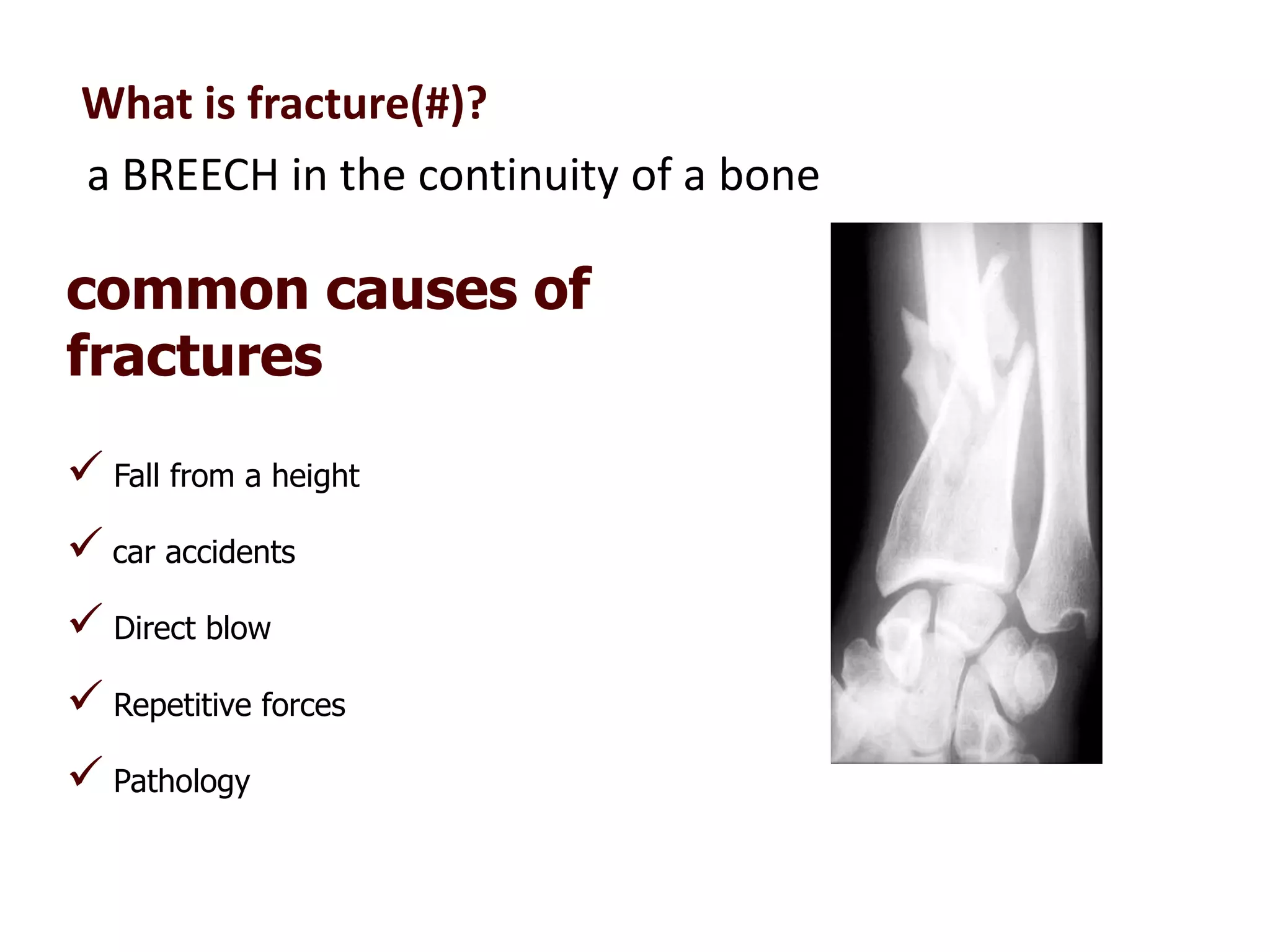
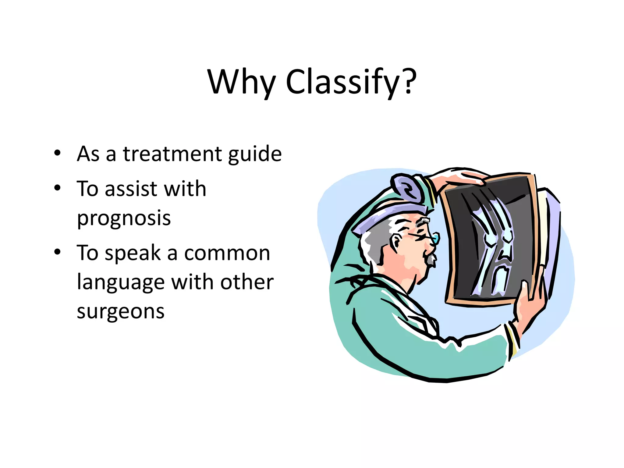
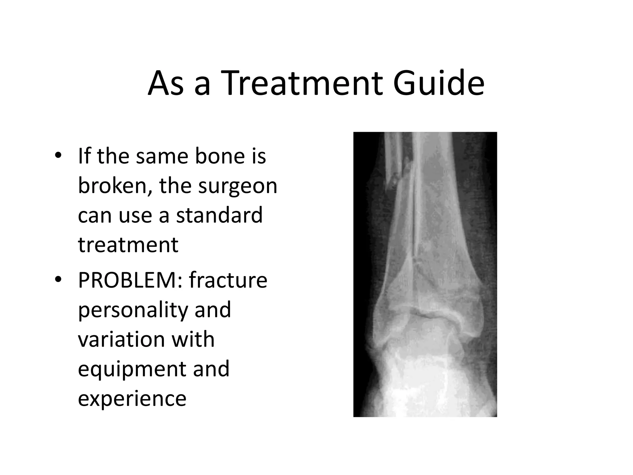
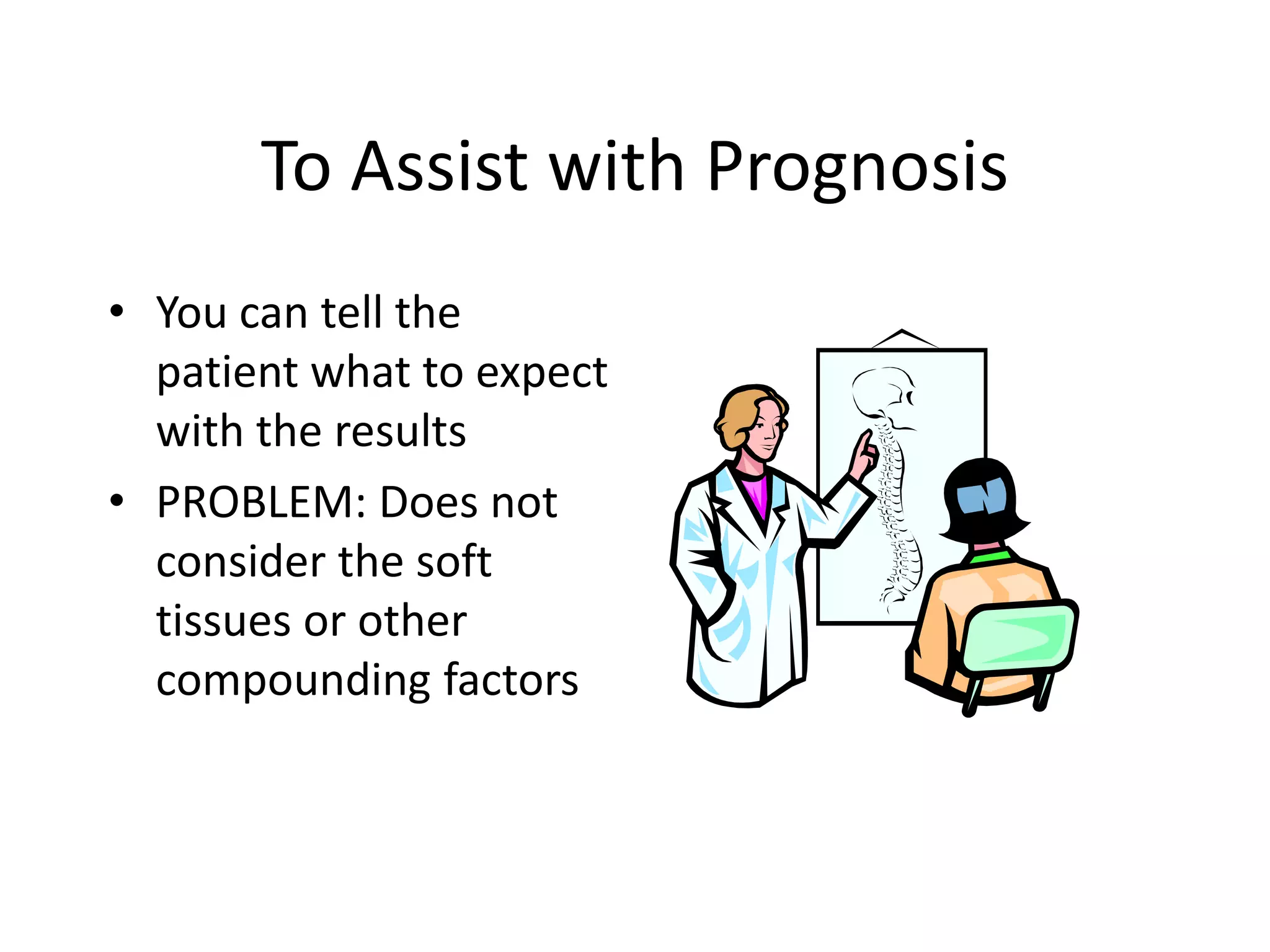
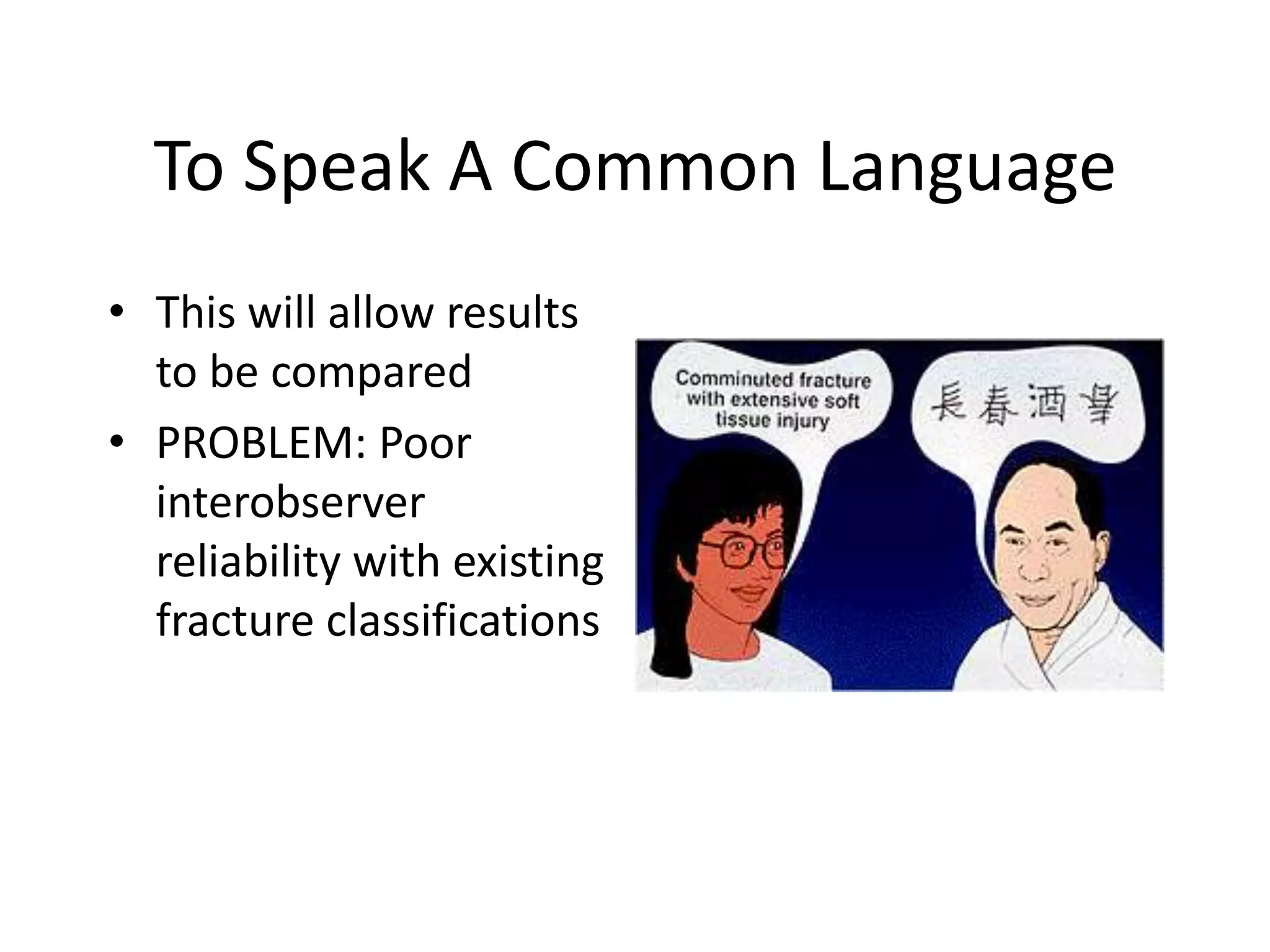
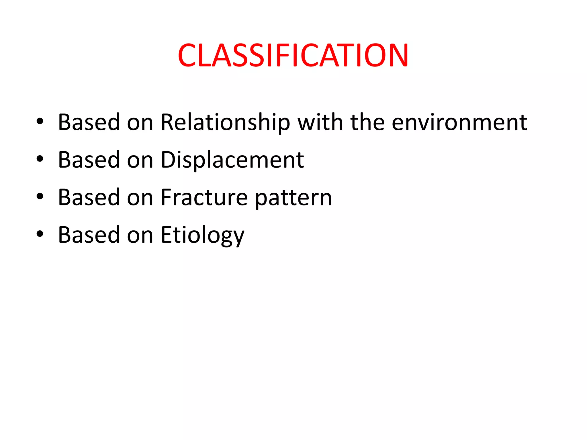
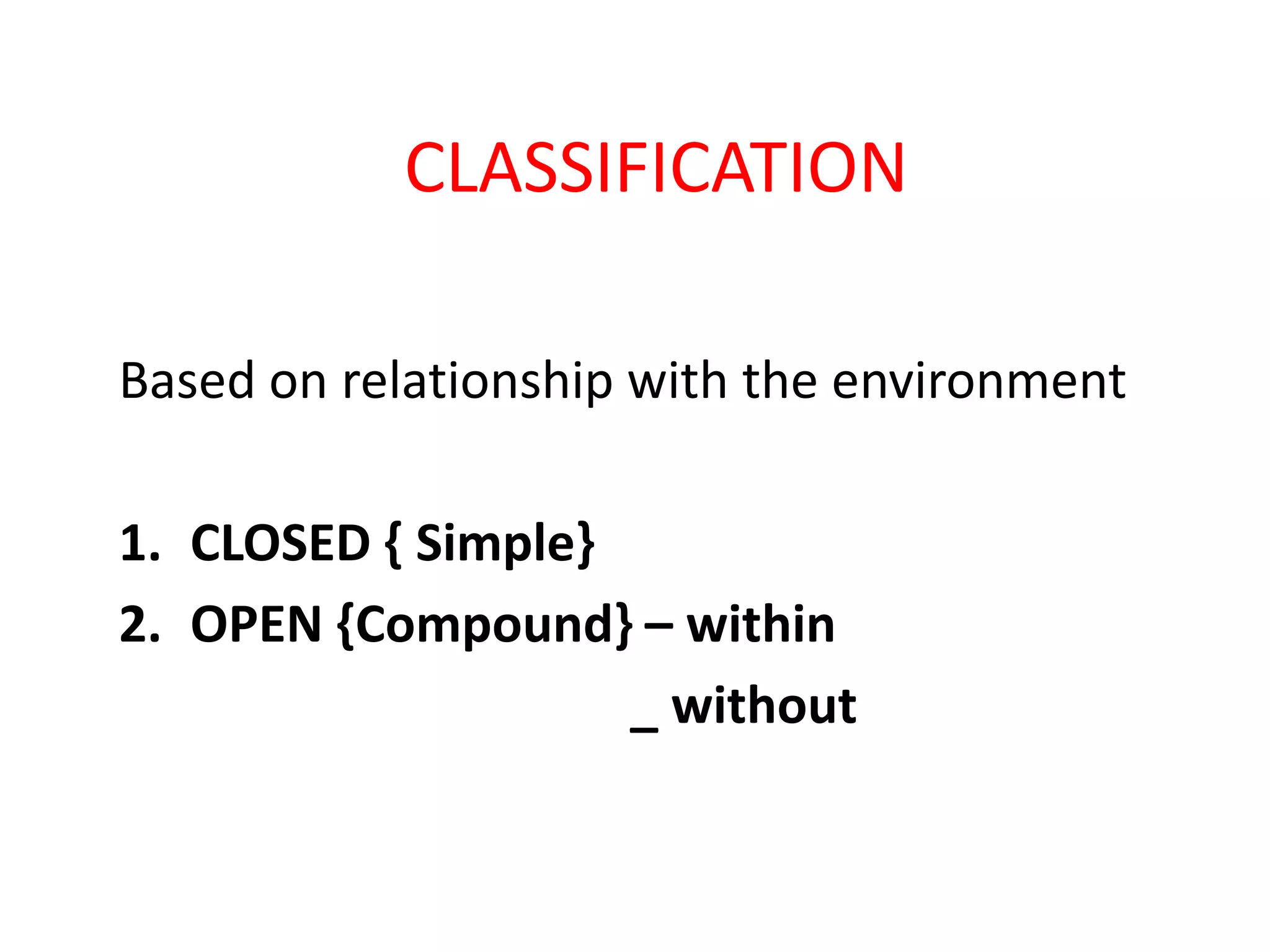
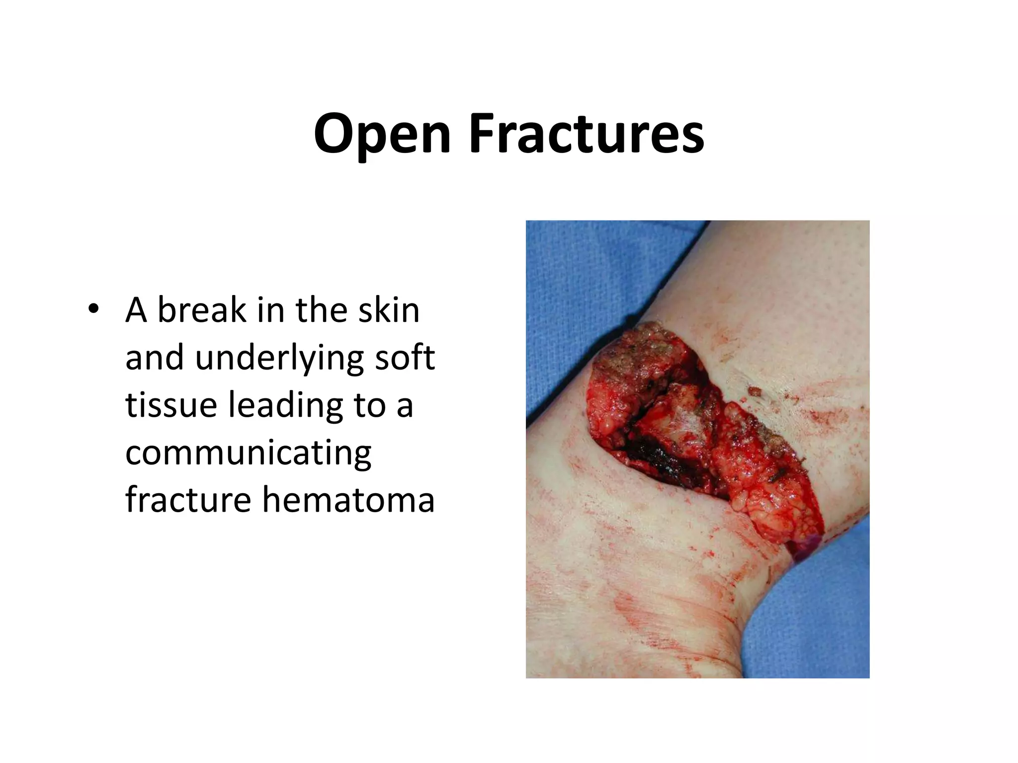
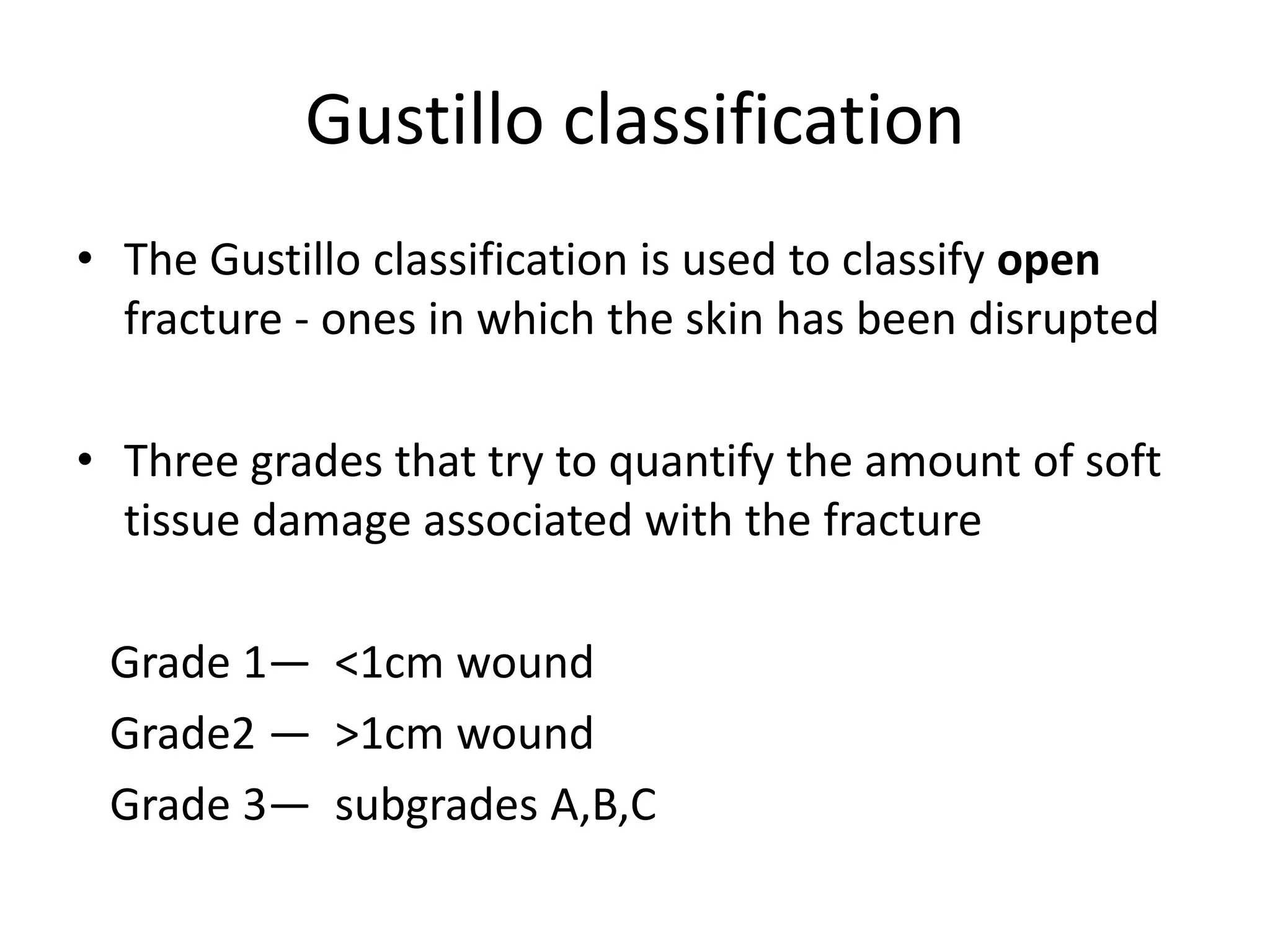
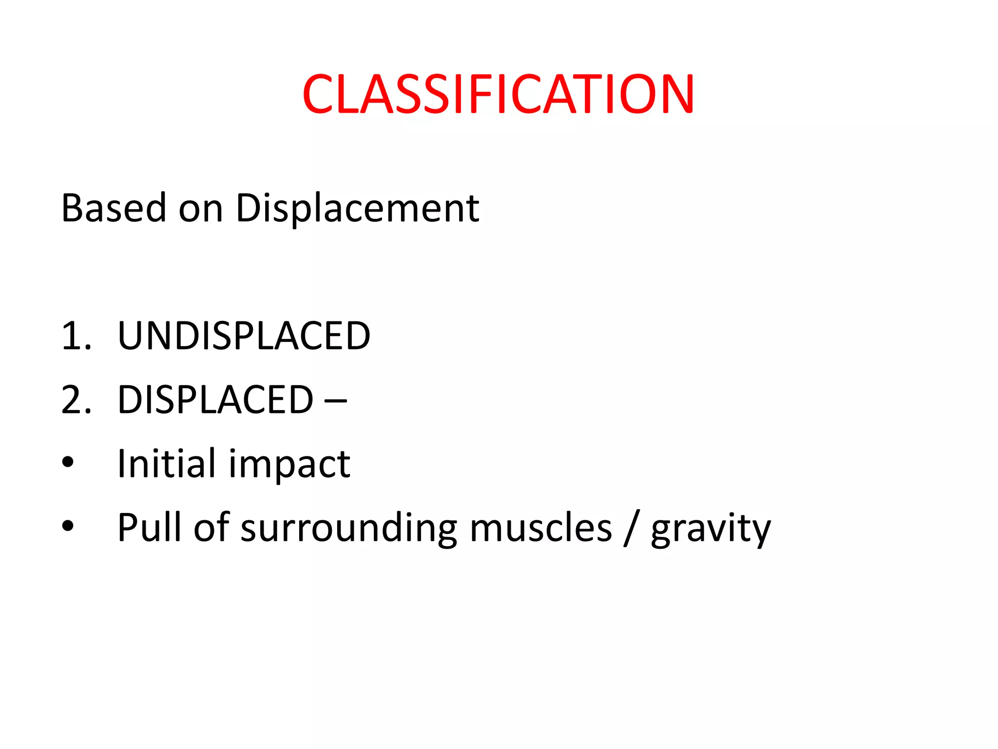
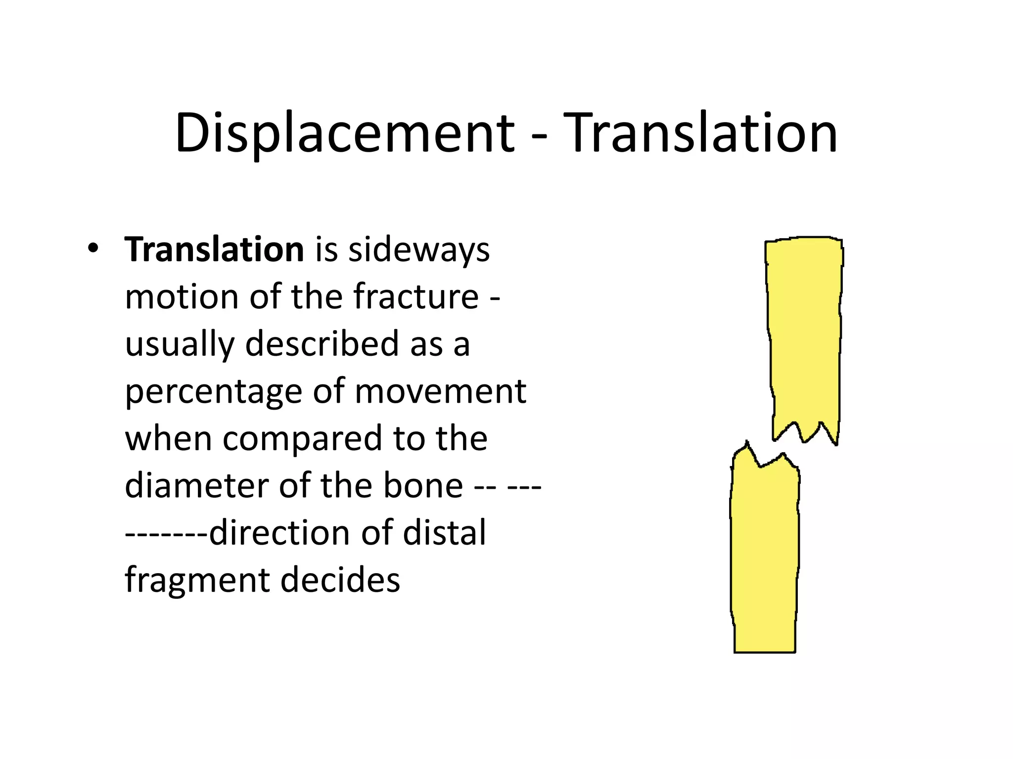
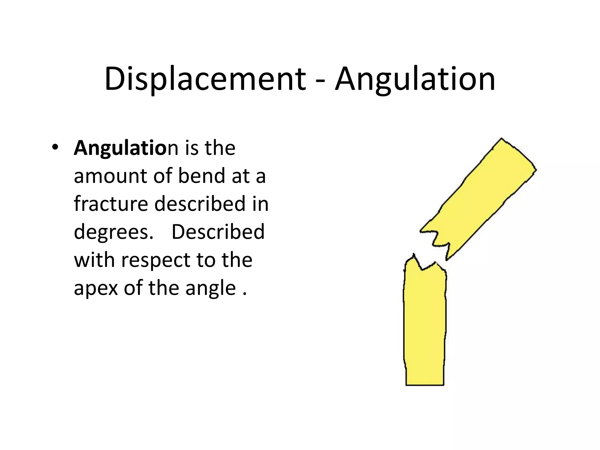
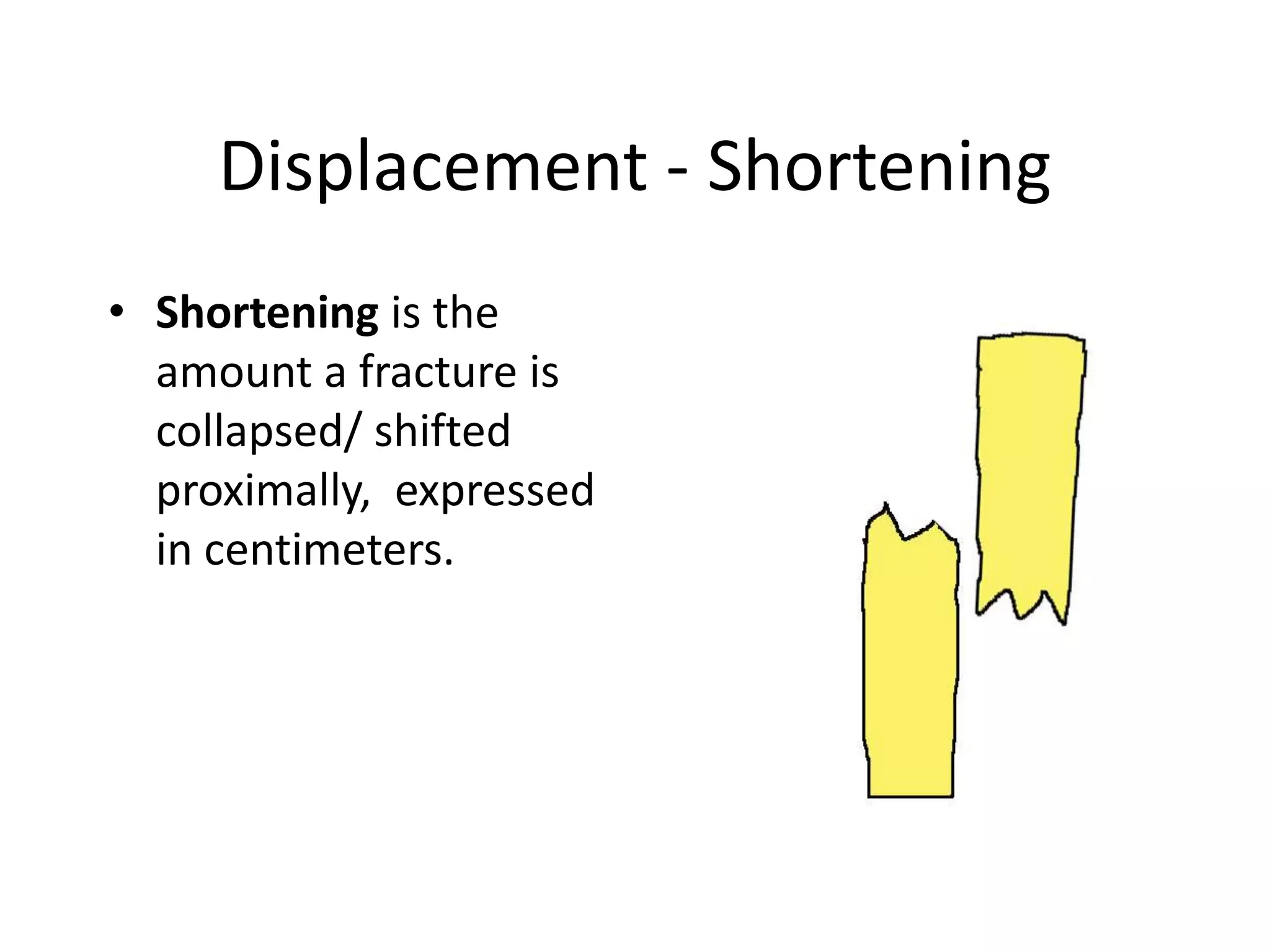
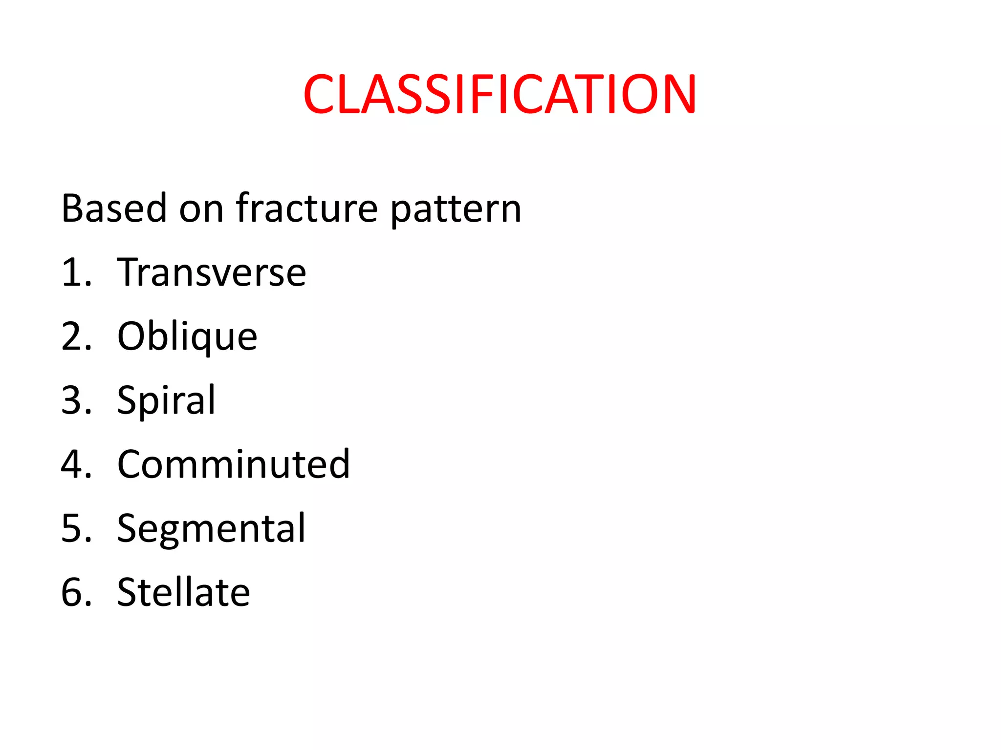
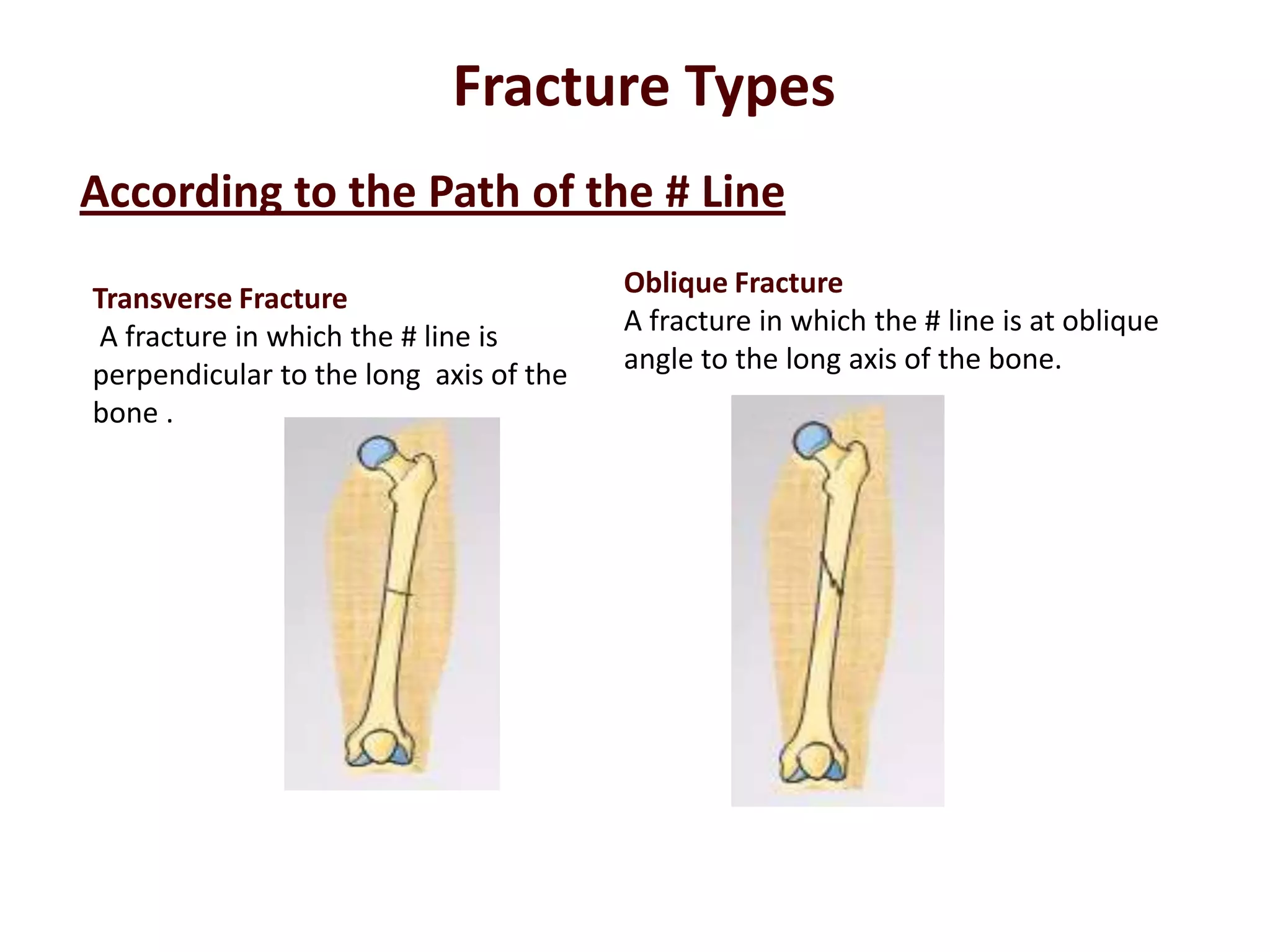
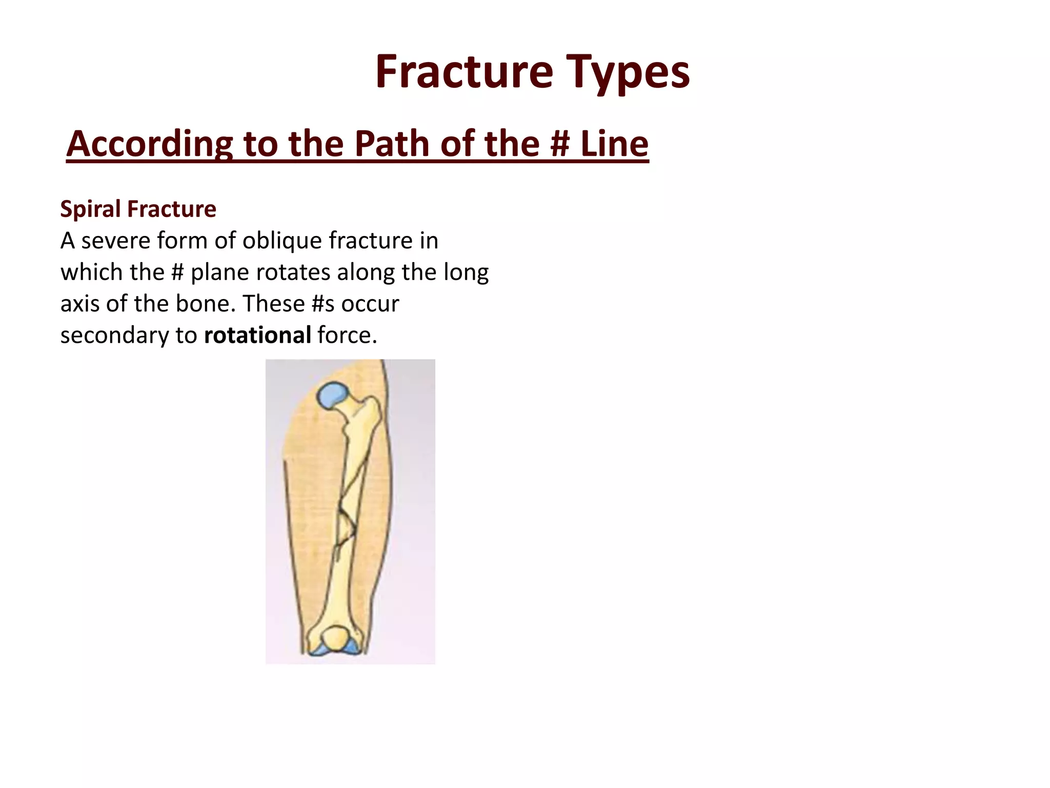
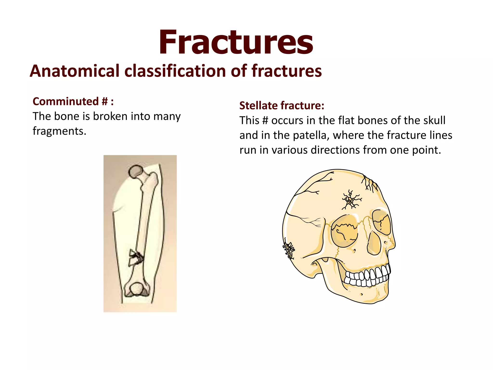
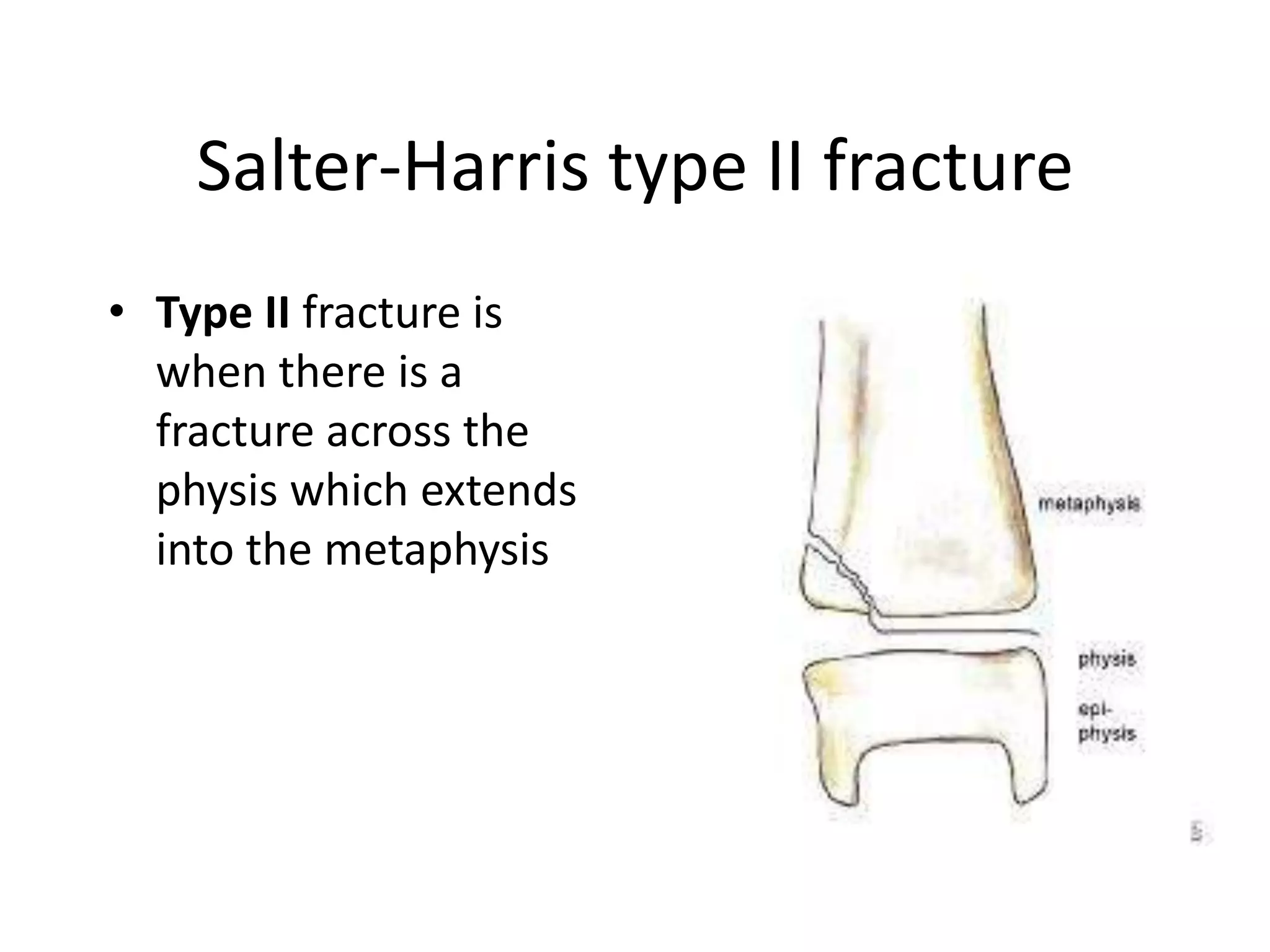
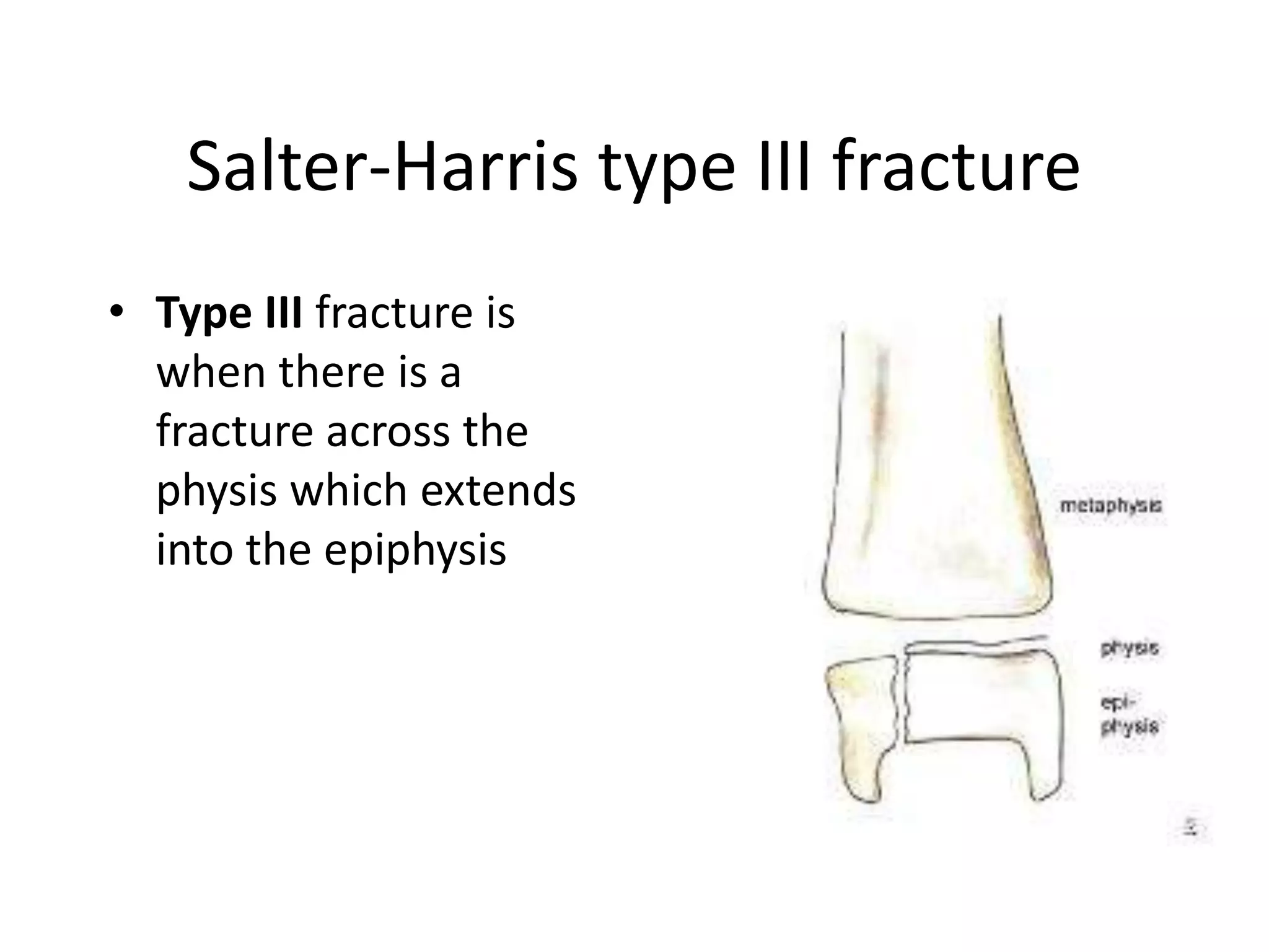
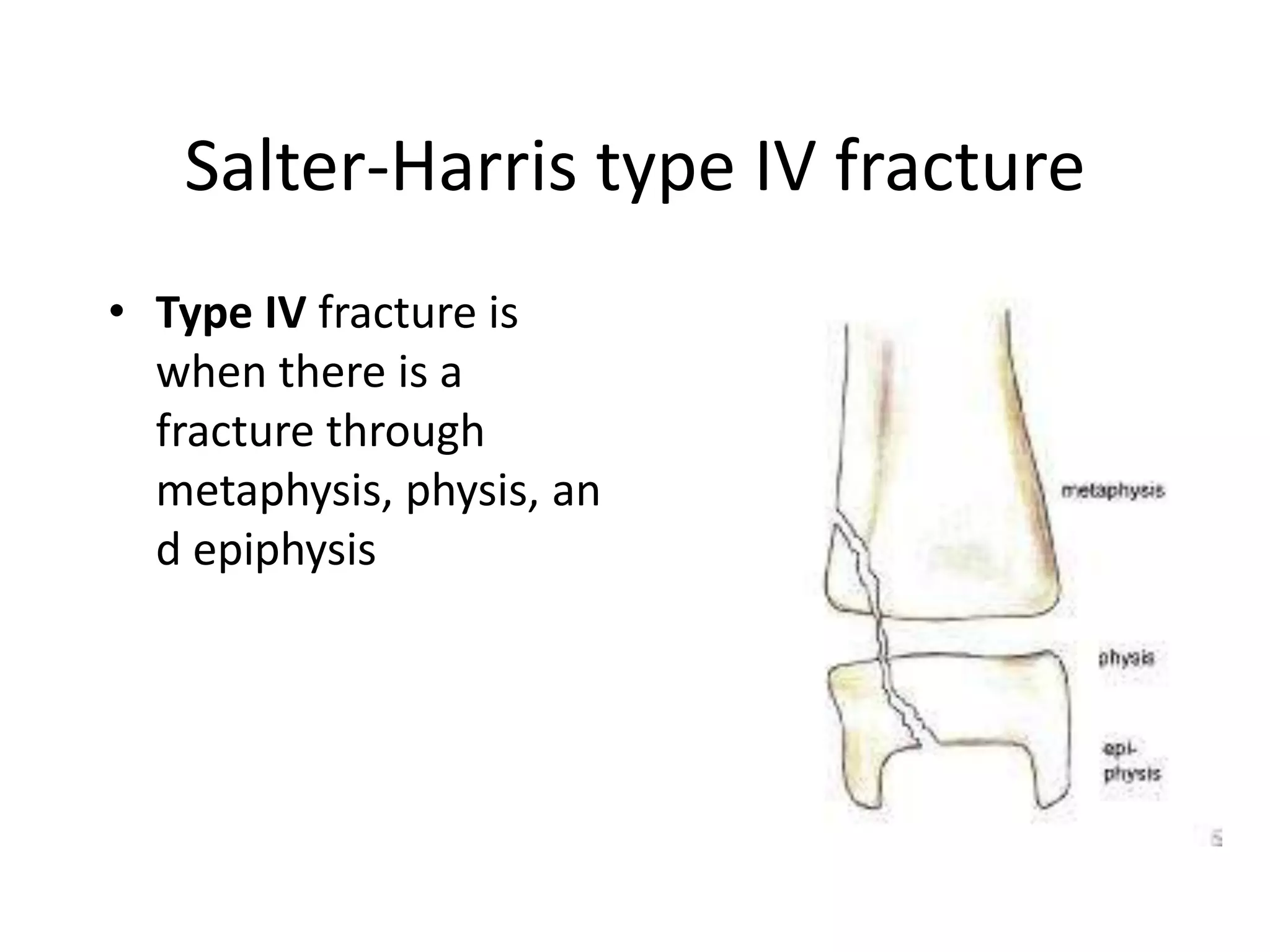
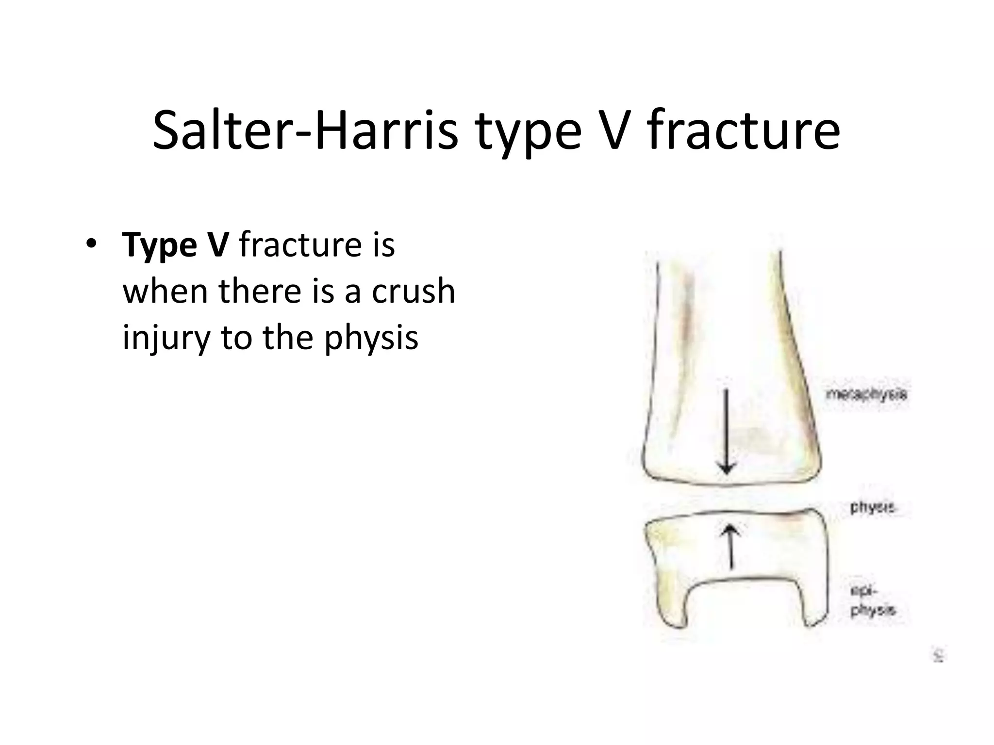
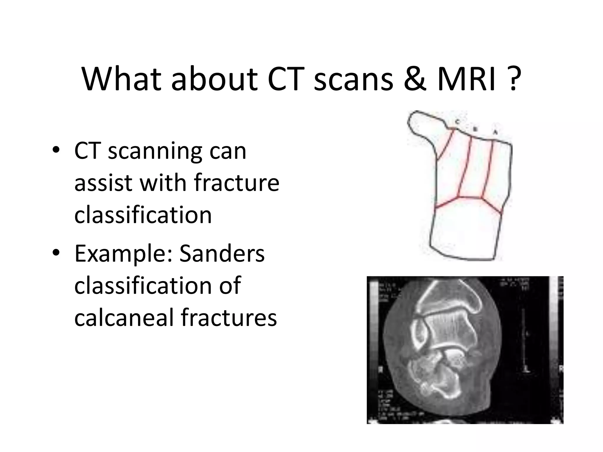
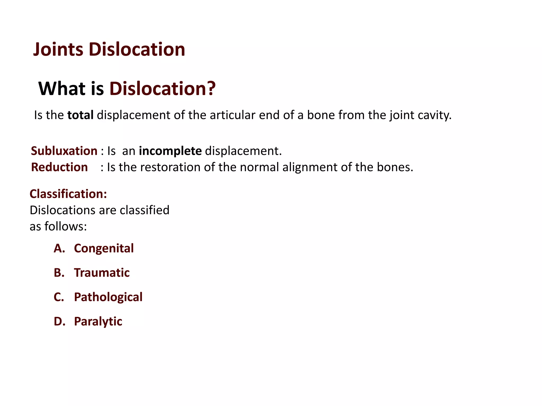
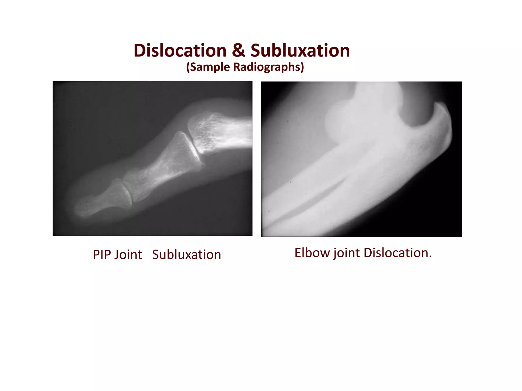
 Define term the term fracture, dislocation and Subluxation
 Identify the general causes, signs , symptoms of fractures
 Classify the different types of fractures
What is fracture(#)?
common causes of
fractures
 Fall from a height
 car accidents
 Direct blow
 Repetitive forces
 Pathology
a BREECH in the continuity of a bone
 Swelling or OEDEMA
 pain and tenderness
 Numbness
 Bleeding
 Broken skin with bone protruding
Limitation to move a limb
Signs and Symptoms
Why Classify?
• As a treatment guide
• To assist with
prognosis
• To speak a common
language with other
surgeons
As a Treatment Guide
• If the same bone is
broken, the surgeon
can use a standard
treatment
• PROBLEM: fracture
personality and
variation with
equipment and
experience
To Assist with Prognosis
• You can tell the
patient what to expect
with the results
• PROBLEM: Does not
consider the soft
tissues or other
compounding factors
To Speak A Common Language
• This will allow results
to be compared
• PROBLEM: Poor
interobserver
reliability with existing
fracture classifications
Intraobserver Reliability
For a given fracture, each physician
should produce the same classification
CLASSIFICATION
• Based on Relationship with the environment
• Based on Displacement
• Based on Fracture pattern
• Based on Etiology
CLASSIFICATION
Based on relationship with the environment
1. CLOSED { Simple}
2. OPEN {Compound} – within
_ without
Closed Fractures
• Fracture is not exposed to the environment
• All fractures have some degree of soft tissue
injury
• Don’t underestimate the soft tissue injury as
this affects treatment and outcome!
Open Fractures
• A break in the skin
and underlying soft
tissue leading to a
communicating
fracture hematoma
Gustillo classification
• The Gustillo classification is used to classify open
fracture - ones in which the skin has been disrupted
• Three grades that try to quantify the amount of soft
tissue damage associated with the fracture
Grade 1— <1cm wound
Grade2 — >1cm wound
Grade 3— subgrades A,B,C
CLASSIFICATION
Based on Displacement
1. UNDISPLACED
2. DISPLACED –
• Initial impact
• Pull of surrounding muscles / gravity
Displacement - Translation
• Translation is sideways
motion of the fracture -
usually described as a
percentage of movement
when compared to the
diameter of the bone -- ---
-------direction of distal
fragment decides
Displacement - Angulation
• Angulation is the
amount of bend at a
fracture described in
degrees. Described
with respect to the
apex of the angle .
Displacement - Shortening
• Shortening is the
amount a fracture is
collapsed/ shifted
proximally, expressed
in centimeters.
CLASSIFICATION
Based on fracture pattern
1. Transverse
2. Oblique
3. Spiral
4. Comminuted
5. Segmental
6. Stellate
Transverse Fracture
A fracture in which the # line is
perpendicular to the long axis of the
bone .
Oblique Fracture
A fracture in which the # line is at oblique
angle to the long axis of the bone.
According to the Path of the # Line
Fracture Types
Spiral Fracture
A severe form of oblique fracture in
which the # plane rotates along the long
axis of the bone. These #s occur
secondary to rotational force.
According to the Path of the # Line
Fracture Types
Anatomical classification of fractures
Fractures
Comminuted # :
The bone is broken into many
fragments.
Stellate fracture:
This # occurs in the flat bones of the skull
and in the patella, where the fracture lines
run in various directions from one point.
CLASSIFICATION
Based on etiology
1. TRAUMATIC
2. PATHOLOGICAL
CLASSIFICATION
PATHOLOGICAL
• --- Tumors
• --- Bone cysts
• --- Osteomyelitis
• --- Osteoporosis
• --- Osteogenesis imperfecta
• --- Rickets etc
AO/OTA Classification
• Describing the fracture localization: bones
and segments
• 1Humerus
• 2Radius/ulna
• 3Femur
• 4Tibia/fibula
AO/OTA Classification
• The identification of the respective segment
needs a little more consideration. Each long
bone has three segments:
• 1 Proximal end segment
• 2 Diaphyseal segment
• 3 Distal end segment
CLASSIFICATION
Based on eponyms :
• Colles’ # :: Dinner fork deformity
• Monteggia # D/L :: # proximal 1/3 shaft ulna with
dislocation of head of radius
• Galeazzi # D/L :: # Distal 1/3 shaft Radius with
dislocation of distal radioulnar joint
• Jone’s # :: Avulsion # of base of 5th metatarsal
• Green stick # :: unicortical # with bent bones in
children
Anatomical classification of fractures
Impacted fracture:
This # where a vertical force drives the
distal fragment of the fracture into the
proximal fragment.
Fracture Types
Depressed fracture:
This # occurs in the skull where a
segment of bone gets depressed into
the cranium.
Avulsion fracture:
This is one, where a chip of bone is avulsed by the sudden and unexpected contraction
of a powerful muscle from its point of insertion,
Examples
1. ASIS Avulsion
2. JONE’S 5th MT base Avulsion
Fracture Types
Anatomical classification of fractures
• Stress fracture :
• It is a fracture occurring at a site in the bone subject to
repeated minor stresses over a period of time.
• Birth fracture:
• It is a fracture in the new born
children due to injury during
birth
History of Fracture Classification
• 18th & 19th century
– History based on
clinical appearance
of limb alone
Colles Fracture Dinner Fork Deformity
Salter-Harris Classification
• Only used for pediatric fractures that involve
the growth plate (physis)
• Five types (I-V)
Salter-Harris type I fracture
• Type I fracture is
when there is a
fracture across the
physis with no
metaphysial or
epiphysial injury
Salter-Harris type II fracture
• Type II fracture is
when there is a
fracture across the
physis which extends
into the metaphysis
Salter-Harris type III fracture
• Type III fracture is
when there is a
fracture across the
physis which extends
into the epiphysis
Salter-Harris type IV fracture
• Type IV fracture is
when there is a
fracture through
metaphysis, physis, an
d epiphysis
Salter-Harris type V fracture
• Type V fracture is
when there is a crush
injury to the physis
What about CT scans & MRI ?
• CT scanning can
assist with fracture
classification
• Example: Sanders
classification of
calcaneal fractures
What is Dislocation?
Joints Dislocation
Is the total displacement of the articular end of a bone from the joint cavity.
Subluxation : Is an incomplete displacement.
Reduction : Is the restoration of the normal alignment of the bones.
Classification:
Dislocations are classified
as follows:
A. Congenital
B. Traumatic
C. Pathological
D. Paralytic
PIP Joint Subluxation Elbow joint Dislocation.
Dislocation & Subluxation
(Sample Radiographs)
Thank You!

Classification of fractures in general

  • 1.
     Define termthe term fracture, dislocation and Subluxation  Identify the general causes, signs , symptoms of fractures  Classify the different types of fractures
  • 2.
    What is fracture(#)? commoncauses of fractures  Fall from a height  car accidents  Direct blow  Repetitive forces  Pathology a BREECH in the continuity of a bone
  • 3.
     Swelling orOEDEMA  pain and tenderness  Numbness  Bleeding  Broken skin with bone protruding Limitation to move a limb Signs and Symptoms
  • 5.
    Why Classify? • Asa treatment guide • To assist with prognosis • To speak a common language with other surgeons
  • 6.
    As a TreatmentGuide • If the same bone is broken, the surgeon can use a standard treatment • PROBLEM: fracture personality and variation with equipment and experience
  • 7.
    To Assist withPrognosis • You can tell the patient what to expect with the results • PROBLEM: Does not consider the soft tissues or other compounding factors
  • 8.
    To Speak ACommon Language • This will allow results to be compared • PROBLEM: Poor interobserver reliability with existing fracture classifications
  • 9.
    Intraobserver Reliability For agiven fracture, each physician should produce the same classification
  • 10.
    CLASSIFICATION • Based onRelationship with the environment • Based on Displacement • Based on Fracture pattern • Based on Etiology
  • 11.
    CLASSIFICATION Based on relationshipwith the environment 1. CLOSED { Simple} 2. OPEN {Compound} – within _ without
  • 12.
    Closed Fractures • Fractureis not exposed to the environment • All fractures have some degree of soft tissue injury • Don’t underestimate the soft tissue injury as this affects treatment and outcome!
  • 13.
    Open Fractures • Abreak in the skin and underlying soft tissue leading to a communicating fracture hematoma
  • 14.
    Gustillo classification • TheGustillo classification is used to classify open fracture - ones in which the skin has been disrupted • Three grades that try to quantify the amount of soft tissue damage associated with the fracture Grade 1— <1cm wound Grade2 — >1cm wound Grade 3— subgrades A,B,C
  • 15.
    CLASSIFICATION Based on Displacement 1.UNDISPLACED 2. DISPLACED – • Initial impact • Pull of surrounding muscles / gravity
  • 16.
    Displacement - Translation •Translation is sideways motion of the fracture - usually described as a percentage of movement when compared to the diameter of the bone -- --- -------direction of distal fragment decides
  • 17.
    Displacement - Angulation •Angulation is the amount of bend at a fracture described in degrees. Described with respect to the apex of the angle .
  • 18.
    Displacement - Shortening •Shortening is the amount a fracture is collapsed/ shifted proximally, expressed in centimeters.
  • 19.
    CLASSIFICATION Based on fracturepattern 1. Transverse 2. Oblique 3. Spiral 4. Comminuted 5. Segmental 6. Stellate
  • 20.
    Transverse Fracture A fracturein which the # line is perpendicular to the long axis of the bone . Oblique Fracture A fracture in which the # line is at oblique angle to the long axis of the bone. According to the Path of the # Line Fracture Types
  • 21.
    Spiral Fracture A severeform of oblique fracture in which the # plane rotates along the long axis of the bone. These #s occur secondary to rotational force. According to the Path of the # Line Fracture Types
  • 22.
    Anatomical classification offractures Fractures Comminuted # : The bone is broken into many fragments. Stellate fracture: This # occurs in the flat bones of the skull and in the patella, where the fracture lines run in various directions from one point.
  • 23.
    CLASSIFICATION Based on etiology 1.TRAUMATIC 2. PATHOLOGICAL
  • 24.
    CLASSIFICATION PATHOLOGICAL • --- Tumors •--- Bone cysts • --- Osteomyelitis • --- Osteoporosis • --- Osteogenesis imperfecta • --- Rickets etc
  • 25.
    AO/OTA Classification • Describingthe fracture localization: bones and segments • 1Humerus • 2Radius/ulna • 3Femur • 4Tibia/fibula
  • 26.
    AO/OTA Classification • Theidentification of the respective segment needs a little more consideration. Each long bone has three segments: • 1 Proximal end segment • 2 Diaphyseal segment • 3 Distal end segment
  • 28.
    CLASSIFICATION Based on eponyms: • Colles’ # :: Dinner fork deformity • Monteggia # D/L :: # proximal 1/3 shaft ulna with dislocation of head of radius • Galeazzi # D/L :: # Distal 1/3 shaft Radius with dislocation of distal radioulnar joint • Jone’s # :: Avulsion # of base of 5th metatarsal • Green stick # :: unicortical # with bent bones in children
  • 29.
    Anatomical classification offractures Impacted fracture: This # where a vertical force drives the distal fragment of the fracture into the proximal fragment. Fracture Types Depressed fracture: This # occurs in the skull where a segment of bone gets depressed into the cranium.
  • 30.
    Avulsion fracture: This isone, where a chip of bone is avulsed by the sudden and unexpected contraction of a powerful muscle from its point of insertion, Examples 1. ASIS Avulsion 2. JONE’S 5th MT base Avulsion Fracture Types Anatomical classification of fractures
  • 31.
    • Stress fracture: • It is a fracture occurring at a site in the bone subject to repeated minor stresses over a period of time. • Birth fracture: • It is a fracture in the new born children due to injury during birth
  • 32.
    History of FractureClassification • 18th & 19th century – History based on clinical appearance of limb alone Colles Fracture Dinner Fork Deformity
  • 33.
    Salter-Harris Classification • Onlyused for pediatric fractures that involve the growth plate (physis) • Five types (I-V)
  • 34.
    Salter-Harris type Ifracture • Type I fracture is when there is a fracture across the physis with no metaphysial or epiphysial injury
  • 35.
    Salter-Harris type IIfracture • Type II fracture is when there is a fracture across the physis which extends into the metaphysis
  • 36.
    Salter-Harris type IIIfracture • Type III fracture is when there is a fracture across the physis which extends into the epiphysis
  • 37.
    Salter-Harris type IVfracture • Type IV fracture is when there is a fracture through metaphysis, physis, an d epiphysis
  • 38.
    Salter-Harris type Vfracture • Type V fracture is when there is a crush injury to the physis
  • 39.
    What about CTscans & MRI ? • CT scanning can assist with fracture classification • Example: Sanders classification of calcaneal fractures
  • 40.
    What is Dislocation? JointsDislocation Is the total displacement of the articular end of a bone from the joint cavity. Subluxation : Is an incomplete displacement. Reduction : Is the restoration of the normal alignment of the bones. Classification: Dislocations are classified as follows: A. Congenital B. Traumatic C. Pathological D. Paralytic
  • 41.
    PIP Joint SubluxationElbow joint Dislocation. Dislocation & Subluxation (Sample Radiographs)
  • 42.