Introduction
Gram positiveanaerobic or aerotolerant rods ,producing
endospores which are wider than the bacillary bodies giving the
characteristics spindle shape ,hence the name
Clostridium( kloster- spindle)
Most of them are motile except C. perfringens, C.tetani type IV
Noncapsulated except C.perfringens and C. butyricum
Proteolytic and saccharolytic
118 species
Found normally in intestine of human and animals
Responsible for three major diseases- C. perfringens
C. tetani
C. botulism
C. difficile
3.
Classification:
1. Based onlocation and shape of spores:
a) Spindle shaped :central spore.
e.g. C. bifermentans
b) Club shaped: Subterminal round spore.
e.g. C. perfringens
c) Tennis racket shaped: Oval and terminal.
e.g. C. tertium, C. choclearum.
d) Drumstick shaped: spherical and terminal
e.g.C.tetani,C.
tetanomorphum,C.sphenoides
4.
2. Based onthe site of infection:
a) Histotoxic clostridia
e.g. C. perfringens type A, C. novyi, C.
septicum, .
b) Enteropathogenic clostridia.
e.g. C. perfringens type A2, and type C.
C. difficile
c) Neurotoxic clostridia
1. C. tetani
2.C. botulinum
5.
3. Based onthe disease they produced:
a. Gas gangrene group:
e.g. C. perfringens, C. septicum, C. novyi,
C. fallax, C. sordeli, C. bifermentans.
b. Tetanus: C. tetani
c. Food poisoning
Gastroenteritis C. perfringens type A
Botulism C. botulinum
Necrotising enteritis C. perfringens type C
d. Acute colitis
C. difficile
6.
4. Based onthe biochemical reactions:
a. Both proteolytic and saccharolytic
1. Proteolytic predominating
- C. botulinum A,B,F
- Cl. bifermentans
2. Saccharolytic predominating
- Cl. perfringens
- Cl. difficile
b. Slightly proteolytic but not saccharolytic
- Cl. Tetani
c. Saccharolytic but not proteolytic
- Cl. Botulinum C,D,E
d. Neither proteolytic nor saccharolytic
- Cl. cochlearum
History
First cultivatedby Achalme(1891) and later
described in detail by Welch & Nuttal (1892)
who isolated it from blood and organs of
cadaver.
commonly known as C. welchi in U.K.
Most important organism causing gas gangrene
, also produces food poisoning ( C. perfringens
type A2) and necrotic enteritis( C. perfringens
type C).
Habitat:
Normal inhabitantof large intestine of human
and animals. (10 4
/gm faeces).
Found in faeces and contaminate skin of
perineum, buttocks and thighs.
Spores are commonly found in soil, dust and
air.
11.
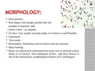
MORPHOLOGY:
Gram positive,
Rod shaped with straight, parallel side and
rounded or truncated ends
( Brick or Box - car shaped).
4-6 um x 1um, usually occuring singly or in chains or small bundles.
Capsulated,
Non motile
Pleomorphic, filamentous and involution form are common.
Spore forming
Spores are spherical & subterminal but rarely seen in artificial culture
media or in material from pathogenic lesions , and their absence is
one of the characteristic morphological features of Cl. perfringens
12.
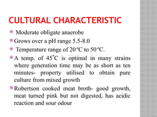
CULTURAL CHARACTERISTIC
Moderateobligate anaerobe
Grows over a pH range 5.5-8.0
Temperature range of 20 ºC to 50ºC.
A temp. of 45˚C is optimal in many strains
where generation time may be as short as ten
minutes- property utilised to obtain pure
culture from mixed growth
Robertson cooked meat broth- good growth,
meat turned pink but not digested, has acidic
reaction and sour odour
13.
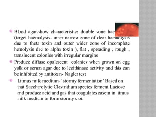
Blood agar-showcharacteristics double zone haemolysis
(target haemolysis- inner narrow zone of clear haemolysis
due to theta toxin and outer wider zone of incomplete
hemolysis due to alpha toxin ), flat , spreading , rough ,
translucent colonies with irregular margins
Produce diffuse opalescent colonies when grown on egg
yolk or serum agar due to lecithinase activity and this can
be inhibited by antitoxin- Nagler test
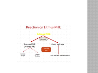
Litmus milk medium- ‘stormy fermentation’ Based on
that Saccharolytic Clostridium species ferment Lactose
and produce acid and gas that coagulates casein in litmus
milk medium to form stormy clot.
15.
Gelatin –hydrolyses gelatin
Selective media- made by incorporation of
neomycin, polymyxin and crystal violet
16.
BIOCHEMICAL REACTION
Fermentsglucose ,lactose,maltose,sucrose
and other sugars with production of acid and
gas
Indole negative
Urease negative
MR positive, VP negative
H2S formed abundantly
Liquefy gelatin
Most strains reduces Nitrate to Nitrite
17.
RESISTANCE
Spores areusually destroyed with in 5
minutes by boiling but those of ‘food
poisoning’ strains of Type A and Type C resist
boiling for 1-3 hrs
Autoclaving for 121C for 15 minutes is lethal
Spores resistant to antiseptics and
disinfectant in common use
18.
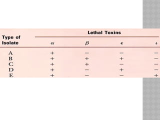
CLASSIFICATION
On thebasis of type of toxin they produce
Classified into five types- A to E
Typing based on 4 major toxins- alpha, beta,
epsilon and iota
Typing done by neutralisation tests with
specific antitoxins by intracutaneous
injection in guinea pig or iv injection in mice
Gene probes or PCR could be used for typing
20.
Type Aand C- human pathogens
Type A- gas gangrene, wound infections,
septicemia, food poisoning
Type C- necrotising enteritis
21.
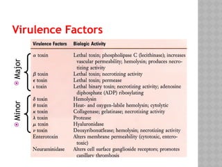
Virulence Factors
Toxins– 12 distinct toxins, four major toxins-
(alpha, beta, epsilon and iota) and
minor toxins
Enzymes
Biologically active soluble substances
22.
Major toxins
1. Alphatoxin
- produced by all types
- in large amount by type A
- is phospholipidase or lecithinase C-splits
lecithin into phosphoryl choline and
diglyceride in presence of Mg and Ca ions
- most important toxin biologically
23.
- responsible forprofound toxaemia of gas
gangrene .
-is lethal, dermonecrotic,and hemolytic
- resposible for lecithinase reaction in egg
yolk agar and hazy zone of haemolysis on
blood
agar
-- lysis is of hot –cold variety- best seen after
incubation at 37C and chilling at 4C
- relatively heat stable
24.
2. Beta toxin-
-major lethal toxin of type B and C
- responsible for lesion of necrotising
enteritis
3. Epsilon toxin-
- have lethal and necrotising property
- activated by proteolytic enzyme
- produced by type B and D
- increases the permeability of intestine-
thus enhancing its own uptake- and acts
systemically as a lethal toxin
25.
4. Iota toxin(i toxin)
- have lethal and necrotising property
- binary toxin consist of two subunits-a and
b (immunologically and biologically distinct
- a- ADP ribosylates
- b- recognises the binding site on a cell
membrane- binds to the site-interact with
a unit to facilitate its entry
26.
Enterotoxin
- protein innature
- responsible for food borne diarrhoea
- occurs after consumption of food containing
large number of vegetative organism
- found in type A,C and D
27.
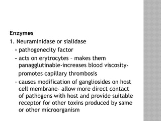
Enzymes
1. Neuraminidase orsialidase
- pathogenecity factor
- acts on erytrocytes – makes them
panagglutinable-increases blood viscosity-
promotes capillary thrombosis
- causes modification of gangliosides on host
cell membrane- allow more direct contact
of pathogens with host and provide suitable
receptor for other toxins produced by same
or other microorganism
28.
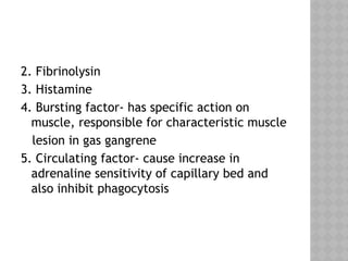
2. Fibrinolysin
3. Histamine
4.Bursting factor- has specific action on
muscle, responsible for characteristic muscle
lesion in gas gangrene
5. Circulating factor- cause increase in
adrenaline sensitivity of capillary bed and
also inhibit phagocytosis
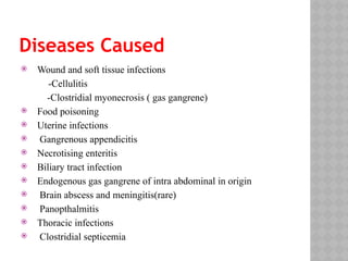
Diseases Caused
Woundand soft tissue infections
-Cellulitis
-Clostridial myonecrosis ( gas gangrene)
Food poisoning
Uterine infections
Gangrenous appendicitis
Necrotising enteritis
Biliary tract infection
Endogenous gas gangrene of intra abdominal in origin
Brain abscess and meningitis(rare)
Panopthalmitis
Thoracic infections
Clostridial septicemia
31.
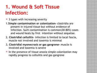
1. Wound &Soft Tissue
Infection:
3 types with increasing severity
1.Simple contamination or colonization- clostridia are
present in injured tissue but without evidence of
infection. Such contamination is common(30-80%) cases
and wound heals by first intention without sequelae.
2. Clostridial cellulitis- infection is limited to local fascia,
muscle not involved and toxemia is minimal
3. Clostridial myonecrosis or gas gangrene- muscle is
involved and toxemia is severe
In the presence of tissue anoxia simple colonization may
rapidly progress to cellulitis and gas gangrene
32.
Factors whichfavours the active multiplication of
Clostridial species.
1. Disrupted or impaired blood supply to the tissues
due to trauma, pressure of tourniquets, casts or dressings,
presence of foreign bodies in wound, presence of necrotic
tissues , haemorrhage in wound due to trauma. action of
necrotizing agents found in soil e.g.CaCl2 etc.
2.Lowering of PH to about 6.8.
3.Utilisation of Oxygen by co -infecting aerobic bacteria.
4. Autolysis of some tissue proteins making aminoacids
e.g. cysteine , tryptophan available – most necessary for
initiation of growth.
33.
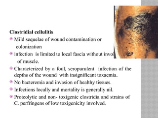
Clostridial cellulitis
Mildsequelae of wound contamination or
colonization
infection is limited to local fascia without involvement
of muscle.
Characterized by a foul, seropurulent infection of the
depths of the wound with insignificant toxaemia.
No bacteremia and invasion of healthy tissues.
Infections locally and mortality is generally nil.
Proteolytic and non- toxigenic clostridia and strains of
C. perfringens of low toxigenicity involved.
34.
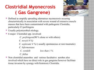
Clostridial Myonecrosis
( GasGangrene)
Defined as arapidly spreading edematous myonecrosis occuring
chracteristically in association with severe wound of extensive muscle
masses that have been contaminated with pathogenic Clostridia,
particularly Cl perfringens
Usually polymicrobial etiology.
6 major Clostridial spp. involved:
C. perfringens(90 % alone or with others)
C. novyi( 8 %)
C. septicum( 4 %) ( usually spontaneous or non traumatic)
C. bifermentans
C. sordelli less than 1 %
C. fallax
Non clostridial anaerobes and various facultative aerobes also
involved which have no direct role in gas gangrene however facilitate
tissue invasion by synergy with histotoxic Clostridia.
35.
Pathogenesis
Route ofentry
a. Exogenous infection- organism enters along
with implanted foreign material, soil, dust,
bits of clothing etc.
Endogenous infection-
36.
Clostridia cannotmultiply and produce disease in
normal tissues because the high oxidation–reduction
potential (Eh) of the circulating blood (+126–+246 mV)
and of the tissues is above that necessary for the
initiation of anaerobic bacterial growth (+74 mV for C.
perfringens)
A series of changes occurs in the damaged and anoxic
tissues that lead to a rapidly falling Eh and establish an
ideal environment for the growth of clostridia
Clostridia multiply and produce toxins
The production of bacterial toxins and products of
bacterial metabolism promotes the growth of the
organisms so that gas gangrene becomes established.
37.
Defenses arefurther compromised, since
neither phagocytes nor antibodies can
enter the necrotic zone, and absence of
perfusion prevents antimicrobial agents
from reaching the affected tissues
Infection spread from the original site
Rapid invasion and destruction of the
healthy tissue
If untreated release of toxin in circulation
Toxaemia and circulatory failure
Death
38.
Clinical Presentation
Incubation period- 7 h to 7 days.
Symptoms –
H/O trauma
Pain develops early in the region of the wound and increases in
intensity
progressive swelling and edema.
The wound is edematous, tender, and exudes a profuse serous or
serosanguinous discharge which is foul smelling
Muscle becomes black and noncontractile
As the disease progresses, bubbles of gas appear in the
discharge, crepitus may become evident in the tissues, and
the skin becomes white and marbled.
39.
The pulserate increases markedly, is feeble,
and often impalpable and there is a mild to
moderate pyrexia.
The patient is collapsed, profoundly toxemic,
and shocked, but remains
mentally alert and anxious.
The blood pressure falls and peripheral
venous collapse often makes venepuncture
impossible.
The syndrome usually terminates with
sudden death due to circulatory failure.
40.
Food Poisoning
Foodborne gastroenteritis due to
consumption of food contaminated with large
number of vegetative cells of
enteropathogenic Clostridia.
Caused by C. perfringens type A
Spores of these are markedly heat reistance
Produces heat labile enterotoxin
The enterotoxin causes marked
hypersecretion in jejunum and ileum.
Food poisoning usually caused by a cold or
warmed up meat dish
41.
When contaminatedmeat is cooked,spores
in the interior may survive-germinate and
multiply during storage or warming in the
anaerobic environment in the cooked meat-
produces enterotoxin- food poisoning
Incubation period: 8-24 hours after ingestion
Characterized by –abdominal pain diarrhaoea
and vomitting
Short duration of illness and symptoms
usually disappear within 10-24 hrs. May be
severe in debillitated person.
42.
Necrotizing Enteritis:
byβ-toxin of C. perfringens type C.
Sporadically occurs among populations in New Guinea known
as “Pig bell” and was recognized as “Drambrand”in Germany.
Organism acquired from consumption of pork from animals
carrying it.
Increase consumption of sweet potato and yam inhibits
protease in GI tract and β-toxin by bacteria got protected and
responsible for disease.
The disease is characterized by acute abdominal pain,
abdominal diarrhoea, vomiting, ulceration of the small
intestine and perforation of the intestine and acute toxemia.
Common cause of death in the native children of Papua New
Guinea.
43.
Clostridial Septicemia:
inmost of the cases , consequences of gas
gangrene following trauma or injury to soft
tissues.(exogenous)
In non penetrating injuiry it may be associated
with intestinal malignancy and involve a
localized myonecrosis in addition to a
fulminating septicemia.
Apparently migrate out of the patient intestine
as a consequence of malignant process or any
surgery. (Usually endogenous)
44.
Endophthalmitis
• C perfringensis a rare cause of
endophthalmitis,
• causes rapid destruction of the ocular tissues.
• result from perforating injury
45.
Urogenital Infection:
Oftenpresent in vagina as flora and may
contaminate perineum, buttocks, thigh
leading to infections under certain
predisposing factors such as malignancy,
threatened abortion etc.
46.
Laboratory Diagnosis :
Laboratory diagnosis of gas gangrene
Laboratory diagnosis of food poisoning
47.
Laboratory Diagnosis OfGas
Gangrene
Specimen collection:
- collect the exudate from the deeper part of wound
- Necrotic tissues, pus or exudate should place in sterile
screw capped bottle.
- Two or three swabbed specimen should be collected . One
for film preperation, other for anaerobic culture.
Transport of specimen:
- cooked meat broth
- Thioglycollate broth
-if collect in syringe then probe the needle into rubber cork
or bend it.
- transported anaerobically on commercially available
anaerobic transport devices.
48.
Direct examination
-gram staining
Culture
-Media used: Cooked meat broth, Blood agar, Lactose
egg yolk agar, Litmus milk agar
- Identification- microscopy, cultural character,
biochemical reaction
Nagler reaction
Rapid enzymological test- for the
identification of clostridial products in wound exudates
a. A species-specific sialidase-inhibition test gives results
in 2–6 h and agrees well with the results of
bacteriological examination for C. perfringens infection,
49.
Laboratory Diagnosis Offood
poisoning
1. Semi quantitative Culture:
- can be done on faeces and food in the basis --
>10 cfu
⁵ /g of C. perfringens in implicated food
- >10 /g of
⁶ C. perfringens in faeces
2) Direct detection of enterotoxin in faeces, food
or culture supernates.
-reversed passive latex agglutination test.
- ELISA
- CIE
- western immunoblotting