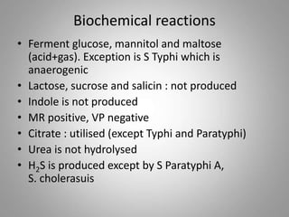
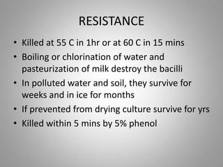
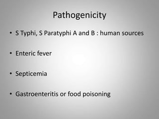
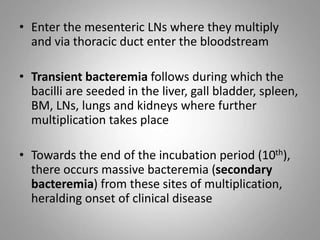
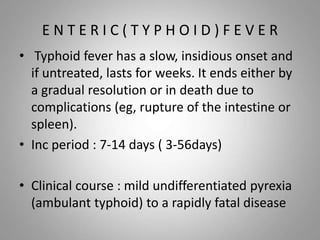
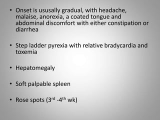
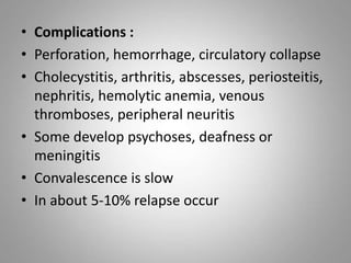
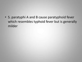
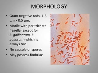
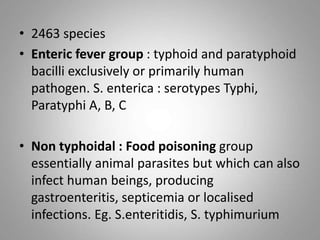
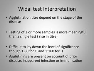
SALMONELLA
MORPHOLOGY
• Gram negative rods, 1-3
µm x 0.5 µm,
• Motile with peritrichate
flagella (except for
S. gallinarum, S
pullorum) which is
always NM
• No capsule or spores
• May possess fimbriae
• 2463 species
• Enteric fever group : typhoid and paratyphoid
bacilli exclusively or primarily human
pathogen. S. enterica : serotypes Typhi,
Paratyphi A, B, C
• Non typhoidal : Food poisoning group
essentially animal parasites but which can also
infect human beings, producing
gastroenteritis, septicemia or localised
infections. Eg. S.enteritidis, S. typhimurium
Cultural Characteristics
• Aerobic and facultative anaerobic
• Media of pH 6.8
• Temp ranges from 15-40° C (opt 37°C)
• Colonies are large, 2-3mm in diameter, circular,
low convex and smooth
• More translucent than coliform colonies
• MA, DCA – NLF colourless colonies
• Wilson and Blair bismuth sulphite media : black
colonies with a metallic sheen due to production
of H2S ( S paratyphi A gives green colonies)
Biochemical reactions
• Ferment glucose, mannitol and maltose
(acid+gas). Exception is S Typhi which is
anaerogenic
• Lactose, sucrose and salicin : not produced
• Indole is not produced
• MR positive, VP negative
• Citrate : utilised (except Typhi and Paratyphi)
• Urea is not hydrolysed
• H2S is produced except by S Paratyphi A,
S. cholerasuis
RESISTANCE
• Killed at 55 C in 1hr or at 60 C in 15 mins
• Boiling or chlorination of water and
pasteurization of milk destroy the bacilli
• In polluted water and soil, they survive for
weeks and in ice for months
• If prevented from drying culture survive for yrs
• Killed within 5 mins by 5% phenol
ANTIGENIC STRUCTURE
• Flagellar antigen : H
• Somatic antigen : O
• Surface antigen : Vi
H ANTIGEN
• Present on the flagella
• Heat labile
• Destroyed by boiling or by treatment with
alcohol but not by formaldehyde
• When mixed with antisera, large loose fluffy
clumps of agglutinate
• Immunogenic, antibody in high titre following
infection or immunisation
O Antigen
• Somatic O antigen is a phospholipid-protein-
polysachharide complex
• Integral part of the cell wall
• Identical to endotoxin
• extracted by treatment with trichloroacetic acid
• Unaffected by boiling, alcohol or weak acids
• When mixed with antisera, suspension forms
compact chalky, granular clumps
• Less immunogenic and the titre of antibody is
generally lower
• Classified into no of groups based on the
characteristics O antigens on the bacterial surface
Vi antigen
• Surface Polysachharide antigen enveloping the
O ag
• Heat labile analogus to the K antigens of
coliform
• bacilli inagglutinable with the O antiserum
become agglutinable after boiling or heating
at 60 C for 1 hr
• Unaffected by alcohol or 0.2% formal
Vi Antigen
• Tends to be lost on serial subculture
• Acts as a virulence factor by inhibiting
phagocytosis, resisting complement activation
and bacterial lysis by the alternative pathway
and peroxidase mediated killing
• Poorly immunogenic
• Persistence of Vi antibody indicates the
development of the carrier state.
Pathogenicity
• S Typhi, S Paratyphi A and B : human sources
• Enteric fever
• Septicemia
• Gastroenteritis or food poisoning
Virulence factors
• Type III secterion system (bacterial proteins):
mediate initial invasion
• O antigen: endotoxin
• Vi antigen
• Invasins: adherence, penetration
ENTERIC FEVER
• Infection acquired by ingestion
• Infective dose : 103 to 106 bacilli
• Incubation period: 7-14 days (3-56)
• On reaching the gut, the bacilli attach
themselves to the lamina propia and
submucosa
• Phagocytosed by macrophages and polymorphs
• Ability to resist intracellular killing and to
multiply within these cells
• Enter the mesenteric LNs where they multiply
and via thoracic duct enter the bloodstream
• Transient bacteremia follows during which the
bacilli are seeded in the liver, gall bladder, spleen,
BM, LNs, lungs and kidneys where further
multiplication takes place
• Towards the end of the incubation period (10th),
there occurs massive bacteremia (secondary
bacteremia) from these sites of multiplication,
heralding onset of clinical disease
• Bile : multiplies abundantly in the GB and is
discharged continuously into the intestine
where it involves the Peyers patches and
lymphoid follicles of the ileum
• They become inflamed, undergo necrosis and
slough off, leaving behind the characteristic
typhoid ulcers
• Ulceration : intestinal perforation and
hemorrhage
E N T E R I C ( T Y P H O I D ) F E V E R
• Typhoid fever has a slow, insidious onset and
if untreated, lasts for weeks. It ends either by
a gradual resolution or in death due to
complications (eg, rupture of the intestine or
spleen).
• Inc period : 7-14 days ( 3-56days)
• Clinical course : mild undifferentiated pyrexia
(ambulant typhoid) to a rapidly fatal disease
• Onset is ususally gradual, with headache,
malaise, anorexia, a coated tongue and
abdominal discomfort with either constipation or
diarrhea
• Step ladder pyrexia with relative bradycardia and
toxemia
• Hepatomegaly
• Soft palpable spleen
• Rose spots (3rd -4th wk)
• Complications :
• Perforation, hemorrhage, circulatory collapse
• Cholecystitis, arthritis, abscesses, periosteitis,
nephritis, hemolytic anemia, venous
thromboses, peripheral neuritis
• Some develop psychoses, deafness or
meningitis
• Convalescence is slow
• In about 5-10% relapse occur
• S. paratyphi A and B cause paratyphoid fever
which resembles typhoid fever but is generally
milder
EPIDEMIOLOGY
• Typhoid fever : still an important cause of
morbidity and mortality worldwide
• Strictly a human disease
• Chronic carriers of serotype Typhi are the
primary reservoir.
• The pathogen can be transmitted in the water
supply in developing endemic areas or where
defects in any system allow sewage from
carriers to contaminate drinking water.
• Transmission is by the fecal–oral route.
• Pt who continue to shed typhoid bacilli in
feces for 3 wks to 3 months after clinical cure :
convalescent carriers
• Those who shed the bacilli for more than 3
mths but less than 1 yr : temporary carriers
• Over a yr : chronic carriers (2-4%)
• Some persons may become carrier following
inapparent infn : symptomless excretors
• Food handlers or cooks who become carriers
are particularly dangerous
• Mary Mallon (Typhoid Mary) : over a period of
15 yrs caused at least 7 outbreaks affecting
over 200 persons
Laboratory Diagnosis
• Isolation of the organism from the patient
– Blood culture
– Stool culture
– Urine culture
– Others- bile, duodenal juice
• Demonstration of antibodies
• Demonstration of antigen in blood or urine
BLOOD CULTURE
• +ve in 90% of the cases in 1st wk, 75 % in 2nd
wk, 60% in 3rd wk and 25% in 4th wk. Becomes
negative on treatment with antibiotics
• 5-10ml of blood in 50-100ml of bile broth (
BHI broth, glucose broth)
• Sodium polyanethol sulphonate counteracts
the bactericidal action of blood
• Incubation at 37C overnight , subcultured on
MacConkey agar
• Pale NLF colonies may appear
• Motility and biochemical test
• Identification of the isolate is confirmed by
slide agglutination
SLIDE AGGLUTINATION
• A loopful of the growth from an agar slope is
emulsified in 2 drops of saline on a slide
• If S typhi is suspected, a loopful of typhoid O
antiserum ( factor 9/group D) is added
• Agglutination is looked for
• If suspected of S paratyphi, then agglutination is
done with O and H antisera
• If not isolated from 1st subculture from bile
broth, subcultures should be repeated every
other day till growth is obtained
• Cultures should be declared negative only
after the incubation for 10 days
• Castaneda’s method : to avoid contamination
and also for economy and safety
• Clot culture yields higher rate of isolation
FECES CULTURE
• Org. shed throughout the disease and also
during convalescent
• Almost as valuable as blood culture
• Use of enrichment (tetrathionate or selenite
broth) and selective media
• Usually helpful in isolation of the patient with
antibiotics
• Plated to MacConkey, DCA and Wilson Blair
media
URINE CULTURES
• Salmonella are shed in urine infrequently and
irregularly
• Less useful
• Cultures are positive only in 2nd and 3rd wk
• Repeated sampling improves the rate of
isolation
• Clean voided urine samples are centrifuged
and the deposit inoculated into enrichement
and selective media
Demonstration of Antibodies
(WIDAL TEST)
• Test for the measurement of O and H
agglutinins for typhoid and paratyphoid bacilli
in the patients sera
• 2 types of tubes :
• Dreyers tube : a narrow tube with a conical
bottom for the H agglutination
• Felix tube : short round bottomed tube for O
agglutination
Dreyers tube
Widal test
• Equal volumes of serial dilution of serum and the
H and O Ag are mixed and incubated in a water
bath at 37 C overnight
• Agglutination titre of the serum are read
• H agglutination : loose, cottony woolly clumps
• O agglutination : disc like pattern at the bottom
of the tube
• Supernatant is clear in both
• Antigens used are O and H antigens of S typhi and
H antigens of S Paratyphi A and B
Widal test Interpretation
• Agglutination titre depend on the stage of the
disease
• Testing of 2 or more samples is more meaningful
than a single test ( rise in titre)
• Difficult to lay down the level of significance
though 1:80 for O and 1:160 for H
• Agglutinins are present on account of prior
disease, inapparent infection or immunisation
• Persons who have had prior infection or
immunisation may develop an anamnestic
response during an unrelated fever. This may
be differentiated by repetition of the test after
a wk. (transient rise)
• Bacterial suspensions used as antigens should
be free from fimbriae ( False positive results)
• Cases treated early with chloramphenicol may
show a poor agglutinin response
Demonstration of Circulating Antigen
• Sensitised Staphylococcal coagglutination test
• Staph aureus (Cowan I strain) containing protein
A is stabilised with formaldehyde and coated with
S typhi antibody
• When 1% suspension of such sensitised
staphylococcal cells are mixed with serum from pt
in the 1st wk of the disease = visible
agglutionation within 2 minutes
Prophylaxis and Treatment
• Improvements in sanitation
• Detection of carriers
• Provision of protected water supply
• Chloramphenicol
• Fluoroquinolones
• Third generation cephalosporins
Vaccines
1. Killed whole cell vaccine :
– Heat killed, phenol preserved, whole cell
containing mixture of S. Typhi and S Paratyphi A
and B (TAB)
– Injected s/c in 2 doses of 0.5ml each at an
interval of 4-6 wk followed by a booster dose
every 3 yrs
– Effication 60-70% protection for 3-7 years
2. Vaccines of purified Vi antigen :
– Contains purified Vi ag which is injected
intramuscularly in a dose of 25µg (typhim- vi)-
75%
3. Live vaccine:
• live avirulent vaccine from a mutant strain of S. typhi
(Ty2 1a)
• to children
• Efficacy : 65- 96%
• 3 oral doses on alternate day one hour before food
with a glass of milk
Salmonella Gastroenteritis
• Food poisoning caused by any Salmonella
except S Typhi
• Source of infection : animal products
• S Typhimurium, S enteritidis, S. anatum, S
haldar, S heidelberg
• Results from contaminated food
• Eggs, egg products, meat, milk and milk
products
Gastroenteritis
• Disease develops after a short inc period of 24
hrs or less, with diarrhea, vomiting, abdominal
pain and fever.
• May vary in severity from the passage of one or
two loose stools to an acute cholera like disease
• Usually subsides in 2-4 days but in some cases a
more prolonged enteritis develops, with passage
of mucus and pus in feces, resembling dysentery
Laboratory diagnosis
• Isolating the organism from the feces
• In outbreaks, causative article of food can
often be identified by taking a proper history
• Isolation of salmonella from the article of food
confirms the diagnosis
Control
• Prevention of food contamination
• Proper cooking destroys salmonella
• Large problem in developed countries largely
because of their food habits and living
conditions
Salmonella septicemia
• S choleraesuis may cause septicemic disease
with focal suppurative lesions such as
osteomyelitis, deep abscesses, endocarditis,
pneumonia and meningitis
• Can be isolated from blood or pus
• Should be treated with chloramphenicol or
other antibiotics as determined by sensitivity
tests.
THANK YOU

Salmonella

  • 1.
  • 2.
    MORPHOLOGY • Gram negativerods, 1-3 µm x 0.5 µm, • Motile with peritrichate flagella (except for S. gallinarum, S pullorum) which is always NM • No capsule or spores • May possess fimbriae
  • 3.
    • 2463 species •Enteric fever group : typhoid and paratyphoid bacilli exclusively or primarily human pathogen. S. enterica : serotypes Typhi, Paratyphi A, B, C • Non typhoidal : Food poisoning group essentially animal parasites but which can also infect human beings, producing gastroenteritis, septicemia or localised infections. Eg. S.enteritidis, S. typhimurium
  • 4.
    Cultural Characteristics • Aerobicand facultative anaerobic • Media of pH 6.8 • Temp ranges from 15-40° C (opt 37°C) • Colonies are large, 2-3mm in diameter, circular, low convex and smooth • More translucent than coliform colonies • MA, DCA – NLF colourless colonies • Wilson and Blair bismuth sulphite media : black colonies with a metallic sheen due to production of H2S ( S paratyphi A gives green colonies)
  • 6.
    Biochemical reactions • Fermentglucose, mannitol and maltose (acid+gas). Exception is S Typhi which is anaerogenic • Lactose, sucrose and salicin : not produced • Indole is not produced • MR positive, VP negative • Citrate : utilised (except Typhi and Paratyphi) • Urea is not hydrolysed • H2S is produced except by S Paratyphi A, S. cholerasuis
  • 7.
    RESISTANCE • Killed at55 C in 1hr or at 60 C in 15 mins • Boiling or chlorination of water and pasteurization of milk destroy the bacilli • In polluted water and soil, they survive for weeks and in ice for months • If prevented from drying culture survive for yrs • Killed within 5 mins by 5% phenol
  • 8.
    ANTIGENIC STRUCTURE • Flagellarantigen : H • Somatic antigen : O • Surface antigen : Vi
  • 9.
    H ANTIGEN • Presenton the flagella • Heat labile • Destroyed by boiling or by treatment with alcohol but not by formaldehyde • When mixed with antisera, large loose fluffy clumps of agglutinate • Immunogenic, antibody in high titre following infection or immunisation
  • 10.
    O Antigen • SomaticO antigen is a phospholipid-protein- polysachharide complex • Integral part of the cell wall • Identical to endotoxin • extracted by treatment with trichloroacetic acid • Unaffected by boiling, alcohol or weak acids • When mixed with antisera, suspension forms compact chalky, granular clumps • Less immunogenic and the titre of antibody is generally lower • Classified into no of groups based on the characteristics O antigens on the bacterial surface
  • 11.
    Vi antigen • SurfacePolysachharide antigen enveloping the O ag • Heat labile analogus to the K antigens of coliform • bacilli inagglutinable with the O antiserum become agglutinable after boiling or heating at 60 C for 1 hr • Unaffected by alcohol or 0.2% formal
  • 12.
    Vi Antigen • Tendsto be lost on serial subculture • Acts as a virulence factor by inhibiting phagocytosis, resisting complement activation and bacterial lysis by the alternative pathway and peroxidase mediated killing • Poorly immunogenic • Persistence of Vi antibody indicates the development of the carrier state.
  • 13.
    Pathogenicity • S Typhi,S Paratyphi A and B : human sources • Enteric fever • Septicemia • Gastroenteritis or food poisoning
  • 14.
    Virulence factors • TypeIII secterion system (bacterial proteins): mediate initial invasion • O antigen: endotoxin • Vi antigen • Invasins: adherence, penetration
  • 15.
    ENTERIC FEVER • Infectionacquired by ingestion • Infective dose : 103 to 106 bacilli • Incubation period: 7-14 days (3-56) • On reaching the gut, the bacilli attach themselves to the lamina propia and submucosa • Phagocytosed by macrophages and polymorphs • Ability to resist intracellular killing and to multiply within these cells
  • 16.
    • Enter themesenteric LNs where they multiply and via thoracic duct enter the bloodstream • Transient bacteremia follows during which the bacilli are seeded in the liver, gall bladder, spleen, BM, LNs, lungs and kidneys where further multiplication takes place • Towards the end of the incubation period (10th), there occurs massive bacteremia (secondary bacteremia) from these sites of multiplication, heralding onset of clinical disease
  • 17.
    • Bile :multiplies abundantly in the GB and is discharged continuously into the intestine where it involves the Peyers patches and lymphoid follicles of the ileum • They become inflamed, undergo necrosis and slough off, leaving behind the characteristic typhoid ulcers • Ulceration : intestinal perforation and hemorrhage
  • 18.
    E N TE R I C ( T Y P H O I D ) F E V E R • Typhoid fever has a slow, insidious onset and if untreated, lasts for weeks. It ends either by a gradual resolution or in death due to complications (eg, rupture of the intestine or spleen). • Inc period : 7-14 days ( 3-56days) • Clinical course : mild undifferentiated pyrexia (ambulant typhoid) to a rapidly fatal disease
  • 19.
    • Onset isususally gradual, with headache, malaise, anorexia, a coated tongue and abdominal discomfort with either constipation or diarrhea • Step ladder pyrexia with relative bradycardia and toxemia • Hepatomegaly • Soft palpable spleen • Rose spots (3rd -4th wk)
  • 20.
    • Complications : •Perforation, hemorrhage, circulatory collapse • Cholecystitis, arthritis, abscesses, periosteitis, nephritis, hemolytic anemia, venous thromboses, peripheral neuritis • Some develop psychoses, deafness or meningitis • Convalescence is slow • In about 5-10% relapse occur
  • 21.
    • S. paratyphiA and B cause paratyphoid fever which resembles typhoid fever but is generally milder
  • 22.
    EPIDEMIOLOGY • Typhoid fever: still an important cause of morbidity and mortality worldwide • Strictly a human disease • Chronic carriers of serotype Typhi are the primary reservoir. • The pathogen can be transmitted in the water supply in developing endemic areas or where defects in any system allow sewage from carriers to contaminate drinking water. • Transmission is by the fecal–oral route.
  • 23.
    • Pt whocontinue to shed typhoid bacilli in feces for 3 wks to 3 months after clinical cure : convalescent carriers • Those who shed the bacilli for more than 3 mths but less than 1 yr : temporary carriers • Over a yr : chronic carriers (2-4%) • Some persons may become carrier following inapparent infn : symptomless excretors
  • 24.
    • Food handlersor cooks who become carriers are particularly dangerous • Mary Mallon (Typhoid Mary) : over a period of 15 yrs caused at least 7 outbreaks affecting over 200 persons
  • 26.
    Laboratory Diagnosis • Isolationof the organism from the patient – Blood culture – Stool culture – Urine culture – Others- bile, duodenal juice • Demonstration of antibodies • Demonstration of antigen in blood or urine
  • 27.
    BLOOD CULTURE • +vein 90% of the cases in 1st wk, 75 % in 2nd wk, 60% in 3rd wk and 25% in 4th wk. Becomes negative on treatment with antibiotics • 5-10ml of blood in 50-100ml of bile broth ( BHI broth, glucose broth) • Sodium polyanethol sulphonate counteracts the bactericidal action of blood
  • 28.
    • Incubation at37C overnight , subcultured on MacConkey agar • Pale NLF colonies may appear • Motility and biochemical test • Identification of the isolate is confirmed by slide agglutination
  • 29.
    SLIDE AGGLUTINATION • Aloopful of the growth from an agar slope is emulsified in 2 drops of saline on a slide • If S typhi is suspected, a loopful of typhoid O antiserum ( factor 9/group D) is added • Agglutination is looked for • If suspected of S paratyphi, then agglutination is done with O and H antisera
  • 30.
    • If notisolated from 1st subculture from bile broth, subcultures should be repeated every other day till growth is obtained • Cultures should be declared negative only after the incubation for 10 days • Castaneda’s method : to avoid contamination and also for economy and safety • Clot culture yields higher rate of isolation
  • 31.
    FECES CULTURE • Org.shed throughout the disease and also during convalescent • Almost as valuable as blood culture • Use of enrichment (tetrathionate or selenite broth) and selective media • Usually helpful in isolation of the patient with antibiotics • Plated to MacConkey, DCA and Wilson Blair media
  • 32.
    URINE CULTURES • Salmonellaare shed in urine infrequently and irregularly • Less useful • Cultures are positive only in 2nd and 3rd wk • Repeated sampling improves the rate of isolation • Clean voided urine samples are centrifuged and the deposit inoculated into enrichement and selective media
  • 33.
    Demonstration of Antibodies (WIDALTEST) • Test for the measurement of O and H agglutinins for typhoid and paratyphoid bacilli in the patients sera • 2 types of tubes : • Dreyers tube : a narrow tube with a conical bottom for the H agglutination • Felix tube : short round bottomed tube for O agglutination
  • 34.
  • 35.
    Widal test • Equalvolumes of serial dilution of serum and the H and O Ag are mixed and incubated in a water bath at 37 C overnight • Agglutination titre of the serum are read • H agglutination : loose, cottony woolly clumps • O agglutination : disc like pattern at the bottom of the tube • Supernatant is clear in both • Antigens used are O and H antigens of S typhi and H antigens of S Paratyphi A and B
  • 37.
    Widal test Interpretation •Agglutination titre depend on the stage of the disease • Testing of 2 or more samples is more meaningful than a single test ( rise in titre) • Difficult to lay down the level of significance though 1:80 for O and 1:160 for H • Agglutinins are present on account of prior disease, inapparent infection or immunisation
  • 38.
    • Persons whohave had prior infection or immunisation may develop an anamnestic response during an unrelated fever. This may be differentiated by repetition of the test after a wk. (transient rise) • Bacterial suspensions used as antigens should be free from fimbriae ( False positive results) • Cases treated early with chloramphenicol may show a poor agglutinin response
  • 39.
    Demonstration of CirculatingAntigen • Sensitised Staphylococcal coagglutination test • Staph aureus (Cowan I strain) containing protein A is stabilised with formaldehyde and coated with S typhi antibody • When 1% suspension of such sensitised staphylococcal cells are mixed with serum from pt in the 1st wk of the disease = visible agglutionation within 2 minutes
  • 40.
    Prophylaxis and Treatment •Improvements in sanitation • Detection of carriers • Provision of protected water supply • Chloramphenicol • Fluoroquinolones • Third generation cephalosporins
  • 41.
    Vaccines 1. Killed wholecell vaccine : – Heat killed, phenol preserved, whole cell containing mixture of S. Typhi and S Paratyphi A and B (TAB) – Injected s/c in 2 doses of 0.5ml each at an interval of 4-6 wk followed by a booster dose every 3 yrs – Effication 60-70% protection for 3-7 years 2. Vaccines of purified Vi antigen : – Contains purified Vi ag which is injected intramuscularly in a dose of 25µg (typhim- vi)- 75%
  • 42.
    3. Live vaccine: •live avirulent vaccine from a mutant strain of S. typhi (Ty2 1a) • to children • Efficacy : 65- 96% • 3 oral doses on alternate day one hour before food with a glass of milk
  • 43.
    Salmonella Gastroenteritis • Foodpoisoning caused by any Salmonella except S Typhi • Source of infection : animal products • S Typhimurium, S enteritidis, S. anatum, S haldar, S heidelberg • Results from contaminated food • Eggs, egg products, meat, milk and milk products
  • 44.
    Gastroenteritis • Disease developsafter a short inc period of 24 hrs or less, with diarrhea, vomiting, abdominal pain and fever. • May vary in severity from the passage of one or two loose stools to an acute cholera like disease • Usually subsides in 2-4 days but in some cases a more prolonged enteritis develops, with passage of mucus and pus in feces, resembling dysentery
  • 45.
    Laboratory diagnosis • Isolatingthe organism from the feces • In outbreaks, causative article of food can often be identified by taking a proper history • Isolation of salmonella from the article of food confirms the diagnosis
  • 46.
    Control • Prevention offood contamination • Proper cooking destroys salmonella • Large problem in developed countries largely because of their food habits and living conditions
  • 47.
    Salmonella septicemia • Scholeraesuis may cause septicemic disease with focal suppurative lesions such as osteomyelitis, deep abscesses, endocarditis, pneumonia and meningitis • Can be isolated from blood or pus • Should be treated with chloramphenicol or other antibiotics as determined by sensitivity tests.
  • 48.