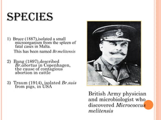
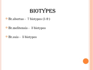
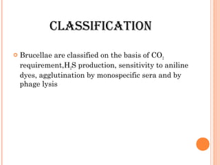
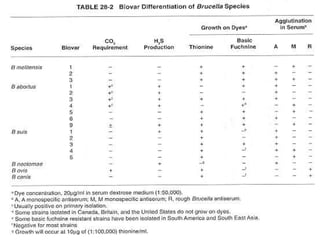
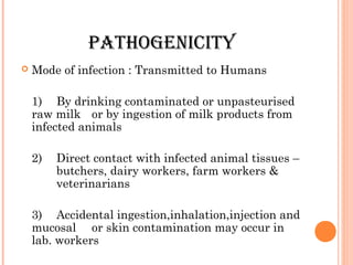
BRUCELLA
Dr. V. S. Vatkar
Asso Prof
Microbiology Department
BRUCELLA
 Kingdom: BACtERiA
 PhyLUm: PRotEoBACtERiA
 CLAss:
ALPhA PRotEoBACtERiA
 oRdER: RhizoBiALEs
 FAmiLy: BRUCELLACEAE
 gEnUs: BRUCELLA
intRodUCtion
 Genus Brucella consists of
very small, aerobic ,non-motile, gram
negative coccobacilli, poorly grow in ordinary
media
Strict parasites of animals
i.e. goats, sheep, cattle, pigs, buffaloes, infection
transmitted to human by contact with infected
animal.
sPECiEs
1) Bruce (1887),isolated a small
microorganism from the spleen of
fatal cases in Malta.
This has been named Br.melitensis
2) Bang (1897),described
Br.abortus in Copenhagen,
the cause of contagious
abortion in cattle
3) Traum (1914), isolated Br.suis
from pigs, in USA
British Army physician
and microbiologist who
discovered Micrococcus
melitensis
AdditionAL sPECiEs
→Br.neotomae – isoltd.from desert wood rats in
USA
→Br.ovis – isoltd.from sheep in Australia
→Br.canis – isoltd.from dogs in USA
moRPhoLogy
 Gram negative coccobacilli
0.5 – 0.7 µ x 0.6 – 1.5 µ
motile
Non sporing
capsulated
Arranged singly or in short chains, non acid fast
CULtURAL
ChARACtERistiCs
 Strict aerobes
Optimum temp. 37°C ranging from 20°C -40°C
Optimum Ph is 6.6 to 7.4
 Can grow on ordinary media though growth is slow and
scanty
 Growth is improved by addition of serum, blood, glucose or
liver extract
 Br.abortus is capnophilic
 Br.suis is unaffected by CO2
CULtURAL ChARACtERistiCs
The media used are :
serum - dextrose agar
serum - potato – infusion agar
trypticase soy agar
tryptose agar
 The addition of bacitracin, cycloheximide, polymyxin B or
vancomycin make the media selective
 On solid media, colonies are small,moist,translucent and
glistening
 Mucoid,smooth and rough type of colonies appear,asso.
with change in antigenic structure & virulence
CULtURAL ChARACtERistiCs
 Brucellae grow on chorioallantoic
membrane of chick embryo & cause their
death in few days
 All grow intracellularly –
Br.melitensis in ectodermal cells
Br.abortus & Br.suis in cells of
mesodermal origin
Erythritol has a specially stimulating
effect on the growth of most strains of
Brucella
CULtiVAtion in PREsEnCE oF
dyEs
 The 3 species are differentiated on the
basis of their ability to grow in media
containing aniline dyes, like basic fuchsin
and thionin
→Br.melitensis is not inhibited by any of
the dyes
→Br.abortus is inhibited by thionin and not
by basic fuchsin
→Br.suis is inhibited by basic fuchsin and
not by thionin
BioChEmiCAL REACtions
 Catalase +
 Oxidase +
 Urease +
 Citrate –
 Indole –
 M.R. & V.P -
 Reduce nitrates to nitrites
 No carbohydrates are fermented though they
have oxidative capacity
RESISTANCE
Killed
 by heat at 60°C for 10 mins
 by pasteurization
 by 1% phenol in 15 mins
Survive
 in soil & manure for several weeks
 in refrigerated milk for 10 days, a month
in ice-cream & for months in butter
 Sensitive to direct sunlight and acid
ANTIGENIC STRUCTURE
 Two main somatic Antigens, A and M,are present
in varying amounts in the three major species
Antigen A is dominant in Br.abortus
Antigen M is dominant in Br.melitensis
Br.suis shows an intermediate antigenic pattern
ANTIGENIC STRUCTURE
The antigenic structure is more complex
Some strains behave biochemically as Br.abortus and some
serologically as Br.melitensis and vice-versa
A large no. of antigens have been identified by gel
ppt.technique
Antigenic cross reactions exist between –
 Vibrio cholera
 E.coli O:116 , O: 157
 Salmonella serotype N (O:30 antigen)
 Ps.maltophila
 F.tularensis
 Y.enterocolitica
BIOTYPES
 Br.abortus - 7 biotypes (1-9 )
 Br.melitensis - 3 biotypes
 Br.suis - 5 biotypes
PHAGE TYPING
 Several bacteriophages that lyse Brucella strains
have been isolated. All these phages are
serologically similar
 The Tblisi (Tb) reference phage is used
 At RTD and at 10000 RTD,it lyses Br.abortus
 Br.suis is lysed only at 10000 RTD
 Br.melitensis is not lysed at all
CLASSIFICATION
 Brucellae are classified on the basis of CO2
requirement,H2S production, sensitivity to aniline
dyes, agglutination by monospecific sera and by
phage lysis
PATHOGENICITY
 Br.melitensis is the most pathogenic
 Br.suis of intermediate pathogenicity
 Br.abortus is the least pathogenic
 Disease caused is Brucellosis
 Incubation period: 10-30 days but sometimes
prolonged.
PATHOGENICITY
 Mode of infection : Transmitted to Humans
1) By drinking contaminated or unpasteurised
raw milk or by ingestion of milk products from
infected animals
2) Direct contact with infected animal tissues –
butchers, dairy workers, farm workers &
veterinarians
3) Accidental ingestion,inhalation,injection and
mucosal or skin contamination may occur in
lab. workers
SOURCES OF BRUCELLA
INFECTION
PATHOGENICITY
 Types of infection :
1) Subclinical or latent infection - only serological
but no clinical evidence
2) Acute brucellosis – Known as undulant fever or
Malta fever. Mostly due to Br.melitensis & is
asso.with prolonged bacteraemia. Symptoms include
muscular & articular pains, nocturnal sweats,asthamatic
attacks,exhaustion,anorexia & constipation
3) Chronic brucellosis – Non-bacteraemic, symptoms
are related to a state of hypersensitivity. There is min.or
no fever at all but shows manifestations like sweating,
lassitude and joint pains
COURSE OF DISEASE
Bacilli
Lymphatic channels
Regional lymph nodes
Thoracic duct
Blood stream
Bacteraemia
PATHOGENICITY
 Primararily a disease of the RES
 May be demo. inside the phagocytic cells
 This accounts for their refractoriness to
chemotherapy & the coexistence of viable bacilli
with high levels of circulating antibodies
PATHOGENICITY
 Cell mediated immunity plays an imp. role in
recovery from brucellosis
 Brucella have a special predilection for placenta,
due to presence of Erythritol in the placenta,
which has a stimulating effect on brucella , in
culture
SPREAD OF BRUCELLAE IN THE
BODY
EPIDEMIOLOGY
 Brucellosis is worldwide in distribution
 Endemic in Mediterranean countries
 All Human infections in diff.parts of India
are due to Br.melitensis,acquired from
goats & sheep
 Serological survey in Haryana
LAB. DIAGNOSIS
 Varied clinical manifestations,
hence clinical diagnosis is very
difficult and sometimes
impossible
1) Culture
2) Serology &
3) Hypersensitivity tests
Specimens include :
Blood, bone marrow, liver ,
lymph node
Brucellae can occasionally be
isolated from
CSF,urine,sputum,breast
milk,vaginal discharge &
seminal fluid
LAB. DIAGNOSIS
 1) Blood culture :
Most definitive method for diagnosis
Blood collected during pyrexial phase.
5-10 ml blood is inoculated into 50-100 ml of
serum dextrose broth or trypticase soy broth or
liver infusion broth – incubated at 37°C ,in
presence of 5-10% CO2
Subcultures are made on solid media every 3-5
days, starting on the 4th
day
Growth is often delayed, so cultures should not
be declared negative in less than 6-8 weeks
LAB. DIAGNOSIS
 Castaneda’s method of
blood culture :
Contains both solid
(Trypticase soy agar)
and liquid (trypticase
soy broth) media in
the same bottle
LAB. DIAGNOSIS
 2) Serology :
Antibodies,IgG and IgM, appear in 7-10 days
after clinical infection. As the disease progresses,
IgM antibodies decline while IgG antibodies
persist or increase
LAB. DIAGNOSIS
a. Tube agglutination test :
Identifies mainly the IgM antibodies
Equal vol.of serial dilutions of patients serum
and the antigen (killed suspension of std.strain
of Br.abortus) are mixed & incubated at 37°C x
24 hrs
A single titre of more than 1:160 is evident of
brucella infection
LAB. DIAGNOSIS
 Several sources of error :
Sera often contains Blocking or Non-agglutinating
antibodies
Removed by heating the serum at 55°C for 30 mins or by
using 4% saline, as a diluent
Most imp. method for removing the blocking effect &
detecting the incomplete antibodies is ‘Antiglobulin test’ or
Coomb’s test
Several serum dilutions have to be tested as Prozone
phenomenon to high titres is frequent in brucellosis
LAB. DIAGNOSIS
b. Complement fixation test
( CFT) :
More useful in chronic
cases as it detects both
IgG and IgM antibodies
c. Castaneda strip test :
A surface fixation test
The antibody is able to
‘fix’ a coloured brucella
antigen, on a filter paper
strip, and prevent its
movement along the
paper
LAB. DIAGNOSIS
d. Indirect immunofluorescence :
Specific & a sensitive method for detecting
the antibodies
It may be positive even after the
agglutination test is negative
The procedure is too complex for its routine
use
e. ELISA & RIA :
Very sensitive & useful for screening of
antibodies & also for differentiation between
acute and chronic brucellosis
ELISA detects IgM and IgG separately
LAB. DIAGNOSIS
f. Huddleson’s opsonocytophagic test :
Based on the opsonic activity of brucella
antibodies but is not suitable for diagnostic
testing
LAB. DIAGNOSIS
 3) Hypersensitivity (Brucellin) test :
Delayed Type IV
An intradermal test using protein extract of the organisms
called Brucellin
Positive reaction is indicated by an erythema and
induration of 6 mm diameter, within 24 hrs.
Not useful for acute infections, as the test may remain
positive for years after acquiring infection
Brucellergen – is the antigen used for this test which is
commercially available
TESTS TO DETECT INFECTION IN
MILK
1) Rapid plate agglutination test
2) Rose Bengal card test
3) Milk ring test
4) Whey agglutination test
 MILK rING TEST :
Detects the Brucella antibodies in milk of infected dairy cattle
A sample of whole milk is mixed with a drop of stained brucella
antigen ,and incubated in water bath at 70°C for 50 mins
If antibodies are present in milk ,the bacilli are agglutinated and
rise with the cream to form a blue ring at the top, leaving the
milk unstained
If antibodies are absent ,no coloured ring is formed and the milk
remains blue
GENOME STrUCTUrE
 The genome of Brucella melitensis strain 16M is comprised
of 3,294,931 bp in two circular chromosomes. Chromosome
I has 2,117,144 bp and chromosome II has 1,177,787 bp;.
Genes encoding for DNA replication, protein synthesis,
core metabolism, and cell-wall biosynthesis can be found on
both chromosomes. It has been speculated that the second,
smaller chromosome might have evolved from a
megaplasmid
Brucella abortus has two circular chromosomes. The first
chromosome is 2,124,241 bp long with a G+C content of
57%.
The second chromosome is 1,162,204 with a 57% G+C
content. This species is able to cross the species barrier and
affects both livestock and humans. In livestock it causes
billions of dollars in losses due to miscarriages of cattle, in
humans it causes a undulant fever that has to be treated
with antibiotics. It is listed as a civilian, military, and
agricultural bioterrorism agent.
PrOPHYLAXIS
1. Persons handling the animals,
should use protective clothing &
gloves
2. Pasteurization of milk is done
3. Unimmunised infected animals
should be slaughtered
4. Vaccines :
Cattle are vaccinated with Live
attenuated Br.abortus strain 19
vaccine
Goats and sheep are vaccinated
with Br.melitensis Rev.1 vaccine or
killed H38 adjuvant vaccine
Br.abortus strain 19-BA – a more
attenuated variant of strain 19,has
been widely employed for Human
immunization in Russia, given
intradermally
TrEATMENT
 Tetracycline + streptomycin for 3 weeks
 WHO
Rifampicin 900 mg + Doxycycline 200 mg
single daily dose for 6 weeks
7.brucella 1

7.brucella 1

  • 1.
    BRUCELLA Dr. V. S.Vatkar Asso Prof Microbiology Department
  • 2.
    BRUCELLA  Kingdom: BACtERiA PhyLUm: PRotEoBACtERiA  CLAss: ALPhA PRotEoBACtERiA  oRdER: RhizoBiALEs  FAmiLy: BRUCELLACEAE  gEnUs: BRUCELLA
  • 3.
    intRodUCtion  Genus Brucellaconsists of very small, aerobic ,non-motile, gram negative coccobacilli, poorly grow in ordinary media Strict parasites of animals i.e. goats, sheep, cattle, pigs, buffaloes, infection transmitted to human by contact with infected animal.
  • 4.
    sPECiEs 1) Bruce (1887),isolateda small microorganism from the spleen of fatal cases in Malta. This has been named Br.melitensis 2) Bang (1897),described Br.abortus in Copenhagen, the cause of contagious abortion in cattle 3) Traum (1914), isolated Br.suis from pigs, in USA British Army physician and microbiologist who discovered Micrococcus melitensis
  • 5.
    AdditionAL sPECiEs →Br.neotomae –isoltd.from desert wood rats in USA →Br.ovis – isoltd.from sheep in Australia →Br.canis – isoltd.from dogs in USA
  • 6.
    moRPhoLogy  Gram negativecoccobacilli 0.5 – 0.7 µ x 0.6 – 1.5 µ motile Non sporing capsulated Arranged singly or in short chains, non acid fast
  • 7.
    CULtURAL ChARACtERistiCs  Strict aerobes Optimumtemp. 37°C ranging from 20°C -40°C Optimum Ph is 6.6 to 7.4  Can grow on ordinary media though growth is slow and scanty  Growth is improved by addition of serum, blood, glucose or liver extract  Br.abortus is capnophilic  Br.suis is unaffected by CO2
  • 8.
    CULtURAL ChARACtERistiCs The mediaused are : serum - dextrose agar serum - potato – infusion agar trypticase soy agar tryptose agar  The addition of bacitracin, cycloheximide, polymyxin B or vancomycin make the media selective  On solid media, colonies are small,moist,translucent and glistening  Mucoid,smooth and rough type of colonies appear,asso. with change in antigenic structure & virulence
  • 9.
    CULtURAL ChARACtERistiCs  Brucellaegrow on chorioallantoic membrane of chick embryo & cause their death in few days  All grow intracellularly – Br.melitensis in ectodermal cells Br.abortus & Br.suis in cells of mesodermal origin Erythritol has a specially stimulating effect on the growth of most strains of Brucella
  • 10.
    CULtiVAtion in PREsEnCEoF dyEs  The 3 species are differentiated on the basis of their ability to grow in media containing aniline dyes, like basic fuchsin and thionin →Br.melitensis is not inhibited by any of the dyes →Br.abortus is inhibited by thionin and not by basic fuchsin →Br.suis is inhibited by basic fuchsin and not by thionin
  • 11.
    BioChEmiCAL REACtions  Catalase+  Oxidase +  Urease +  Citrate –  Indole –  M.R. & V.P -  Reduce nitrates to nitrites  No carbohydrates are fermented though they have oxidative capacity
  • 12.
    RESISTANCE Killed  by heatat 60°C for 10 mins  by pasteurization  by 1% phenol in 15 mins Survive  in soil & manure for several weeks  in refrigerated milk for 10 days, a month in ice-cream & for months in butter  Sensitive to direct sunlight and acid
  • 13.
    ANTIGENIC STRUCTURE  Twomain somatic Antigens, A and M,are present in varying amounts in the three major species Antigen A is dominant in Br.abortus Antigen M is dominant in Br.melitensis Br.suis shows an intermediate antigenic pattern
  • 14.
    ANTIGENIC STRUCTURE The antigenicstructure is more complex Some strains behave biochemically as Br.abortus and some serologically as Br.melitensis and vice-versa A large no. of antigens have been identified by gel ppt.technique Antigenic cross reactions exist between –  Vibrio cholera  E.coli O:116 , O: 157  Salmonella serotype N (O:30 antigen)  Ps.maltophila  F.tularensis  Y.enterocolitica
  • 15.
    BIOTYPES  Br.abortus -7 biotypes (1-9 )  Br.melitensis - 3 biotypes  Br.suis - 5 biotypes
  • 16.
    PHAGE TYPING  Severalbacteriophages that lyse Brucella strains have been isolated. All these phages are serologically similar  The Tblisi (Tb) reference phage is used  At RTD and at 10000 RTD,it lyses Br.abortus  Br.suis is lysed only at 10000 RTD  Br.melitensis is not lysed at all
  • 17.
    CLASSIFICATION  Brucellae areclassified on the basis of CO2 requirement,H2S production, sensitivity to aniline dyes, agglutination by monospecific sera and by phage lysis
  • 19.
    PATHOGENICITY  Br.melitensis isthe most pathogenic  Br.suis of intermediate pathogenicity  Br.abortus is the least pathogenic  Disease caused is Brucellosis  Incubation period: 10-30 days but sometimes prolonged.
  • 20.
    PATHOGENICITY  Mode ofinfection : Transmitted to Humans 1) By drinking contaminated or unpasteurised raw milk or by ingestion of milk products from infected animals 2) Direct contact with infected animal tissues – butchers, dairy workers, farm workers & veterinarians 3) Accidental ingestion,inhalation,injection and mucosal or skin contamination may occur in lab. workers
  • 21.
  • 22.
    PATHOGENICITY  Types ofinfection : 1) Subclinical or latent infection - only serological but no clinical evidence 2) Acute brucellosis – Known as undulant fever or Malta fever. Mostly due to Br.melitensis & is asso.with prolonged bacteraemia. Symptoms include muscular & articular pains, nocturnal sweats,asthamatic attacks,exhaustion,anorexia & constipation 3) Chronic brucellosis – Non-bacteraemic, symptoms are related to a state of hypersensitivity. There is min.or no fever at all but shows manifestations like sweating, lassitude and joint pains
  • 23.
    COURSE OF DISEASE Bacilli Lymphaticchannels Regional lymph nodes Thoracic duct Blood stream Bacteraemia
  • 24.
    PATHOGENICITY  Primararily adisease of the RES  May be demo. inside the phagocytic cells  This accounts for their refractoriness to chemotherapy & the coexistence of viable bacilli with high levels of circulating antibodies
  • 25.
    PATHOGENICITY  Cell mediatedimmunity plays an imp. role in recovery from brucellosis  Brucella have a special predilection for placenta, due to presence of Erythritol in the placenta, which has a stimulating effect on brucella , in culture
  • 26.
  • 27.
    EPIDEMIOLOGY  Brucellosis isworldwide in distribution  Endemic in Mediterranean countries  All Human infections in diff.parts of India are due to Br.melitensis,acquired from goats & sheep  Serological survey in Haryana
  • 28.
    LAB. DIAGNOSIS  Variedclinical manifestations, hence clinical diagnosis is very difficult and sometimes impossible 1) Culture 2) Serology & 3) Hypersensitivity tests Specimens include : Blood, bone marrow, liver , lymph node Brucellae can occasionally be isolated from CSF,urine,sputum,breast milk,vaginal discharge & seminal fluid
  • 29.
    LAB. DIAGNOSIS  1)Blood culture : Most definitive method for diagnosis Blood collected during pyrexial phase. 5-10 ml blood is inoculated into 50-100 ml of serum dextrose broth or trypticase soy broth or liver infusion broth – incubated at 37°C ,in presence of 5-10% CO2 Subcultures are made on solid media every 3-5 days, starting on the 4th day Growth is often delayed, so cultures should not be declared negative in less than 6-8 weeks
  • 30.
    LAB. DIAGNOSIS  Castaneda’smethod of blood culture : Contains both solid (Trypticase soy agar) and liquid (trypticase soy broth) media in the same bottle
  • 31.
    LAB. DIAGNOSIS  2)Serology : Antibodies,IgG and IgM, appear in 7-10 days after clinical infection. As the disease progresses, IgM antibodies decline while IgG antibodies persist or increase
  • 32.
    LAB. DIAGNOSIS a. Tubeagglutination test : Identifies mainly the IgM antibodies Equal vol.of serial dilutions of patients serum and the antigen (killed suspension of std.strain of Br.abortus) are mixed & incubated at 37°C x 24 hrs A single titre of more than 1:160 is evident of brucella infection
  • 33.
    LAB. DIAGNOSIS  Severalsources of error : Sera often contains Blocking or Non-agglutinating antibodies Removed by heating the serum at 55°C for 30 mins or by using 4% saline, as a diluent Most imp. method for removing the blocking effect & detecting the incomplete antibodies is ‘Antiglobulin test’ or Coomb’s test Several serum dilutions have to be tested as Prozone phenomenon to high titres is frequent in brucellosis
  • 34.
    LAB. DIAGNOSIS b. Complementfixation test ( CFT) : More useful in chronic cases as it detects both IgG and IgM antibodies c. Castaneda strip test : A surface fixation test The antibody is able to ‘fix’ a coloured brucella antigen, on a filter paper strip, and prevent its movement along the paper
  • 35.
    LAB. DIAGNOSIS d. Indirectimmunofluorescence : Specific & a sensitive method for detecting the antibodies It may be positive even after the agglutination test is negative The procedure is too complex for its routine use e. ELISA & RIA : Very sensitive & useful for screening of antibodies & also for differentiation between acute and chronic brucellosis ELISA detects IgM and IgG separately
  • 36.
    LAB. DIAGNOSIS f. Huddleson’sopsonocytophagic test : Based on the opsonic activity of brucella antibodies but is not suitable for diagnostic testing
  • 37.
    LAB. DIAGNOSIS  3)Hypersensitivity (Brucellin) test : Delayed Type IV An intradermal test using protein extract of the organisms called Brucellin Positive reaction is indicated by an erythema and induration of 6 mm diameter, within 24 hrs. Not useful for acute infections, as the test may remain positive for years after acquiring infection Brucellergen – is the antigen used for this test which is commercially available
  • 38.
    TESTS TO DETECTINFECTION IN MILK 1) Rapid plate agglutination test 2) Rose Bengal card test 3) Milk ring test 4) Whey agglutination test
  • 39.
     MILK rINGTEST : Detects the Brucella antibodies in milk of infected dairy cattle A sample of whole milk is mixed with a drop of stained brucella antigen ,and incubated in water bath at 70°C for 50 mins If antibodies are present in milk ,the bacilli are agglutinated and rise with the cream to form a blue ring at the top, leaving the milk unstained If antibodies are absent ,no coloured ring is formed and the milk remains blue
  • 40.
    GENOME STrUCTUrE  Thegenome of Brucella melitensis strain 16M is comprised of 3,294,931 bp in two circular chromosomes. Chromosome I has 2,117,144 bp and chromosome II has 1,177,787 bp;. Genes encoding for DNA replication, protein synthesis, core metabolism, and cell-wall biosynthesis can be found on both chromosomes. It has been speculated that the second, smaller chromosome might have evolved from a megaplasmid Brucella abortus has two circular chromosomes. The first chromosome is 2,124,241 bp long with a G+C content of 57%. The second chromosome is 1,162,204 with a 57% G+C content. This species is able to cross the species barrier and affects both livestock and humans. In livestock it causes billions of dollars in losses due to miscarriages of cattle, in humans it causes a undulant fever that has to be treated with antibiotics. It is listed as a civilian, military, and agricultural bioterrorism agent.
  • 41.
    PrOPHYLAXIS 1. Persons handlingthe animals, should use protective clothing & gloves 2. Pasteurization of milk is done 3. Unimmunised infected animals should be slaughtered 4. Vaccines : Cattle are vaccinated with Live attenuated Br.abortus strain 19 vaccine Goats and sheep are vaccinated with Br.melitensis Rev.1 vaccine or killed H38 adjuvant vaccine Br.abortus strain 19-BA – a more attenuated variant of strain 19,has been widely employed for Human immunization in Russia, given intradermally
  • 42.
    TrEATMENT  Tetracycline +streptomycin for 3 weeks  WHO Rifampicin 900 mg + Doxycycline 200 mg single daily dose for 6 weeks