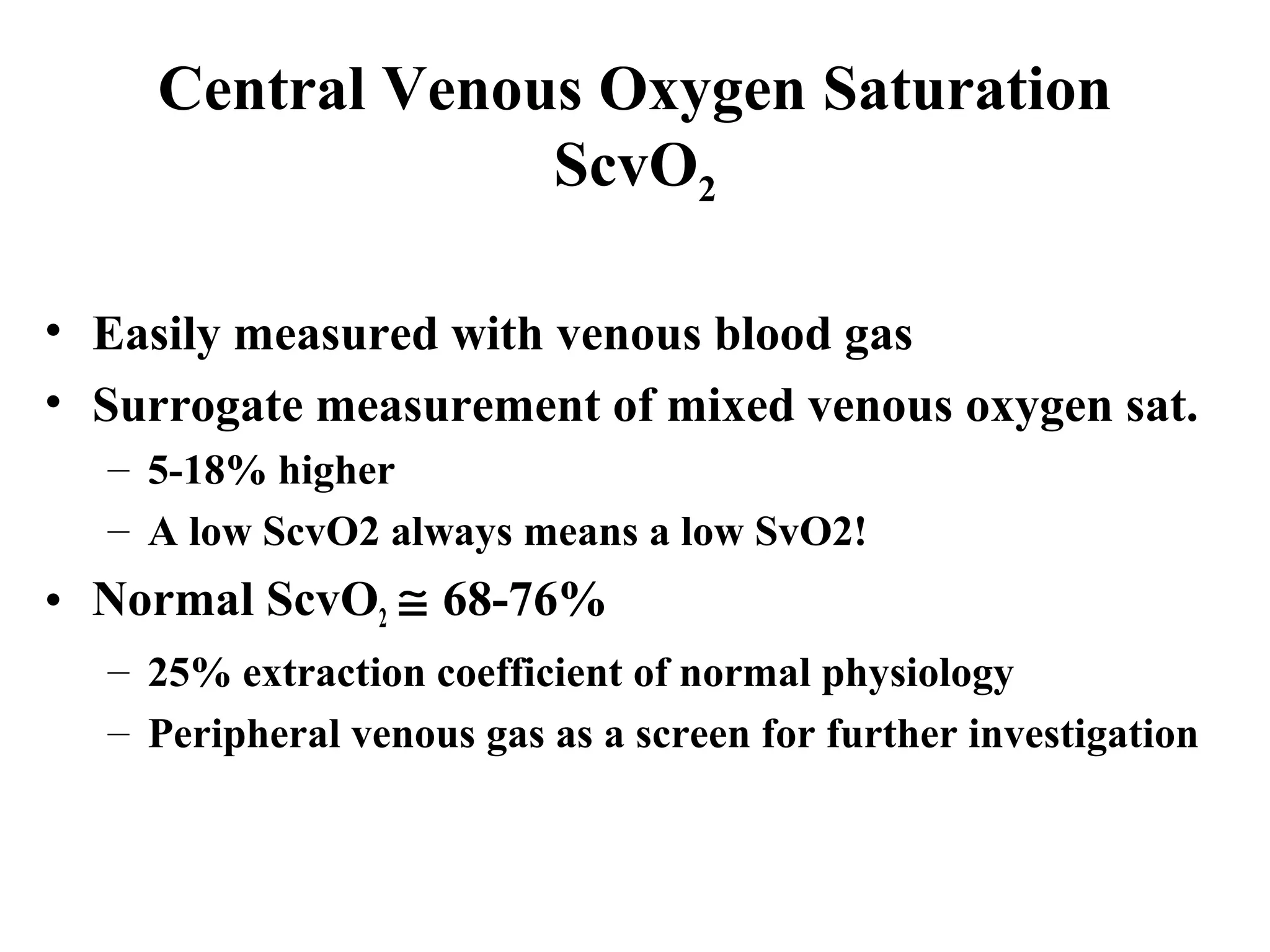
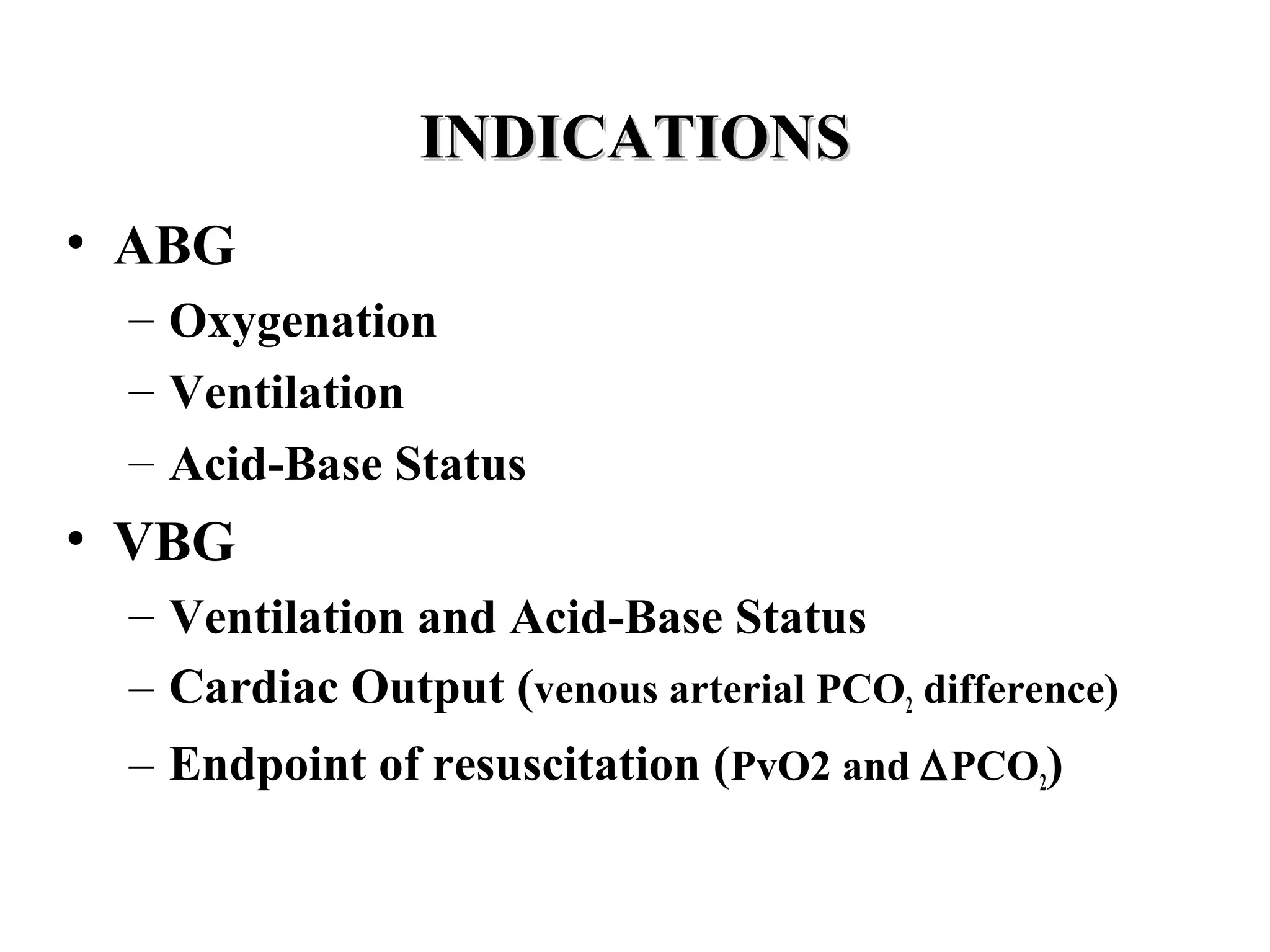
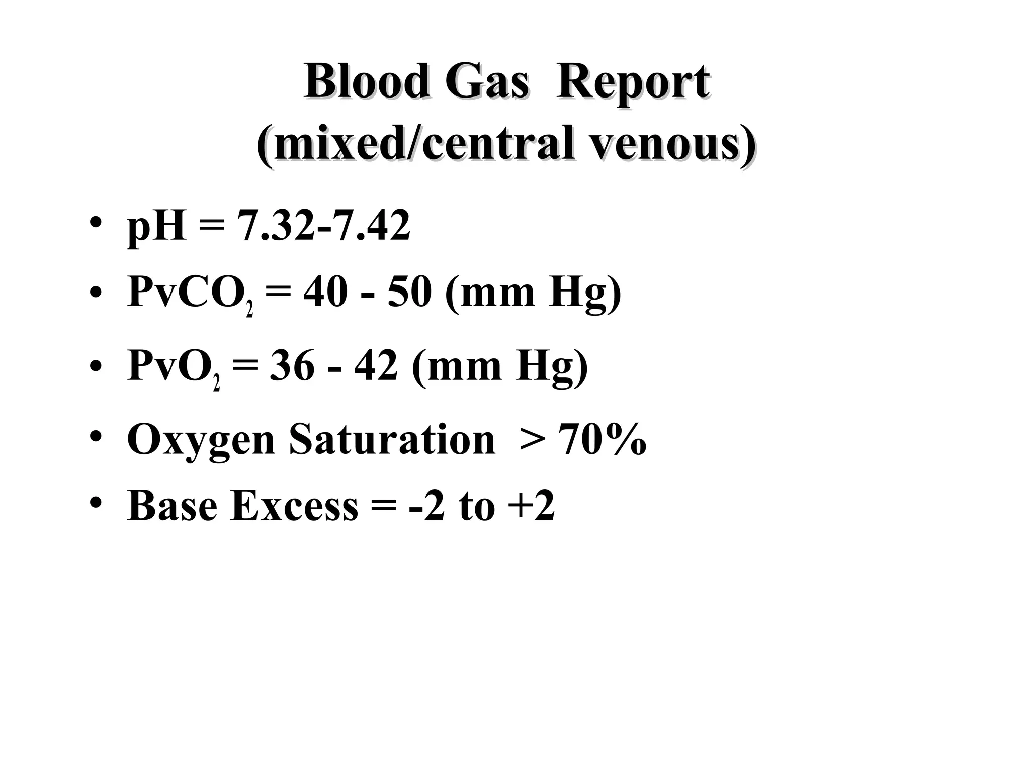
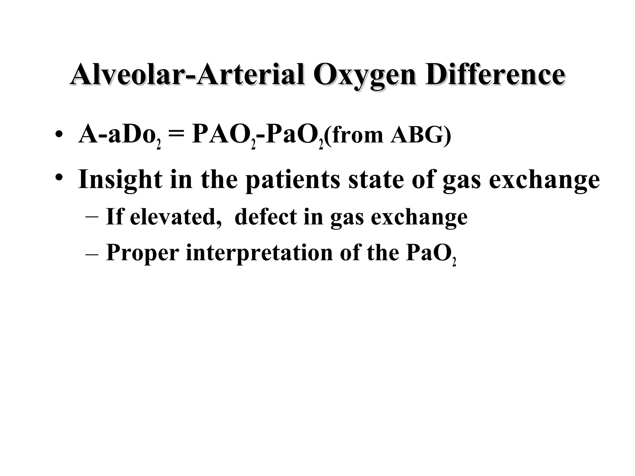
Arterial And Venous BloodArterial And Venous Blood
Gas AnalysisGas Analysis
ByBy
Edward Omron MD MPH FCCPEdward Omron MD MPH FCCP
Pulmonary Critical Care MedicinePulmonary Critical Care Medicine
Morgan Hill, CAMorgan Hill, CA
www.docomron.comwww.docomron.com
INDICATIONSINDICATIONS
• ABG
– Oxygenation
– Ventilation
– Acid-Base Status
• VBG
– Ventilation and Acid-Base Status
– Cardiac Output (venous arterial PCO2 difference)
– Endpoint of resuscitation (PvO2 and ∆PCO2)
Blood Gas Report(Blood Gas Report(ArterialArterial))
• pH (No Units) 7.35-7.45
• PaCO2 (mm Hg) 35-45
• PaO2 (mm Hg) 110 - 0.5(age)
• HCO3
-
(mmol/L): calc. 22-26
• B.E. (mmol/L) -2 to 2
• O2 saturation: calc. >90%
Blood Gas ReportBlood Gas Report
(mixed/central venous)(mixed/central venous)
• pH = 7.32-7.42
• PvCO2 = 40 - 50 (mm Hg)
• PvO2 = 36 - 42 (mm Hg)
• Oxygen Saturation > 70%
• Base Excess = -2 to +2
ANALYSIS OF OXYGENATIONANALYSIS OF OXYGENATION
• Alveolar Gas Equation
– PAO2 = FIO2(PB - 47) - 1.2(PaCO2)
– PAO2 defines upper limit of PaO2
– FIO2 is 21% at all altitudes
– Factor 1.2 determined by RQ
– Water vapor pressure = 47 mm Hg
– PAO2= 150 - 1.2(PaCO2) at room air
– PAO2 = 102
Alveolar-Arterial Oxygen DifferenceAlveolar-Arterial Oxygen Difference
• A-aDo2 = PAO2-PaO2(from ABG)
• Insight in the patients state of gas exchange
– If elevated, defect in gas exchange
– Proper interpretation of the PaO2
Causes of a low PaOCauses of a low PaO22 and A-a Doand A-a Do22
• P(B): Altitude
• Alveolar Hypoventilation(Nl A-a Do2)
• V/Q mismatch
• Shunt
• Diffusion Impairment
• Decreased mixed / central venous O2
content
Alveolar HypoventilationAlveolar Hypoventilation
• No increase in P(A-a)O2 gradient
• Muscle weakness
• Neuromuscular Junction Disease
• Reduced Respiratory Drive (drugs)
• Chest wall elastic loads
• Brainstem injury
Approach To HypoxemiaApproach To Hypoxemia
PaO2 A-a Do2 pCO2
N
FIO2
N
Alv. Hypo.
100% O2 Corrects V/Q Mis.
No Correction Shunt
Diffusion
Changes in P(A-a)O2 with FIO2
Acute Lung Injury
• PaO2/FIO2 ratio
– Normal = 90 mm Hg/0.2 = 450
– Acute lung injury < 300
– Acute Respiratory Distress syndrome
<200
OXIMETRYOXIMETRY
• Binding sites for O2 are heme groups
• OXYGEN SATURATION
– % of all heme sites saturated with O2
• Measures the difference in the light absorbance
characeteristics between Oxy Hb and Deoxy Hb
• SpO2 = Oxy Hb x 100
Oxy Hb + Deoxy Hb
• ABG SaO2 is a calculated value from PaO2
Pulse OximetryPulse Oximetry
Whole Blood
PaO2
SaO2
SpO2:
Pulse Oximetry
OximetryOximetry
• 54 yo WM with headaches, dyspnea and
a Kerosene heater at home
– ABG: PaO2 = 89, PaCO2 = 38, pH = 7.43
– SaO2 = 98%
– Whats the problem?
OximetryOximetry
• Carboxyhemoglobin: Hb +CO
– Does not affect PaO2 only SaO2
– Pulse oximetry reads CO-HB as OxyHb
• Follow Up:
– PaO2 = 79, PaCO2 = 31, SpO2 = 53%, pH =7.36
– CO-Hb46%
Correlation of symptoms and signs with carbon monoxide level
Percent of CO in inspired
air
Percent of HbCO in blood Signs and symptoms
0.007 10
Common in cigarette smokers; dyspnea
during vigorousexertion; occasional
tightness in forehead; dilation of cutaneous
blood vessels
0.012 20
Dyspnea during moderate exertion;
occasional throbbing headache in temples
0.022 30
Severe headache; irritability; easy
fatigability; disturbed judgment; possible
dizziness and possible dimness of vision
0.035 40+ Headache; confusion; fainting on exertion
0.080 60+
Unconsciousness; intermittent convulsions;
respiratory failure; death if exposure
prolonged
0.195 80 Fatal
Modified from Winter, P.M., and Miller, J.N.: JAMA 236:1503, 1976, Copyright 1976, American Medical
Association.
ProblemProblem
• 42 yo HIV pt with fevers,chills, SOB,cough
– Taking Dapsone for PCP prophylaxis
– ABG: PaO2 82.5, PaCO2 35.2, pH 7.43, SaO2
89%
– PCP Pneumonia, started onPrimaquine, Clinda,
and Prednisone
– ABG: PaO2 378, PaCO2 of 35, pH 7.42, SaO2
80%
– Whats Happening?
MethemoglobinMethemoglobin
• Oxidation of Fe++ to Fe+++ state
• Unlike CO-Hb, Met-Hb does depress the
SpO2 reading
• Both Dapsone and Primaquine are
oxidants
• Met-Hb depresses the SpO2 to 80’s
– Further increaeses in Met-Hb do not depress
SpO2
• Methylene Blue administration is Rx
Some drugs implicated in causing methemoglobinemia
Generic name Use
Dapsone Skin protectant
Benzocaine Local anesthetic
Metoclopramide Gastric stasis
Nitroglycerin Angina
Phenazopyridine Urinary tract analgesic
Prilocaine Local anesthetic
Primaquine Malaria prophylaxis and treatment
Trimethoprim Urinary antibacterial
Amyl nitrite
Rarely used clinically; often used by drug
abusers
ANALYSIS OF VENTILATONANALYSIS OF VENTILATON
• PaCO2 = VCO2 x K
VA
Hypercapnea > 45 mm Hg (Hypoventilation)
Respiratory Acidosis
Hypocapnea < 35 mm Hg (Hyperventilation)
Respiratory Alkalosis
Respiratory Acid-Base Status
• Respiratory Disturbances
– CO2+H20 H2CO3 H+
+ HCO3
– Acute changes:
• Delta 10 mm Hg PaCO2, pH changes by 0.08
• Chronic change: 40 + B.E
– Alveolar Ventilation
• VA CO2 pH
• Respiratory Acidosis pCO2 > 45
• Respiratory Alkalosis pCO2 < 35
BASE EXCESS (B.E.)BASE EXCESS (B.E.)
• Base Excess
• Quantity of acid or base needed to restore plasma
pH to 7.4, at a PCO2 equillibrated to 40 mm Hg
• Positive value, excess base, metabolic alkalosis
• Negative value, excess acid, metabolic acidosis
• Metabolic component of acid-base status
• PCO2 independent
• Estimated by BE = (Total CO2 – 24)
Problem Solving
1. LOOK AT THE pH
– Whatever side of pH 7.4 is the primary disorder
2. Look at pH, PCO2direction
– Both decrease or increase, then metabolic
– If move in opposite directions, respiratory
3. Respiration: acute or chronic?
– Acute: 10 mm Hg / 0.08 change in pH
– Chronic: 40+Base Excess
• Calculate the Adjusted Anion Gap
– High vs normal ANG differential
– 2.8 mmol of acid /gram serum albumin
– Law of Electrical Neutrality
• Positive charges = negative charges or
• Positive charges - negative charges = 0
– [Na+
] - [Cl-
] -[HCO3
-
] - [Albumin-
] = 0
– [Na+
] - [Cl-
] -[HCO3
-
] = [Albumin-
]
– 140 - 104 -24 = 4.4 gm/dL* 2.8 ≅ 12
– Normal ANG = 12 = 2.8 * [Albumin-
]
– Adjusted ANG = ANG + 2.8(4.4 -Albumin)
Arterial Draw:
• pH = 7.28, PaCO2 = 34, HCO3 = 16
• Na = 153 Cl = 106 Total CO2 = 17
• Alb = 3 g/dL
• Primary Acid-Base Disturbance?
• ANG and Adjusted ANG
• Metabolic Acid-Base Status
74 yo male found unresponsive and pulseless
• Primary Disorder
– Acidosis and acidemia (pH < 7.4)
• pH and PCO2direction
– Both down: Metabolic Acidosis
• Base Excess
– 16 – 24 = -8 mmols/L
• Adjusted Anion Gap
– ANG + 2.8*(4.4 - 3) or 30 + 4 = 34
– Anion Gap Acidosis
• Compensation?
– 40 + BE or 32 mm Hg
Venous Draw
• pH = 7.08, pCO2 = 75, HCO3 = 21
• Na = 145, Cl = 103, Total CO2 =22
• Alb = 3 g/dL
• Primary Acid-Base Disorder?
• ANG and expected ANG?
• Metabolic Acid-Base Status?
• Primary Disorder
– pH < 7.4, acidosis and acidemia
• pH and PCO2 direction
– Opposite therefore RESPIRATORY acidosis
• Base Excess
– 22 – 24 = -2 mmol/L
• Adjusted Anion Gap
– ANG + 2.8(4.4 -3) = 20+4 =24
– Anion Gap Acidosis
• 40 + BE rule ≠ Comp in VBG
74 yo male found unresponsive and pulseless
• Why a metabolic acidosis in arterial bed
and respiratory acidosis in venous bed?
– Venous arterial PCO2 difference?
– PaCO2 = 34 and PvCO2 = 75
– PvCO2 – PaCO2 ∝ 1 / cardiac index
• Venous vs Arterial saturation difference?
– PaO2 = 50 mm Hg, saturation = 84%
– PvO2 =18, Venous Saturation = 20%
– Increased oxygen extraction from circulatory
failure
Central Venous Oxygen Saturation
ScvO2
• Easily measured with venous blood gas
• Surrogate measurement of mixed venous oxygen sat.
– 5-18% higher
– A low ScvO2 always means a low SvO2!
• Normal ScvO2 ≅ 68-76%
– 25% extraction coefficient of normal physiology
– Peripheral venous gas as a screen for further investigation
PaO2 vs PvO2 in Cardiogenic Shock
Paradoxical Respiratory Acidosis of Cardiopulmonary Arrest
Four Determinants of Central Venous Oximetry
ScvOScvO22 ≅≅ SvOSvO22 = SaO= SaO22 - VO- VO22 / C.O. x Hgb x 1.36/ C.O. x Hgb x 1.36
ScvO2 = Central venous saturation (%)
SvO2 = Mixed venous saturation (%)
SaO2 = Arterial oxygen saturation (%)
VO2 = Oxygen consumption mL (O2/min)
Hgb = Hemoglobin concentration (g/dL)
Cardiac Output (C.O.) = dL/min
Effect of changes in PaO2 on SvO2
600
200
100
80
60
40
0
0.1
0.2
0.3
0.4
0.5
0.6
0.7
0.8
0.9
1
0 100 200 300 400 500 600 700
PaO2 (mm Hg)
SvO2
SvO2
Effect of changes in Hgn on SvO2
13
10
7.5
5
0
0.1
0.2
0.3
0.4
0.5
0.6
0.7
0.8
0.9
0 2 4 6 8 10 12 14
Hemoglobin (g/dL)
SvO2
SvO2
The Effects of Cardiac Output on SvO2
0.87
0.83
0.73
0.66
0.55
0.31
0
0.1
0.2
0.3
0.4
0.5
0.6
0.7
0.8
0.9
1
0 2 4 6 8 10 12
Cardiac Output (L/min)
SvO2
SvO2
Effect of Oxygen Consumption (VO2) on SvO2
0.85
0.79
0.74
0.68
0.57
0.46
0
0.1
0.2
0.3
0.4
0.5
0.6
0.7
0.8
0.9
0 100 200 300 400 500 600
Oxygen Consumption (VO2)
SvO2
SvO2
Venous Arterial CO2 Difference
• Circulatory Failure
– Associated with Tissue Hypercarbic Acidosis
– Hypovolemia, sepsis, shock …
• Cardiac Index = e (1.787–0.151(v-aCO2))
– Endpoint of Resuscitation
• PvO2
– Enpoint of Resuscitation
SvO2 vs SaO2 response in Septic Shock "Golden Hour"
27
51
68
73
100 100 98
91
0
10
20
30
40
50
60
70
80
90
100
1 2 3 4
Days
Percentage
SvO2
SaO2
REFERENCES
• Current Opinion Critical Care 2001; 7: 204-211
• NEJM 2001; 345: 1368-1377
• Critical Care Medicine 2002; 30: 1686-1692
• Circulation 1969; 40: 165
• Thorax 2002; 57: 170-177
• Academic Emer Med 1999; 6: 421

Arterial and Venous Blood Gas Analysis Edward Omron MD, MPH

  • 1.
    Arterial And VenousBloodArterial And Venous Blood Gas AnalysisGas Analysis ByBy Edward Omron MD MPH FCCPEdward Omron MD MPH FCCP Pulmonary Critical Care MedicinePulmonary Critical Care Medicine Morgan Hill, CAMorgan Hill, CA www.docomron.comwww.docomron.com
  • 2.
    INDICATIONSINDICATIONS • ABG – Oxygenation –Ventilation – Acid-Base Status • VBG – Ventilation and Acid-Base Status – Cardiac Output (venous arterial PCO2 difference) – Endpoint of resuscitation (PvO2 and ∆PCO2)
  • 3.
    Blood Gas Report(BloodGas Report(ArterialArterial)) • pH (No Units) 7.35-7.45 • PaCO2 (mm Hg) 35-45 • PaO2 (mm Hg) 110 - 0.5(age) • HCO3 - (mmol/L): calc. 22-26 • B.E. (mmol/L) -2 to 2 • O2 saturation: calc. >90%
  • 4.
    Blood Gas ReportBloodGas Report (mixed/central venous)(mixed/central venous) • pH = 7.32-7.42 • PvCO2 = 40 - 50 (mm Hg) • PvO2 = 36 - 42 (mm Hg) • Oxygen Saturation > 70% • Base Excess = -2 to +2
  • 6.
    ANALYSIS OF OXYGENATIONANALYSISOF OXYGENATION • Alveolar Gas Equation – PAO2 = FIO2(PB - 47) - 1.2(PaCO2) – PAO2 defines upper limit of PaO2 – FIO2 is 21% at all altitudes – Factor 1.2 determined by RQ – Water vapor pressure = 47 mm Hg – PAO2= 150 - 1.2(PaCO2) at room air – PAO2 = 102
  • 7.
    Alveolar-Arterial Oxygen DifferenceAlveolar-ArterialOxygen Difference • A-aDo2 = PAO2-PaO2(from ABG) • Insight in the patients state of gas exchange – If elevated, defect in gas exchange – Proper interpretation of the PaO2
  • 8.
    Causes of alow PaOCauses of a low PaO22 and A-a Doand A-a Do22 • P(B): Altitude • Alveolar Hypoventilation(Nl A-a Do2) • V/Q mismatch • Shunt • Diffusion Impairment • Decreased mixed / central venous O2 content
  • 9.
    Alveolar HypoventilationAlveolar Hypoventilation •No increase in P(A-a)O2 gradient • Muscle weakness • Neuromuscular Junction Disease • Reduced Respiratory Drive (drugs) • Chest wall elastic loads • Brainstem injury
  • 10.
    Approach To HypoxemiaApproachTo Hypoxemia PaO2 A-a Do2 pCO2 N FIO2 N Alv. Hypo. 100% O2 Corrects V/Q Mis. No Correction Shunt Diffusion
  • 11.
  • 12.
    Acute Lung Injury •PaO2/FIO2 ratio – Normal = 90 mm Hg/0.2 = 450 – Acute lung injury < 300 – Acute Respiratory Distress syndrome <200
  • 13.
    OXIMETRYOXIMETRY • Binding sitesfor O2 are heme groups • OXYGEN SATURATION – % of all heme sites saturated with O2 • Measures the difference in the light absorbance characeteristics between Oxy Hb and Deoxy Hb • SpO2 = Oxy Hb x 100 Oxy Hb + Deoxy Hb • ABG SaO2 is a calculated value from PaO2
  • 14.
    Pulse OximetryPulse Oximetry WholeBlood PaO2 SaO2 SpO2: Pulse Oximetry
  • 15.
    OximetryOximetry • 54 yoWM with headaches, dyspnea and a Kerosene heater at home – ABG: PaO2 = 89, PaCO2 = 38, pH = 7.43 – SaO2 = 98% – Whats the problem?
  • 16.
    OximetryOximetry • Carboxyhemoglobin: Hb+CO – Does not affect PaO2 only SaO2 – Pulse oximetry reads CO-HB as OxyHb • Follow Up: – PaO2 = 79, PaCO2 = 31, SpO2 = 53%, pH =7.36 – CO-Hb46%
  • 17.
    Correlation of symptomsand signs with carbon monoxide level Percent of CO in inspired air Percent of HbCO in blood Signs and symptoms 0.007 10 Common in cigarette smokers; dyspnea during vigorousexertion; occasional tightness in forehead; dilation of cutaneous blood vessels 0.012 20 Dyspnea during moderate exertion; occasional throbbing headache in temples 0.022 30 Severe headache; irritability; easy fatigability; disturbed judgment; possible dizziness and possible dimness of vision 0.035 40+ Headache; confusion; fainting on exertion 0.080 60+ Unconsciousness; intermittent convulsions; respiratory failure; death if exposure prolonged 0.195 80 Fatal Modified from Winter, P.M., and Miller, J.N.: JAMA 236:1503, 1976, Copyright 1976, American Medical Association.
  • 18.
    ProblemProblem • 42 yoHIV pt with fevers,chills, SOB,cough – Taking Dapsone for PCP prophylaxis – ABG: PaO2 82.5, PaCO2 35.2, pH 7.43, SaO2 89% – PCP Pneumonia, started onPrimaquine, Clinda, and Prednisone – ABG: PaO2 378, PaCO2 of 35, pH 7.42, SaO2 80% – Whats Happening?
  • 19.
    MethemoglobinMethemoglobin • Oxidation ofFe++ to Fe+++ state • Unlike CO-Hb, Met-Hb does depress the SpO2 reading • Both Dapsone and Primaquine are oxidants • Met-Hb depresses the SpO2 to 80’s – Further increaeses in Met-Hb do not depress SpO2 • Methylene Blue administration is Rx
  • 20.
    Some drugs implicatedin causing methemoglobinemia Generic name Use Dapsone Skin protectant Benzocaine Local anesthetic Metoclopramide Gastric stasis Nitroglycerin Angina Phenazopyridine Urinary tract analgesic Prilocaine Local anesthetic Primaquine Malaria prophylaxis and treatment Trimethoprim Urinary antibacterial Amyl nitrite Rarely used clinically; often used by drug abusers
  • 21.
    ANALYSIS OF VENTILATONANALYSISOF VENTILATON • PaCO2 = VCO2 x K VA Hypercapnea > 45 mm Hg (Hypoventilation) Respiratory Acidosis Hypocapnea < 35 mm Hg (Hyperventilation) Respiratory Alkalosis
  • 22.
    Respiratory Acid-Base Status •Respiratory Disturbances – CO2+H20 H2CO3 H+ + HCO3 – Acute changes: • Delta 10 mm Hg PaCO2, pH changes by 0.08 • Chronic change: 40 + B.E – Alveolar Ventilation • VA CO2 pH • Respiratory Acidosis pCO2 > 45 • Respiratory Alkalosis pCO2 < 35
  • 23.
    BASE EXCESS (B.E.)BASEEXCESS (B.E.) • Base Excess • Quantity of acid or base needed to restore plasma pH to 7.4, at a PCO2 equillibrated to 40 mm Hg • Positive value, excess base, metabolic alkalosis • Negative value, excess acid, metabolic acidosis • Metabolic component of acid-base status • PCO2 independent • Estimated by BE = (Total CO2 – 24)
  • 24.
    Problem Solving 1. LOOKAT THE pH – Whatever side of pH 7.4 is the primary disorder 2. Look at pH, PCO2direction – Both decrease or increase, then metabolic – If move in opposite directions, respiratory 3. Respiration: acute or chronic? – Acute: 10 mm Hg / 0.08 change in pH – Chronic: 40+Base Excess
  • 25.
    • Calculate theAdjusted Anion Gap – High vs normal ANG differential – 2.8 mmol of acid /gram serum albumin – Law of Electrical Neutrality • Positive charges = negative charges or • Positive charges - negative charges = 0 – [Na+ ] - [Cl- ] -[HCO3 - ] - [Albumin- ] = 0 – [Na+ ] - [Cl- ] -[HCO3 - ] = [Albumin- ] – 140 - 104 -24 = 4.4 gm/dL* 2.8 ≅ 12 – Normal ANG = 12 = 2.8 * [Albumin- ] – Adjusted ANG = ANG + 2.8(4.4 -Albumin)
  • 26.
    Arterial Draw: • pH= 7.28, PaCO2 = 34, HCO3 = 16 • Na = 153 Cl = 106 Total CO2 = 17 • Alb = 3 g/dL • Primary Acid-Base Disturbance? • ANG and Adjusted ANG • Metabolic Acid-Base Status 74 yo male found unresponsive and pulseless
  • 27.
    • Primary Disorder –Acidosis and acidemia (pH < 7.4) • pH and PCO2direction – Both down: Metabolic Acidosis • Base Excess – 16 – 24 = -8 mmols/L • Adjusted Anion Gap – ANG + 2.8*(4.4 - 3) or 30 + 4 = 34 – Anion Gap Acidosis • Compensation? – 40 + BE or 32 mm Hg
  • 28.
    Venous Draw • pH= 7.08, pCO2 = 75, HCO3 = 21 • Na = 145, Cl = 103, Total CO2 =22 • Alb = 3 g/dL • Primary Acid-Base Disorder? • ANG and expected ANG? • Metabolic Acid-Base Status?
  • 29.
    • Primary Disorder –pH < 7.4, acidosis and acidemia • pH and PCO2 direction – Opposite therefore RESPIRATORY acidosis • Base Excess – 22 – 24 = -2 mmol/L • Adjusted Anion Gap – ANG + 2.8(4.4 -3) = 20+4 =24 – Anion Gap Acidosis • 40 + BE rule ≠ Comp in VBG
  • 30.
    74 yo malefound unresponsive and pulseless • Why a metabolic acidosis in arterial bed and respiratory acidosis in venous bed? – Venous arterial PCO2 difference? – PaCO2 = 34 and PvCO2 = 75 – PvCO2 – PaCO2 ∝ 1 / cardiac index • Venous vs Arterial saturation difference? – PaO2 = 50 mm Hg, saturation = 84% – PvO2 =18, Venous Saturation = 20% – Increased oxygen extraction from circulatory failure
  • 31.
    Central Venous OxygenSaturation ScvO2 • Easily measured with venous blood gas • Surrogate measurement of mixed venous oxygen sat. – 5-18% higher – A low ScvO2 always means a low SvO2! • Normal ScvO2 ≅ 68-76% – 25% extraction coefficient of normal physiology – Peripheral venous gas as a screen for further investigation
  • 33.
    PaO2 vs PvO2in Cardiogenic Shock
  • 34.
    Paradoxical Respiratory Acidosisof Cardiopulmonary Arrest
  • 35.
    Four Determinants ofCentral Venous Oximetry ScvOScvO22 ≅≅ SvOSvO22 = SaO= SaO22 - VO- VO22 / C.O. x Hgb x 1.36/ C.O. x Hgb x 1.36 ScvO2 = Central venous saturation (%) SvO2 = Mixed venous saturation (%) SaO2 = Arterial oxygen saturation (%) VO2 = Oxygen consumption mL (O2/min) Hgb = Hemoglobin concentration (g/dL) Cardiac Output (C.O.) = dL/min
  • 36.
    Effect of changesin PaO2 on SvO2 600 200 100 80 60 40 0 0.1 0.2 0.3 0.4 0.5 0.6 0.7 0.8 0.9 1 0 100 200 300 400 500 600 700 PaO2 (mm Hg) SvO2 SvO2
  • 37.
    Effect of changesin Hgn on SvO2 13 10 7.5 5 0 0.1 0.2 0.3 0.4 0.5 0.6 0.7 0.8 0.9 0 2 4 6 8 10 12 14 Hemoglobin (g/dL) SvO2 SvO2
  • 38.
    The Effects ofCardiac Output on SvO2 0.87 0.83 0.73 0.66 0.55 0.31 0 0.1 0.2 0.3 0.4 0.5 0.6 0.7 0.8 0.9 1 0 2 4 6 8 10 12 Cardiac Output (L/min) SvO2 SvO2
  • 39.
    Effect of OxygenConsumption (VO2) on SvO2 0.85 0.79 0.74 0.68 0.57 0.46 0 0.1 0.2 0.3 0.4 0.5 0.6 0.7 0.8 0.9 0 100 200 300 400 500 600 Oxygen Consumption (VO2) SvO2 SvO2
  • 40.
    Venous Arterial CO2Difference • Circulatory Failure – Associated with Tissue Hypercarbic Acidosis – Hypovolemia, sepsis, shock … • Cardiac Index = e (1.787–0.151(v-aCO2)) – Endpoint of Resuscitation • PvO2 – Enpoint of Resuscitation
  • 41.
    SvO2 vs SaO2response in Septic Shock "Golden Hour" 27 51 68 73 100 100 98 91 0 10 20 30 40 50 60 70 80 90 100 1 2 3 4 Days Percentage SvO2 SaO2
  • 42.
    REFERENCES • Current OpinionCritical Care 2001; 7: 204-211 • NEJM 2001; 345: 1368-1377 • Critical Care Medicine 2002; 30: 1686-1692 • Circulation 1969; 40: 165 • Thorax 2002; 57: 170-177 • Academic Emer Med 1999; 6: 421

Editor's Notes

  • #3 There are 3 reasones why one should obtain a blood gas:
  • #4 This is a typical printout from a blood gas report:
  • #7 Recall altitiude Recall RS Allows to assess properly if the lungs are transferring loxygen appropriately into the blood