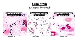
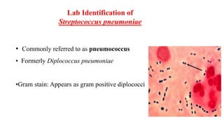
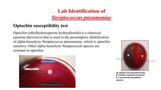
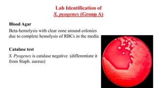
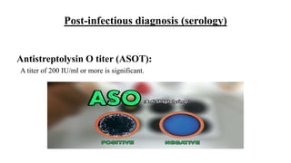
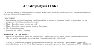
Streptococcus
Lab 3
Prepared by
Dr. Samira Fattah
PhD in Medical Bacteriology
College of Health Sciences-HMU
Sources of the specimen
• Respiratory tract(RT)
• Gastrointestinal tract(GIT)
• Genitourinary tract(GUT)
• Skin
Culture on Blood Agar
Hemolysis on BA
– -hemolysis
Partial hemolysis
Green discoloration around colonies
e.g. S. pneumoniae S. viridans, S. mutans
– -hemolysis
Complete hemolysis
Clear zone of hemolysis around colonies
e.g. Lancefield Group A & B (S. pyogenes & S.
agalactiae)
– -hemolysis
No hemolysis
e.g. Group D (Enterococcus faecalis, E.
faecium)
Gram stain
gram positive cocci
Diplococci long chain short chain
Lab Identification of
Streptococcus pneumoniae
• Commonly referred to as pneumococcus
• Formerly Diplococcus pneumoniae
•Gram stain: Appears as gram positive diplococci
Lab Identification of
Streptococcus pneumoniae
Blood Agar
greenish colonies due to partial hemolysis of RBCs in the media
Optochin susceptibility test
Optochin (ethylhydrocupreine hydrochloride) is a chemical
(quinine derivative) that is used in the presumptive identification
of alpha-hemolytic Streptococcus pneumoniae, which is optochin
sensitive. Other alpha-hemolytic Streptococcal species are
resistant to optochin.
Lab Identification of
Streptococcus pneumoniae
Procedure
• Using an inoculating loop, select a well-isolated colony of the alpha-hemolytic organism to
be tested.
• Streak the isolates onto 5%sheep blood agar plate.
• Using sterile forceps, place an optochin disk onto the inoculated surface of the agar.
• Press disk gently with the sterile forceps or loop so that the disk adheres firmly to the agar
surfaces.
• Incubate the plate at 35-37°C for 18-24 hour in 5 to 10% CO2.
(NOTE: Cultures do not grow as well in ambient air, and larger zone of inhibition occur.)
• Measure the diameter of zone of inhibition including diameter of disk.
Result interpretation
• Sensitive: zone of inhibition ≥ 14mm (15-30mm) around the disk.
• Resistant: No zone of inhibition around the disk.
Lab Identification of
S. pyogenes (Group A)
Gram stain
Gram positive bacteria appear as long chain
Lab Identification of
S. pyogenes (Group A)
Blood Agar
Beta-hemolysis with clear zone around colonies
due to complete hemolysis of RBCs in the media.
Catalase test
S. Pyogenes is catalase negative (differentiate it
from Staph. aureus)
Lab Identification of
S. pyogenes (Group A)
Bacitracin test
Bacitracin is a bactericidal antibiotic used
to distinguish S. pyogenes from other β-
hemolytic streptococci
Procedure of Bacitracin test
• Using an inoculating loop, streak two or three suspect colonies of a pure culture onto
a blood agar plate.
• Using heated forceps, place a bacitracin disk in the first quadrant (area of heaviest
growth). Gently tap the disk to ensure adequate contact with the agar surface.
• Incubate the plate for 18 to 24 hours at 35oC in CO2.
• Look for a zone of inhibition around the disk.
Results:
• Bacitracin sensitive: Any zone of inhibition around the disk. Streptococcus pyogenes
• Bacitracin resistant: No zone of inhibition around the disk. Streptococcus agalactiae
Post-infectious diagnosis (serology)
Antistreptolysin O titer (ASOT):
A titer of 200 IU/ml or more is significant.
The principle is based on the immunological reaction between ASO antibodies with Streptolysin O antigen coated onto latex
particles, to form visible agglutination.
PROCEDURE:
1. Centrifuge the blood specimen in the centrifuge machine at 400rpm for 5 minutes, in order to sediment the red cell
component of the blood and obtain the serum.
2. Place a drop of the serum onto the test card.
3. Place a drop of the reagent onto the serum on the test card.
4. Mix the mixture together.
5. Rotate the test card for 2 minutes.
REPORTING OF THE RESULT:
• Presence of agglutination within the 2 minutes of rocking indicates that the patient’s serum contains ASO antibodies.
This is a positive result and is reported as thus: Concentration of ASO > 200 IU/ml.
• Absence of agglutination within the 2 minutes of rocking indicates that the patient’s serum does not contain ASO
antibodies that are up to 200 IU/ml. This is a negative result and is reported as thus: Concentration of ASO < 200 IU/ml.
Antistreptolysin O titer
API 20 Strep test
All-positive tests (up) and all-negative tests (down)
Streptococcus

Streptococcus

  • 1.
    Streptococcus Lab 3 Prepared by Dr.Samira Fattah PhD in Medical Bacteriology College of Health Sciences-HMU
  • 2.
    Sources of thespecimen • Respiratory tract(RT) • Gastrointestinal tract(GIT) • Genitourinary tract(GUT) • Skin
  • 3.
    Culture on BloodAgar Hemolysis on BA – -hemolysis Partial hemolysis Green discoloration around colonies e.g. S. pneumoniae S. viridans, S. mutans – -hemolysis Complete hemolysis Clear zone of hemolysis around colonies e.g. Lancefield Group A & B (S. pyogenes & S. agalactiae) – -hemolysis No hemolysis e.g. Group D (Enterococcus faecalis, E. faecium)
  • 4.
    Gram stain gram positivecocci Diplococci long chain short chain
  • 5.
    Lab Identification of Streptococcuspneumoniae • Commonly referred to as pneumococcus • Formerly Diplococcus pneumoniae •Gram stain: Appears as gram positive diplococci
  • 6.
    Lab Identification of Streptococcuspneumoniae Blood Agar greenish colonies due to partial hemolysis of RBCs in the media
  • 7.
    Optochin susceptibility test Optochin(ethylhydrocupreine hydrochloride) is a chemical (quinine derivative) that is used in the presumptive identification of alpha-hemolytic Streptococcus pneumoniae, which is optochin sensitive. Other alpha-hemolytic Streptococcal species are resistant to optochin. Lab Identification of Streptococcus pneumoniae
  • 8.
    Procedure • Using aninoculating loop, select a well-isolated colony of the alpha-hemolytic organism to be tested. • Streak the isolates onto 5%sheep blood agar plate. • Using sterile forceps, place an optochin disk onto the inoculated surface of the agar. • Press disk gently with the sterile forceps or loop so that the disk adheres firmly to the agar surfaces. • Incubate the plate at 35-37°C for 18-24 hour in 5 to 10% CO2. (NOTE: Cultures do not grow as well in ambient air, and larger zone of inhibition occur.) • Measure the diameter of zone of inhibition including diameter of disk. Result interpretation • Sensitive: zone of inhibition ≥ 14mm (15-30mm) around the disk. • Resistant: No zone of inhibition around the disk.
  • 9.
    Lab Identification of S.pyogenes (Group A) Gram stain Gram positive bacteria appear as long chain
  • 10.
    Lab Identification of S.pyogenes (Group A) Blood Agar Beta-hemolysis with clear zone around colonies due to complete hemolysis of RBCs in the media. Catalase test S. Pyogenes is catalase negative (differentiate it from Staph. aureus)
  • 11.
    Lab Identification of S.pyogenes (Group A) Bacitracin test Bacitracin is a bactericidal antibiotic used to distinguish S. pyogenes from other β- hemolytic streptococci
  • 12.
    Procedure of Bacitracintest • Using an inoculating loop, streak two or three suspect colonies of a pure culture onto a blood agar plate. • Using heated forceps, place a bacitracin disk in the first quadrant (area of heaviest growth). Gently tap the disk to ensure adequate contact with the agar surface. • Incubate the plate for 18 to 24 hours at 35oC in CO2. • Look for a zone of inhibition around the disk. Results: • Bacitracin sensitive: Any zone of inhibition around the disk. Streptococcus pyogenes • Bacitracin resistant: No zone of inhibition around the disk. Streptococcus agalactiae
  • 13.
    Post-infectious diagnosis (serology) AntistreptolysinO titer (ASOT): A titer of 200 IU/ml or more is significant.
  • 14.
    The principle isbased on the immunological reaction between ASO antibodies with Streptolysin O antigen coated onto latex particles, to form visible agglutination. PROCEDURE: 1. Centrifuge the blood specimen in the centrifuge machine at 400rpm for 5 minutes, in order to sediment the red cell component of the blood and obtain the serum. 2. Place a drop of the serum onto the test card. 3. Place a drop of the reagent onto the serum on the test card. 4. Mix the mixture together. 5. Rotate the test card for 2 minutes. REPORTING OF THE RESULT: • Presence of agglutination within the 2 minutes of rocking indicates that the patient’s serum contains ASO antibodies. This is a positive result and is reported as thus: Concentration of ASO > 200 IU/ml. • Absence of agglutination within the 2 minutes of rocking indicates that the patient’s serum does not contain ASO antibodies that are up to 200 IU/ml. This is a negative result and is reported as thus: Concentration of ASO < 200 IU/ml. Antistreptolysin O titer
  • 15.
    API 20 Streptest All-positive tests (up) and all-negative tests (down)