Prepared by
Fakhre-yar
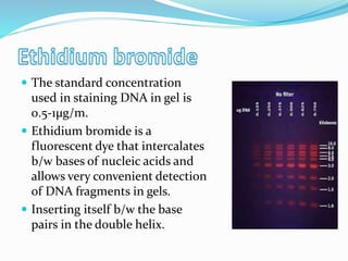
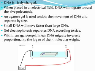
 Agarose gel electrophoresis is a method to separate
DNA or RNA molecule by size.
 This is achieved by moving –ively charged nucleic acid
molecule through an agarose matrix with an electric
field electrophoresis.
 Shorter molecules move faster and migrate faster than
longer ones.
 Powerful separation method frequently used to
analyze DNA fragments generated by restriction
enzymes.
 Convenient analytical method for determining the size
of DNA molecules in the range of 500-30,000 bp.
 Employs electromotive force to move molecules
through a porous gel.
 Separates molecules from each other on the basis of
• Size and/or
• Charge and/or
• Shape
 Basis of separation depends on how the sample and gel
are prepared.
 Electrophoresis chamber
 Agarose gel
 Gel casting tray
 Buffer
 Staining agent(dye)
 A comb
 DNA ladder
 Sample to be separated
 A linear carbohydrate polymer extracted from
seaweed, agarobiose.
 Forms a porous matrix as it gels.
 Shift from random coil in solution to structure in which
chains are bundled into double helices.
 Standard agaarose-LE
 Gel at 35-38℃
 Melt at 90-95℃
 Become opaque at high concentration
 Low melting agarose (NuSieve)
 Gel at 35℃
 Melt at 65℃
 Often used to isolate DNA fragment from gel.
 Intermediate forms or combination of LE and NuSieve
can provide sturdy translucent gels at high agarose
concentrations
 Available in variety of sizes and
composed of UV transparent
plastics.
 The open ends of the trays are
closed with tape while the gel is
being cast, then removed prior to
electrophoresis.
 During electrophoresis water undergoes hydrolysis
H2O ------> H+ + OH⁻
 Buffer prevent the PH from changing by reacting with
the H+ or OH⁻ products.
 Most common buffer used is called TRIS.
[tris(hydroxymethyl) amino methane)
 Another compound is added to make Tris an effective
buffer- either boric or acetic acid.
 Another compound is added to bind metals EDTA.
 The buffer is either TBE or TAE.
 TBE is made with Tris/boric acid/EDTA
 TAE is made with Tris/acetic acid/EDTA
 To make DNA fragments visible after electrophoresis,
the DNA must be stained.
 Favorite-ethidium bromide.
 When bond to DNA it fluoresces under UV light
(reddish-orange color).
 Convenient because it can be added directly to the gel.
 Sensitive-detect 0.1µg of DNA.
 The standard concentration
used in staining DNA in gel is
0.5-1µg/m.
 Ethidium bromide is a
fluorescent dye that intercalates
b/w bases of nucleic acids and
allows very convenient detection
of DNA fragments in gels.
 Inserting itself b/w the base
pairs in the double helix.
 UV absorbance maxima at 300 and 360 nm and
emission maxima at 590nm.
 Detection limit of bound DNA is 0.5-5ng/band.
 Ethidium bromide is mutagenic so care must be taken
while handling the dye.
 Other alternative for Ethidium bromide:
 Methylene blue
 Syber safe
 Xylene cyanol
 Bromphenol blue
 A comb is placed in the
liquid agarose after it has
been poured.
 Removing the comb from
the hardened gel produces a
series of wells used to load
the DNA.
 It is a solution of DNA
molecules of different
length.
 DNA ladder consists of
known DNA sizes used to
determine the size of an
unknown DNA sample.
 The DNA ladder usually
contains regularly spaced
sized samples which when
run on an agarose gel looks
like a ladder.
Method for electrophoresis
Prepare agarose gel
•Melt, cool and add ethidium bromide. Mix thoroughly.
•Pour into casting tray with comb and allow to solidify.
•Add running buffer, load samples and marker.
•Run gel at constant voltage until band separation occurs.
•View DNA on UV light box and show result.
 DNA is –ively charged.
 When placed in an electrical field, DNA will migrate toward
the +ive pole anode.
 An agarose gel is used to slow the movement of DNA and
separate by size.
 Small DNA will move faster than large DNA.
 Gel electrophoresis separates DNA according to size.
 Within an agarose gel, linear DNA migrate inversely
proportional to the log 10 of their molecular weight.
Applied voltage
 The higher the voltage, the more quickly the gel runs.
 But if the voltage is too high the agarose gel melts.
 The best separation will apply voltage at no more than
5v/cm of gel length.

Electrophoresis: agarose

  • 1.
  • 2.
     Agarose gelelectrophoresis is a method to separate DNA or RNA molecule by size.  This is achieved by moving –ively charged nucleic acid molecule through an agarose matrix with an electric field electrophoresis.  Shorter molecules move faster and migrate faster than longer ones.
  • 3.
     Powerful separationmethod frequently used to analyze DNA fragments generated by restriction enzymes.  Convenient analytical method for determining the size of DNA molecules in the range of 500-30,000 bp.  Employs electromotive force to move molecules through a porous gel.
  • 4.
     Separates moleculesfrom each other on the basis of • Size and/or • Charge and/or • Shape  Basis of separation depends on how the sample and gel are prepared.
  • 5.
     Electrophoresis chamber Agarose gel  Gel casting tray  Buffer  Staining agent(dye)  A comb  DNA ladder  Sample to be separated
  • 7.
     A linearcarbohydrate polymer extracted from seaweed, agarobiose.  Forms a porous matrix as it gels.  Shift from random coil in solution to structure in which chains are bundled into double helices.
  • 8.
     Standard agaarose-LE Gel at 35-38℃  Melt at 90-95℃  Become opaque at high concentration  Low melting agarose (NuSieve)  Gel at 35℃  Melt at 65℃  Often used to isolate DNA fragment from gel.  Intermediate forms or combination of LE and NuSieve can provide sturdy translucent gels at high agarose concentrations
  • 9.
     Available invariety of sizes and composed of UV transparent plastics.  The open ends of the trays are closed with tape while the gel is being cast, then removed prior to electrophoresis.
  • 10.
     During electrophoresiswater undergoes hydrolysis H2O ------> H+ + OH⁻  Buffer prevent the PH from changing by reacting with the H+ or OH⁻ products.  Most common buffer used is called TRIS. [tris(hydroxymethyl) amino methane)
  • 11.
     Another compoundis added to make Tris an effective buffer- either boric or acetic acid.  Another compound is added to bind metals EDTA.  The buffer is either TBE or TAE.  TBE is made with Tris/boric acid/EDTA  TAE is made with Tris/acetic acid/EDTA
  • 12.
     To makeDNA fragments visible after electrophoresis, the DNA must be stained.  Favorite-ethidium bromide.  When bond to DNA it fluoresces under UV light (reddish-orange color).  Convenient because it can be added directly to the gel.  Sensitive-detect 0.1µg of DNA.
  • 13.
     The standardconcentration used in staining DNA in gel is 0.5-1µg/m.  Ethidium bromide is a fluorescent dye that intercalates b/w bases of nucleic acids and allows very convenient detection of DNA fragments in gels.  Inserting itself b/w the base pairs in the double helix.
  • 14.
     UV absorbancemaxima at 300 and 360 nm and emission maxima at 590nm.  Detection limit of bound DNA is 0.5-5ng/band.  Ethidium bromide is mutagenic so care must be taken while handling the dye.  Other alternative for Ethidium bromide:  Methylene blue  Syber safe  Xylene cyanol  Bromphenol blue
  • 15.
     A combis placed in the liquid agarose after it has been poured.  Removing the comb from the hardened gel produces a series of wells used to load the DNA.
  • 16.
     It isa solution of DNA molecules of different length.  DNA ladder consists of known DNA sizes used to determine the size of an unknown DNA sample.  The DNA ladder usually contains regularly spaced sized samples which when run on an agarose gel looks like a ladder.
  • 18.
    Method for electrophoresis Prepareagarose gel •Melt, cool and add ethidium bromide. Mix thoroughly. •Pour into casting tray with comb and allow to solidify. •Add running buffer, load samples and marker. •Run gel at constant voltage until band separation occurs. •View DNA on UV light box and show result.
  • 19.
     DNA is–ively charged.  When placed in an electrical field, DNA will migrate toward the +ive pole anode.  An agarose gel is used to slow the movement of DNA and separate by size.  Small DNA will move faster than large DNA.  Gel electrophoresis separates DNA according to size.  Within an agarose gel, linear DNA migrate inversely proportional to the log 10 of their molecular weight.
  • 20.
    Applied voltage  Thehigher the voltage, the more quickly the gel runs.  But if the voltage is too high the agarose gel melts.  The best separation will apply voltage at no more than 5v/cm of gel length.