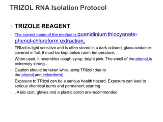
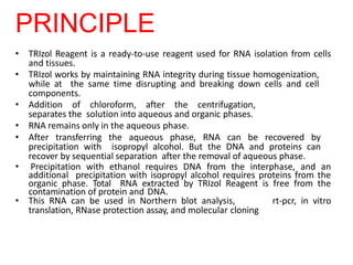
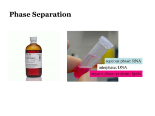
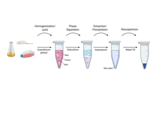
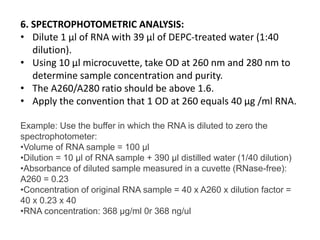
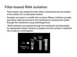
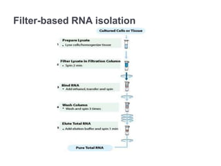
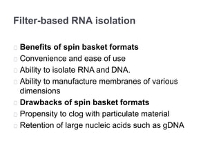
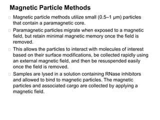
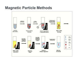
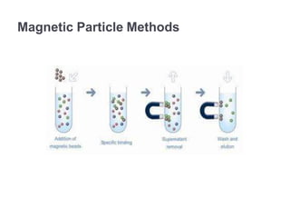
RNA isolation is the purification of RNA from biological samples. It requires strict precautions to avoid degradation by RNases, enzymes that naturally degrade RNA. The TRIzol method uses phenol and chloroform to separate RNA, DNA and proteins during cell lysis and homogenization. Chloroform separates the mixture into aqueous and organic phases, with RNA remaining in the aqueous phase. RNA is then precipitated with isopropanol and washed with ethanol before being re-dissolved in water. RNA concentration and purity is determined spectroscopically. Filter-based and magnetic particle methods provide alternative RNA isolation techniques.
methods of isolation and extraction of RNA by using different source such as plant tissues, bacterial culture, etc. Ribonucleic acid can be isolated from plant tissue for the purpose of:
– mRNA isolation
– In vitro translation
– Northern analysis
– cDNA library construction
Rigorous ribonuclease free environment is to be maintained
All glasswares, plasticwares and reagents made RNAse free (using 0.01% DEPC)
Next day, DEPC is inactivated by autoclaving for 30 min
Total RNA is isolated and separated from DNA and protein after extraction with a solution called as Trizol. Trizol is an acidic solution containing guanidinium thiocyanate (GITC), phenol and chloroform. GITC irreversibly denatures proteins and RNases. This is followed by centrifugation.
There are 'n' number of DNA isolation methods depending on the sample type, final use of DNA product, etc. This presentation gives an overall idea about different methods of DNA isolation in a simplified way.
Techniques of DNA Extraction, Purification and QuantificationBHUMI GAMETI
Introduction
The overall process…
Uses of isolated genomic DNA
Extraction of DNA from plant material
Components of DNA extraction solutions
Cell Lysis or Cell disruption :
Purification of DNA
CTAB Method
Phenol–chloroform extraction
PROTEINASE K
Salting out
Silica adsorption method
Magnetic beads
FTA Paper
Nucleic acid quantification
Agarose Gel Electrophoresis
UV spectroscopy
DNA quantification using NanoDrop
Nucleic Acid Quantification Methods - DNA / RNA Quantificationajithnandanam
Nucleic acids are quantified to check the concentration and purity of DNA/RNA present in the solution mixture.it is important to know the concentration and purity of the nucleic acid for the use in further applications like PCR, restriction digestion etc. Spectrophotometric analysis is the most commonly used method of quantifying DNA, agarose gel electrophoresis can also be used to analyse the DNA sample for purity.
Sanger sequencing is a method of DNA sequencing based on the selective incorporation of chain-terminating dideoxynucleotides by DNA polymerase during in vitro DNA replication.
Isolation and Purification of Chromosomal DNA,Plasmid DNA,Bacteriophage DNA used in Recombinant DNA Technology or Biotechnology to produce Recombinant DNA or Desired DNA
This is technique used widely for protein separation from a mixture and is very easy and less costly method. Slides cover all essential points about EMSA and it is quite interesting to know that how it detect and separate different proteins and their mobility shift assay.
1. CENTRAL DOGMA OF MOLECULAR BIOLOGY
2. NUCLEIC ACID PREPARATION & APPLICATIONS
3. FUNDAMENTAL STEPS IN DNA PURIFICATION
4. ANALYSIS OF NUCLEIC ACIDS
5. STORAGE CONDITIONS
molecular biology techniques -jaypee university of information technology- ra...RAVI RANJAN
molcular biology techniques- ravi ranjan lb-
contents- basic molecular biology techniques - DNA and RNA isolation from plant sample, nanodrop technique, pcr and cloning.
This simple laboratory PPT was designed for UPES-SOHST students as a guide for illustrating the experiment mentioned above, kindly share to help someone learn
The extraction of DNA involves three main steps that are cell lysis, protein separation, and DNA purification. Cell lysis is usually performed by incubation of cell in buffer containing detergent and protease. Cellular proteins are salted out or phase separated using organic solvents. Finally DNA is isolated and purified either by alcohol precipitation or adsorption with silica and elution.
Back to basics: Fundamental Concepts and Special Considerations in RNA IsolationQIAGEN
RNA integrity and quality are critical to obtain meaningful and reliable downstream data. This slidedeck details the challenges and considerations of handling RNA samples, RNA stabilization, the need for quality control analysis and common methods for RNA integrity and quality assessment.
RNA, DNA Isolation and cDNA synthesis.pptxASJADRAZA10
Isolation, quantification of nucleic acids from wheat and synthesis of cDNA.
Introduction
List of Genotypes
DNA Isolation (CTAB method)
Qualitative check of DNA- Gel electrophoresis
Quantitative test of DNA- Spectrophotometer
Protocol for RNA Isolation
RNA Confirmation
Normalization of RNA
cDNA Synthesis
Protocol for DNA Isolation of plant
50-100mg (2-3) young leaves were collected, then washed with tap water followed by distilled water in petri dish.
Leaves were ground using ethanol sterilized mortar pestle for 15-20 sec, by taking 1mL extraction buffer.
1mL (1000μL) of extraction buffer was again added to collect paste from mortar pestle & then transferred to the 2 mL micro centrifuge tube.
The sample in the tube is incubated at 65°C in water bath for 35-45 mins. (Contents in the tube was mixed by inverting at an interval for 5-10 mins)
The tubes were cooled for 10 minutes in ice.
The sample of equal vol (2mL) was centrifuged @14,000 rpm for 10 mins.
After that the supernatant was transferred to new 2 mL centrifuge tube and equal volume (as of sample) of chloroform: Isoamyl alcohol (24:1) was added.
Then mixed gently for 5-7 mins by inverting the tubes.
Again centrifuged for 10 mins @10,000 rpm
After centrifugation, three layers were observed in the tube.
a) aqueous phase i.e. DNA+RNA
b) protein coagulate
c) organic phase i.e. Chloroform
Again the supernatant (aqueous phase) was collected in 1.5mL tube and equal volume of ice-cold isopropanol was added and stored in -20°C overnight.
Following day, tubes were again centrifuged @10,000rpm for 10 mins.
The supernatant was discarded without disturbing the DNA pellet.
70% ethanol is taken and 0.5mL of it was added to the sample and mixed by tapping for 5 mins.
Again centrifuged @10,000rpm for 10 mins and the supernatant was discarded.
Pellet (DNA Precipitate) was air dried for 10 mins.
Then dissolved in 50μL TE-1X Buffer and the sample was stored at -20°C.
1g of analytical grade Agarose was weighed.
100 mL of autoclaved 1X TBE was added in flask.
Now heated on the oven until the solution becomes transparent.
Solution was allowed to cool down to 60℃.
2 μL of Ethidium Bromide (EtBr) is added in the flask.
Melted agarose gel was poured into the casting tray along with comb.
Any bubble in the gel was removed.
After solidification of gel, comb was removed gently and then running buffer was added in the electrophoretic tank.
Once gel got solidified, it was transferred it into gel tank.
A parafilm was taken and on it 2μL loading dye and 3μL sample was taken, gently mixed with the pipette tip only.
Then the mixture (sample +loading dye) was loaded into the well.
Then electrophoretic unit was run at 90 volt for 50-55 mins.
After that gel was put into the Gel Doc to see the DNA band
(using UV light).
Bright colour band were observed as in the figure.
Few (100-150mg) young leaves were ground into fine powder using liquid Nitrogen.
Lung Cancer: Artificial Intelligence, Synergetics, Complex System Analysis, S...Oleg Kshivets
RESULTS: Overall life span (LS) was 2252.1±1742.5 days and cumulative 5-year survival (5YS) reached 73.2%, 10 years – 64.8%, 20 years – 42.5%. 513 LCP lived more than 5 years (LS=3124.6±1525.6 days), 148 LCP – more than 10 years (LS=5054.4±1504.1 days).199 LCP died because of LC (LS=562.7±374.5 days). 5YS of LCP after bi/lobectomies was significantly superior in comparison with LCP after pneumonectomies (78.1% vs.63.7%, P=0.00001 by log-rank test). AT significantly improved 5YS (66.3% vs. 34.8%) (P=0.00000 by log-rank test) only for LCP with N1-2. Cox modeling displayed that 5YS of LCP significantly depended on: phase transition (PT) early-invasive LC in terms of synergetics, PT N0—N12, cell ratio factors (ratio between cancer cells- CC and blood cells subpopulations), G1-3, histology, glucose, AT, blood cell circuit, prothrombin index, heparin tolerance, recalcification time (P=0.000-0.038). Neural networks, genetic algorithm selection and bootstrap simulation revealed relationships between 5YS and PT early-invasive LC (rank=1), PT N0—N12 (rank=2), thrombocytes/CC (3), erythrocytes/CC (4), eosinophils/CC (5), healthy cells/CC (6), lymphocytes/CC (7), segmented neutrophils/CC (8), stick neutrophils/CC (9), monocytes/CC (10); leucocytes/CC (11). Correct prediction of 5YS was 100% by neural networks computing (area under ROC curve=1.0; error=0.0).
CONCLUSIONS: 5YS of LCP after radical procedures significantly depended on: 1) PT early-invasive cancer; 2) PT N0--N12; 3) cell ratio factors; 4) blood cell circuit; 5) biochemical factors; 6) hemostasis system; 7) AT; 8) LC characteristics; 9) LC cell dynamics; 10) surgery type: lobectomy/pneumonectomy; 11) anthropometric data. Optimal diagnosis and treatment strategies for LC are: 1) screening and early detection of LC; 2) availability of experienced thoracic surgeons because of complexity of radical procedures; 3) aggressive en block surgery and adequate lymph node dissection for completeness; 4) precise prediction; 5) adjuvant chemoimmunoradiotherapy for LCP with unfavorable prognosis.
Pulmonary Thromboembolism - etilogy, types, medical- Surgical and nursing man...VarunMahajani
Disruption of blood supply to lung alveoli due to blockage of one or more pulmonary blood vessels is called as Pulmonary thromboembolism. In this presentation we will discuss its causes, types and its management in depth.
Report Back from SGO 2024: What’s the Latest in Cervical Cancer?bkling
Are you curious about what’s new in cervical cancer research or unsure what the findings mean? Join Dr. Emily Ko, a gynecologic oncologist at Penn Medicine, to learn about the latest updates from the Society of Gynecologic Oncology (SGO) 2024 Annual Meeting on Women’s Cancer. Dr. Ko will discuss what the research presented at the conference means for you and answer your questions about the new developments.
Anti ulcer drugs and their Advance pharmacology ||
Anti-ulcer drugs are medications used to prevent and treat ulcers in the stomach and upper part of the small intestine (duodenal ulcers). These ulcers are often caused by an imbalance between stomach acid and the mucosal lining, which protects the stomach lining.
||Scope: Overview of various classes of anti-ulcer drugs, their mechanisms of action, indications, side effects, and clinical considerations.
Title: Sense of Smell
Presenter: Dr. Faiza, Assistant Professor of Physiology
Qualifications:
MBBS (Best Graduate, AIMC Lahore)
FCPS Physiology
ICMT, CHPE, DHPE (STMU)
MPH (GC University, Faisalabad)
MBA (Virtual University of Pakistan)
Learning Objectives:
Describe the primary categories of smells and the concept of odor blindness.
Explain the structure and location of the olfactory membrane and mucosa, including the types and roles of cells involved in olfaction.
Describe the pathway and mechanisms of olfactory signal transmission from the olfactory receptors to the brain.
Illustrate the biochemical cascade triggered by odorant binding to olfactory receptors, including the role of G-proteins and second messengers in generating an action potential.
Identify different types of olfactory disorders such as anosmia, hyposmia, hyperosmia, and dysosmia, including their potential causes.
Key Topics:
Olfactory Genes:
3% of the human genome accounts for olfactory genes.
400 genes for odorant receptors.
Olfactory Membrane:
Located in the superior part of the nasal cavity.
Medially: Folds downward along the superior septum.
Laterally: Folds over the superior turbinate and upper surface of the middle turbinate.
Total surface area: 5-10 square centimeters.
Olfactory Mucosa:
Olfactory Cells: Bipolar nerve cells derived from the CNS (100 million), with 4-25 olfactory cilia per cell.
Sustentacular Cells: Produce mucus and maintain ionic and molecular environment.
Basal Cells: Replace worn-out olfactory cells with an average lifespan of 1-2 months.
Bowman’s Gland: Secretes mucus.
Stimulation of Olfactory Cells:
Odorant dissolves in mucus and attaches to receptors on olfactory cilia.
Involves a cascade effect through G-proteins and second messengers, leading to depolarization and action potential generation in the olfactory nerve.
Quality of a Good Odorant:
Small (3-20 Carbon atoms), volatile, water-soluble, and lipid-soluble.
Facilitated by odorant-binding proteins in mucus.
Membrane Potential and Action Potential:
Resting membrane potential: -55mV.
Action potential frequency in the olfactory nerve increases with odorant strength.
Adaptation Towards the Sense of Smell:
Rapid adaptation within the first second, with further slow adaptation.
Psychological adaptation greater than receptor adaptation, involving feedback inhibition from the central nervous system.
Primary Sensations of Smell:
Camphoraceous, Musky, Floral, Pepperminty, Ethereal, Pungent, Putrid.
Odor Detection Threshold:
Examples: Hydrogen sulfide (0.0005 ppm), Methyl-mercaptan (0.002 ppm).
Some toxic substances are odorless at lethal concentrations.
Characteristics of Smell:
Odor blindness for single substances due to lack of appropriate receptor protein.
Behavioral and emotional influences of smell.
Transmission of Olfactory Signals:
From olfactory cells to glomeruli in the olfactory bulb, involving lateral inhibition.
Primitive, less old, and new olfactory systems with different path
- Video recording of this lecture in English language: https://youtu.be/lK81BzxMqdo
- Video recording of this lecture in Arabic language: https://youtu.be/Ve4P0COk9OI
- Link to download the book free: https://nephrotube.blogspot.com/p/nephrotube-nephrology-books.html
- Link to NephroTube website: www.NephroTube.com
- Link to NephroTube social media accounts: https://nephrotube.blogspot.com/p/join-nephrotube-on-social-media.html
Ethanol (CH3CH2OH), or beverage alcohol, is a two-carbon alcohol
that is rapidly distributed in the body and brain. Ethanol alters many
neurochemical systems and has rewarding and addictive properties. It
is the oldest recreational drug and likely contributes to more morbidity,
mortality, and public health costs than all illicit drugs combined. The
5th edition of the Diagnostic and Statistical Manual of Mental Disorders
(DSM-5) integrates alcohol abuse and alcohol dependence into a single
disorder called alcohol use disorder (AUD), with mild, moderate,
and severe subclassifications (American Psychiatric Association, 2013).
In the DSM-5, all types of substance abuse and dependence have been
combined into a single substance use disorder (SUD) on a continuum
from mild to severe. A diagnosis of AUD requires that at least two of
the 11 DSM-5 behaviors be present within a 12-month period (mild
AUD: 2–3 criteria; moderate AUD: 4–5 criteria; severe AUD: 6–11 criteria).
The four main behavioral effects of AUD are impaired control over
drinking, negative social consequences, risky use, and altered physiological
effects (tolerance, withdrawal). This chapter presents an overview
of the prevalence and harmful consequences of AUD in the U.S.,
the systemic nature of the disease, neurocircuitry and stages of AUD,
comorbidities, fetal alcohol spectrum disorders, genetic risk factors, and
pharmacotherapies for AUD.
micro teaching on communication m.sc nursing.pdfAnurag Sharma
Microteaching is a unique model of practice teaching. It is a viable instrument for the. desired change in the teaching behavior or the behavior potential which, in specified types of real. classroom situations, tends to facilitate the achievement of specified types of objectives.
TEST BANK for Operations Management, 14th Edition by William J. Stevenson, Ve...kevinkariuki227
TEST BANK for Operations Management, 14th Edition by William J. Stevenson, Verified Chapters 1 - 19, Complete Newest Version.pdf
2. RNA extraction
RNA extraction is the purification of RNA from
biological samples. This procedure is complicated by
the presence of ribonuclease enzymes in cells and
tissues.
3. Isolation of RNA
• Requires STRICT precautions to avoid sample
degradation.
• RNA especially labile.
4. RNAses
• RNases are naturally occurring enzymes that degrade
RNA
• Common laboratory contaminant (from bacterial and
human sources)
• Also released from cellular compartments during
isolation of RNA from biological samples
• Can be difficult to inactivate
5. RNAses
• RNAses are enzymes which are small proteins that can
renature and become active.
• MUST be eliminated or inactivated BEFORE isolation.
• CRITICAL to have a separate RNAse free area of lab.
6. Protecting Against RNAse
• Wear gloves at all times
• Use RNase-free tubes and pipet tips
• Use dedicated, RNase-free, chemicals
• Pre-treat materials with extended heat (180 C for several
hours), wash with DEPC-treated water, NaOH or H2O2
• Supplement reactions with RNase inhibitors
7. TRIZOL RNA Isolation Protocol
TRIZOLE REAGENT
The correct name of the method is guanidinium thiocyanate-
phenol-chloroform extraction.
TRIzol is light sensitive and is often stored in a dark-colored, glass container
covered in foil. It must be kept below room temperature.
When used, it resembles cough syrup, bright pink. The smell of the phenol is
extremely strong..
Caution should be taken while using TRIzol (due to
the phenol and chloroform).
Exposure to TRIzol can be a serious health hazard. Exposure can lead to
serious chemical burns and permanent scarring
. A lab coat, gloves and a plastic apron are recommended
8. PRINCIPLE
• TRIzol Reagent is a ready-to-use reagent used for RNA isolation from cells
and tissues.
• TRIzol works by maintaining RNA integrity during tissue homogenization,
while at the same time disrupting and breaking down cells and cell
components.
• Addition of chloroform, after the centrifugation,
separates the solution into aqueous and organic phases.
• RNA remains only in the aqueous phase.
• After transferring the aqueous phase, RNA can be recovered by
precipitation with isopropyl alcohol. But the DNA and proteins can
recover by sequential separation after the removal of aqueous phase.
• Precipitation with ethanol requires DNA from the interphase, and an
additional precipitation with isopropyl alcohol requires proteins from the
organic phase. Total RNA extracted by TRIzol Reagent is free from the
contamination of protein and DNA.
• This RNA can be used in Northern blot analysis, rt-pcr, in vitro
translation, RNase protection assay, and molecular cloning
10. Homogenization:
1. Tissues: Homogenize tissue samples in 1 ml of
TRIZOL reagent per 50 to 100 mg of tissue using
a glass-Teflon or power homogenizer.
2. Cells grown in Monolayer: Rinse cell monolayer
with ice cold PBS once. Lyse cells directly in a
culture dish by adding 1 ml of TRIZOL Reagent
per 3.5 cm diameter dish and scraping with cell
scraper. Pass the cell lysate several times
through a pipette. Vortex thoroughly.
11. 2. Phase Separation
•The homogenized samples were incubated for 5 minutes at 15
to 30°C for the complete dissociation of nucleoprotein
complexes.
•0.2 ml (200 microliters)of chloroform per 0.75 ml of TRIZOL
LS Reagent was added. The tubes were shaked vigorously by
hand for 15 seconds and incubated them at 15 to 30°C for 2
minutes.
•The samples were centrifuged for 15 minutes at no more than
12,000 g (4°C).
•The aqueous phase was transferred to other tubes. ( Following
centrifugation, the mixture separates into a lower red, phenol-
chloroform phase, an interphase, and a colorless upper
aqueous phase. RNA remains only in the aqueous phase. The
volume of the aqueous phase is about 70% of the volume of
TRIZOL Reagent used for homogenization.)
13. 3. RNA Precipitation
•The RNA was precipitated from the aqueous phase
by mixing with 3 microlitre of glycogen and 500
microlitre of isopropyl alcohol.
•The mixture was centrifuged for 30 minutes at
12,000 × g (2 to 8°C).( The RNA precipitate forms
a gel-like pellet on the side of the tube at bottom).
14.
15. 4. RNA Wash
1.The supernatant was removed. The RNA pellet was
washed once with 75% ethanol, adding 900 microlitre
of 75% ethanol per 0.75 ml of TRIZOL Reagent used
for the initial homogenization.
2.The sample were inverted and mixed and
centrifuged at 12,000 rpm for 30 minutes at 4
degree.
16. 5. Redissolving RNA
•The RNA pellet was dried .
•RNA was dissolved in RNase-free water (or 0.5%
SDS solution) by passing the solution through the
pipette tip for a few times, and incubating for 10
minutes at 55 to 60°C.
17.
18. 6. SPECTROPHOTOMETRIC ANALYSIS:
• Dilute 1 μl of RNA with 39 μl of DEPC-treated water (1:40
dilution).
• Using 10 μl microcuvette, take OD at 260 nm and 280 nm to
determine sample concentration and purity.
• The A260/A280 ratio should be above 1.6.
• Apply the convention that 1 OD at 260 equals 40 µg /ml RNA.
Example: Use the buffer in which the RNA is diluted to zero the
spectrophotometer:
•Volume of RNA sample = 100 µl
•Dilution = 10 µl of RNA sample + 390 µl distilled water (1/40 dilution)
•Absorbance of diluted sample measured in a cuvette (RNase-free):
A260 = 0.23
•Concentration of original RNA sample = 40 x A260 x dilution factor =
40 x 0.23 x 40
•RNA concentration: 368 µg/ml 0r 368 ng/ul
19. Other Methods of RNA ISOLATION
Filter-based RNA isolation
Magnetic Particle Methods
20. Filter-based RNA isolation
Filter-based, spin basket formats utilize membranes that are seated
at the bottom of a small plastic basket.
Samples are lysed in a buffer that contains RNase inhibitors (usually
guanidine salts),are bound to the membrane by passing the lysate
through the membrane using centrifugal force.
Wash solutions are passed through the membrane and discarded.
An appropriate elution solution is applied and the sample is collected
into a tube by centrifugation.
22. Filter-based RNA isolation
Benefits of spin basket formats
Convenience and ease of use
Ability to isolate RNA and DNA.
Ability to manufacture membranes of various
dimensions
Drawbacks of spin basket formats
Propensity to clog with particulate material
Retention of large nucleic acids such as gDNA
23. Magnetic Particle Methods
Magnetic particle methods utilize small (0.5–1 µm) particles
that contain a paramagnetic core.
Paramagnetic particles migrate when exposed to a magnetic
field, but retain minimal magnetic memory once the field is
removed.
This allows the particles to interact with molecules of interest
based on their surface modifications, be collected rapidly using
an external magnetic field, and then be resuspended easily
once the field is removed.
Samples are lysed in a solution containing RNase inhibitors
and allowed to bind to magnetic particles. The magnetic
particles and associated cargo are collected by applying a
magnetic field.