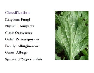
- Albugo candida is a fungus that causes white rust, a disease affecting plants in the mustard family and others like asters, morning glories, and lamb's quarters.
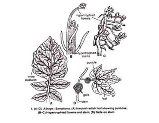
- It infects above-ground plant parts through stomata, causing white irregular patches on leaves and stems that later powder. Flowers and fruits become deformed.
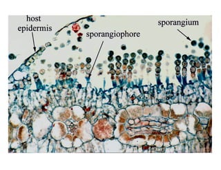
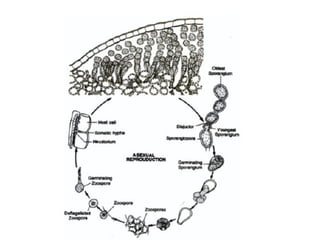
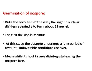
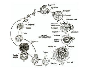
- The fungus reproduces asexually through conidia produced on sporangiophores and sexually through fertilization of female oogonia by male antheridia, forming thick-walled oospores.
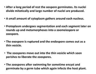
- Oospores can remain dormant for long periods until conditions are suitable for germination into zoospores that can infect new host plants


![Polymer [ बहुलक ] Chemistry Notes PDF - Irfanullah Mehar - JJ Sir Chemistry.pdf](https://cdn.slidesharecdn.com/ss_thumbnails/polymerchemistrynotespdf-irfanullahmehar-jjsirchemistry-260210172118-3f9b37f7-thumbnail.jpg?width=640&height=640&fit=bounds)