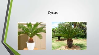
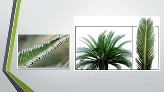



Cycas
Gymnosperms
Cycas
Life cycle ( reproduction
methods)
Vegetative reproduction
Adventitious buds containing reserve food
Present on the base of stem on axil of each
scale
Each bulbil consist of few scales and foliage
leaves
Develop into new plant
Sexual reproduction
• Heteropsorous
• Male plant is rare
• Microspores and mega spores are
produced
• Spores are produced on sporangia
that develop on sporophylls
• Sporophyll arrange to form strobili
or cones
• Male and female cones are formed
on seprate plants
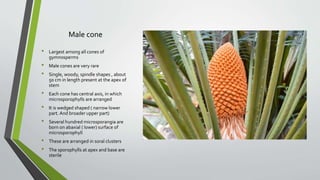
Male cone
• Largest among all cones of
gymnosperms
• Male cones are very rare
• Single, woody, spindle shapes , about
50 cm in length present at the apex of
stem
• Each cone has central axis, in which
microsporophylls are arranged
• It is wedged shaped ( narrow lower
part. And broader upper part)
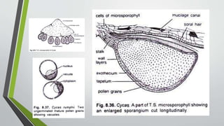
• Several hundred microsporangia are
born on abaxial ( lower) surface of
microsporophyll
• These are arranged in soral clusters
• The sporophylls at apex and base are
sterile
Structure of
micrsoporangium
Each microsporangium Oval in shape
and borne on short stalk
Several layered mass of sporogenous
cells are present inside
The outermost layer isTapetum
Each sporangium contains large
number of microspores
Female cone
Megasporphylls are not organized in to
cone
It consist of loosely arranged
megasporophylls that surrounds the
shoot apex like foliage leaves
Each megasporophyll is pinnate like
Differentiated into upper, broad, lobed
leaf like portion that has pointed end,
And lower stalk like stipe
The ovules are naked and 6-8 ovules are
arranged laterally on stipe
Ovules are hairy , red and 6 cm in length
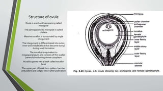
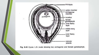
Structure of ovule
Ovule is erect and has opening called
micropyle
The part opposite to micropyle is called
chalaza
Massive nucellus is surrounded by single
integument
The integument is differentiated into outer,
inner and middle( thick that become stony)
during seed formation
The nucellus is equivalent to
megasporangium and consists of thin walled
parenchyma having dense cytoplasm
Nucellus grows into a beak called nucellor
beak
The upper part of beak form pollen chamber
and pollens are lodged into it after pollination
https://www.youtube.com/watch?v=A0hStZf9_R8
https://www.youtube.com/watch?v=mngWYnRt6Yo
https://www.youtube.com/watch?v=lSFiRuiqCGI
Megasporogenesis
A deeply lying cell of nucellus enlarges
and functions as megaspore mother cell
After meiosis it form tetrad of
megaspores
Upper three degenerate and lower most
enlarges to form embryo sac or female
gametophyte
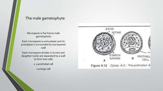
The male gametophyte
Microspore is the future male
gametophyte
Each microspore is uninucleate and its
protoplast is surrounded by two layered
wall
Each microspore divides in to two and
daughter nuclei are separated by a wall
to form two cells
1: a prothalial cell
2:a large cell
Release of pollen grain
Large cell divides to form
1: a tube cell
2: and generative cell
At this stage the sporophylls are separated
,
sporangium walls ruptures
And release 3 celled semi-germinated
male gametophyte called pollen grain
They are then carried to female
gametophyte where further development
completes
Pollination
• Transfer of pollen grains from
microsporangia to micropyle of the
ovule is called pollination
• Some cells at apex from nucellus
disintegrate and form a colloidal
material called pollination droplet
• It protrudes out through micropyle
and attach pollens
• The drop dries , contract and carries
pollens inside pollen chamber,
where they remain dormant for 4-6
months
Pollen tube
During resting period following changed
takes place
Protoplast of each pollen grain protrudes
from upper part of microspore and form
pollen tube
The tube nucleus migrates into pollen tube
and the generative cell divides into
1: stalk cell
2: body cell
The body cell divides to produce two
multiflagellate sperms or antherozoids
Antherozoid
Each sperm cell consists of a large nucleus
surrounded by a thin layer of cytoplasm
The flagella are arranged spirally in a band
like structure
The pollen tube is haustorial in nature and
it digests its way to nucellus to the
archegonia
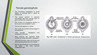
Female gametophyte
• The functional megaspore in ovule
begins to enlarge and is called as
embryo sac
• The tissue around it become
vacuolated and form spongy layer to
give nutrition for megaspore
• The nucleus of megaspore divides to
form 1000 more daughter nuclei
• The vacuole appears in center
• The nuclei are pushed and arranged in
the periphery
• Later vacuole disappears and
cytoplasm is formed
• Now embryo sac contains large
number of nuclei and water cytoplasm
• Cell wall formation stats and at the end
entire gametophyte become cellular
• Mature gametophyte is surrounded by
two layers , the inner become
megaspore wall
Structure of archegonium
A mature archegonium consists of 4
neck cells and an egg ( surrounded by
tough membrane)
The egg in Cycus is largest amongst all
living plants. Two to eight archegonia
are formed in Cycus revoluta.
Seed formation
The embryo enlarges and ovule
increases in size
The outermost layer of integument
develops red color and becomes fleshy
The middle layer become stony and
hard, the inner layer becomes soft
These layers constitute seed coat
Germination of seed
• Seed absorbs water and after that
seed coat ruptures at micropyle end
emerging its radicle
• The plumule is followed
• The cotyledons remains inside
• Foliage leaves are produced by the
activity of apical bud after several
years
• Seed remains attach with young
sporophyte by withered cotyledons
for one or two years
Videos
https://www.youtube.com/watch?v=0_q0b3nncJY