 Kingdom : Mycota
 Division : Eumycota
 Subdivision : Basidiomycotina
 Class : Teliomycetes
 Order : Uredinales
 Family : Pucciniaceae
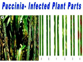
Puccinia infection on plants is commonly known as
rust because of the rusty patches it produces on the
stems and leaves.
It is an obligate parasite, capable of thriving only
upon living hosts , including many cereals and
millets, and it causes heavy financial loss.
• Species of Puccinia are either autoecious or heteroecious.
• P. asparagi, completes its life cycle on a single host, namely
Asparagus. Hence. it is an autoecious fungus.
• P. graminis completes its life cycle on two different host
plants, wheat and barberry. Hence, it is a heteroecious
fungus.
• Three species of Puccinia are known to attack wheat.
• P. graminis which causes the black rust or stem rust,
• P. glumarum which causes yellow rust or stripe rust
• P. triticina which causes brown rust or orange leaf rust
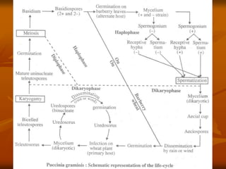
• Puccinia graminis is a heteroecious parasite.
• It’s life cycle is completed on Wheat and Barberry.
• Wheat is the primary host.
• Barberry is the secondary or alternate host.
• The dikaryophase of the life cycle is completed on the
wheat plant and the haplophase is completed on the
barberry plant.
• The dikaryotic mycelium is unable to grow on
barberry and the haplophase is unable to grow on
wheat.
• Thus, the life cycle is completed only when both the
hosts are present.
• Life-cycle
• Wheat is the primary host upon which the dikaryophase of the
pathogen is completed.
• This phase consists of a dikaryotic mycelium and two spore
stages,
– uredineal stage and telial stage.
• The aecidiospore, produced in the aecidial cups on the lower
surface of the barberry leaves, brings about the primary
infection.
• Aecidiospores are binucleate.
• When they fall upon a wheat plant they germinate.
• They put forth a germ tube or primary hypha that enters into
the host through the stoma.
• The mycelium is branched, septate and it ramifies in the
intercellular spaces of the host tissue.
• It produces haustoria for absorbing nutrients.
• The cells of the hypha are binucleate, and the two nuclei
together constitute a dikaryon.
• This dikaryotic mycelium reproduces by producing two kinds
of spores, namely the uredospores and teleutospores.
• The spores are produced on the surface of the host tissue on
specialized pustules or sori, known respectively as uredineal
sori and teleuto sori.
Uredineal Stage
• A few days after infection, the dikaryotic mycelium begins to
form the uredosori.
• The hyphae of the mycelium aggregate to form a hyphal mass
near the surface of the infected leaf, stem or glumes.
• These hyphal mass surrounds isolated host cells which are
called uredia.
• From the uredium arise large numbers of uredospores.
• The group of uredospores thus formed is called uredosorus.
• The spores are produced on long stalks and each spore is
binucleate, somewhat oval to round in outline with a warty
wall and four equatorial germ pores.
• The formation of large numbers of uredospores, and the
elongation of their stalk will lead to the cracking of the host
epidermis to expose the uredospores.
• Under favourable conditions, the uredospores germinate and put forth
the germ tube through one of the germ pores.
• The germ tube, on reaching the stoma, swells up into an elongated
vesicle called appressorium, from which, an infection hypha is
produced.
• It enters the stomatal aperture, grows and ramifies in the intercellular
spaces of the host tissue, and absorbs nourishment through haustoria.
• Later on, it will produce pustules on the infected leaf called
uredosori.
• Uredospores, therefore, serve as an efficient means of secondary
infection.
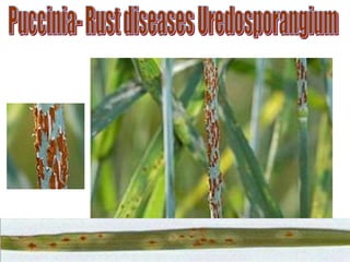
• Uredosori can be seen as reddish brown pustules on the leaves and
stem of wheat plant.
• Hence, the uredineal stage is often called the 'red stage' or the
‘summer stage'.
Puccinia
Uredo-
sporangium
-Wheat
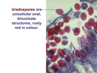
Uredospores are
unicellular oval,
binucleate
structures, rusty
red in colour.
Telial Stage:
 Late in the growing season, the same hyphal mass that gave
rise to uredia or the uredosori produces telia or teleutosori.
 In other words, the 'red stage' or the 'summer stage' is
gradually replaced by the 'black stage' or the 'winter stage'.
 At first, early in the season, teleutospores are produced
among the uredospores in the same sorus.
 Later, more teleutospores are produced and finally the sori
contain only teleutospores.
 These sori are now called teleutosori and the stage is called
telial stage.
 Teleutospores get exposed when the host epidermis breaks
open.
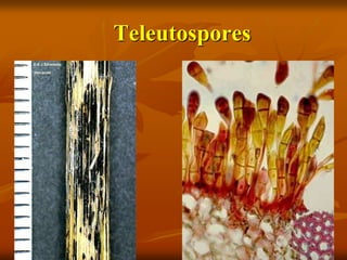
Teleutospores
Dark brown or
black in colour.
stalked, two
celled, spindle
shaped.
The wall is thick,
black and
smooth.
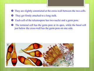
 They are slightly constricted at the cross-wall between the two cells.
 They get firmly attached to a long stalk.
 Each cell of the teleutospore has two nuclei and a germ pore.
 The terminal cell has the germ pore at its apex, while the basal cell
just below the cross-wall has the germ pore on one side.
Teleutospores
 Teleutospores do not germinate readily, but
undergo a period of rest and carry the fungus to the
next growing season.
 During the maturation period, the two nuclei in
each cell of the teleutospore fuse and a diploid
nucleus is formed.
 This represents the diploid phase and in this
condition the spore tides over the winter.
Germination of Teluto spore
 On Soil
 Favourable condition- Proper
Temperature, Moisture
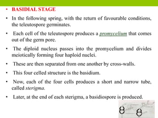
• BASIDIAL STAGE
• In the following spring, with the return of favourable conditions,
the teleutospore germinates.
• Each cell of the teleutospore produces a promycelium that comes
out of the germ pore.
• The diploid nucleus passes into the promycelium and divides
meiotically forming four haploid nuclei.
• These are then separated from one another by cross-walls.
• This four celled structure is the basidium.
• Now, each of the four cells produces a short and narrow tube,
called sterigma.
• Later, at the end of each sterigma, a basidiospore is produced.
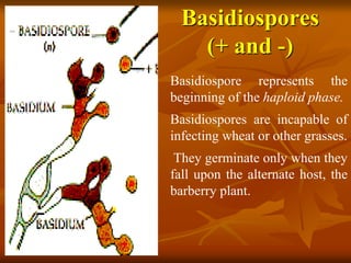
Basidiospores
(+ and -)
Basidiospore represents the
beginning of the haploid phase.
Basidiospores are incapable of
infecting wheat or other grasses.
They germinate only when they
fall upon the alternate host, the
barberry plant.
Stages of Puccinia
on Barberry leaf
 Pycniospores stage: On upper epidermis
 Aecidiospore stage: On lower epidermis
Infection of Puccinia on Barberry
leaf Lower and Upper sides
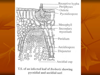
Pycnidial or spermagonial stage
The monokaryotic mycelium now begins to organize itself
beneath the upper epidermis of the barberry leaf and
forms a dense mat.
From this mycelial mat, arise groups of hyphae, which
organize themselves into pycnidium.
Pycnidia also belong to + and - strains like the mycelial
mat from which they are produced.
Pycnidium or spermagonium is a flask-shaped structure
that opens to the outside by a small pore, called ostiole.
Ostiole is guarded by sterile hyphae, called periphyses.
Pycnium on Upper epidermis
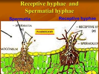
 There are two kinds of hyphae in a pycnidium.
 They are the long and delicate receptive hyphae which
protrude beyond the ostiole and the short spermatial hyphae
arranged in a palisade-like layer lining the cavity.
 Spermatial hyphae bear spermatia or pycniospores at their tip.
 Spermatium is a small, non-motile oval cell with a thin wall
and a relatively large nucleus.
 A pycnidium gives rise to either + or - spermatium
Receptive hyphae and
Spermatial hyphae
Spermatia Receptive hyphae
 The transfer of spermatium from one pycnidium to another is effected by
insects, such as flies, which are regular visitors of the pycnidial cups as
they are attracted by nectar oozing out from the pycnidial cup.
 During their visit, they transfer spermatium of one strain to the pycnidial
cups containing receptive hyphae of the opposite strain.
 A binucleate cell is formed by the dissolution of the intervening wall at the
point of contact between spermatium and receptive hyphae.
 One of the nuclei of this pair is plus strain and the other one is minus
strain.
 They together constitute a dikaryon.
 From this dikaryotic cell, a dikaryotic mycelium is formed.
 The hyphae with dikaryotic cells collect together and form the aecidia,
embedded in the mesophyll of the barberry leaf.
Develop-ment of
Aecidiosporangium
Aecidial stage
From the dikaryotic cell, a dikaryotic mycelium is
organized on the lower surface of the barberry leaf.
The hyphae with dikaryotic cells collect to form the
aecidia.
Aecidia are cup-shaped structures in which
aecidiospores are produced in basipetal succession,
alternating with small intercalary cells.
Aecidiospores are liberated by the disintegration
of the intercalary cells.
A mature aecidiospore is binucleate, thick-
walled and usually subglobose to polyhedral in
shape with many germ pores.
The liberated aecidiospores are carried by
air and they gain access to wheat plant.
Thus, the life cycle is completed.
Aecidiospores are incapable of infecting a
barberry plant.
But, they can infect the wheat plant.
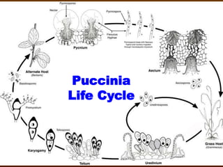
Puccinia life cycle
Puccinia
Life Cycle