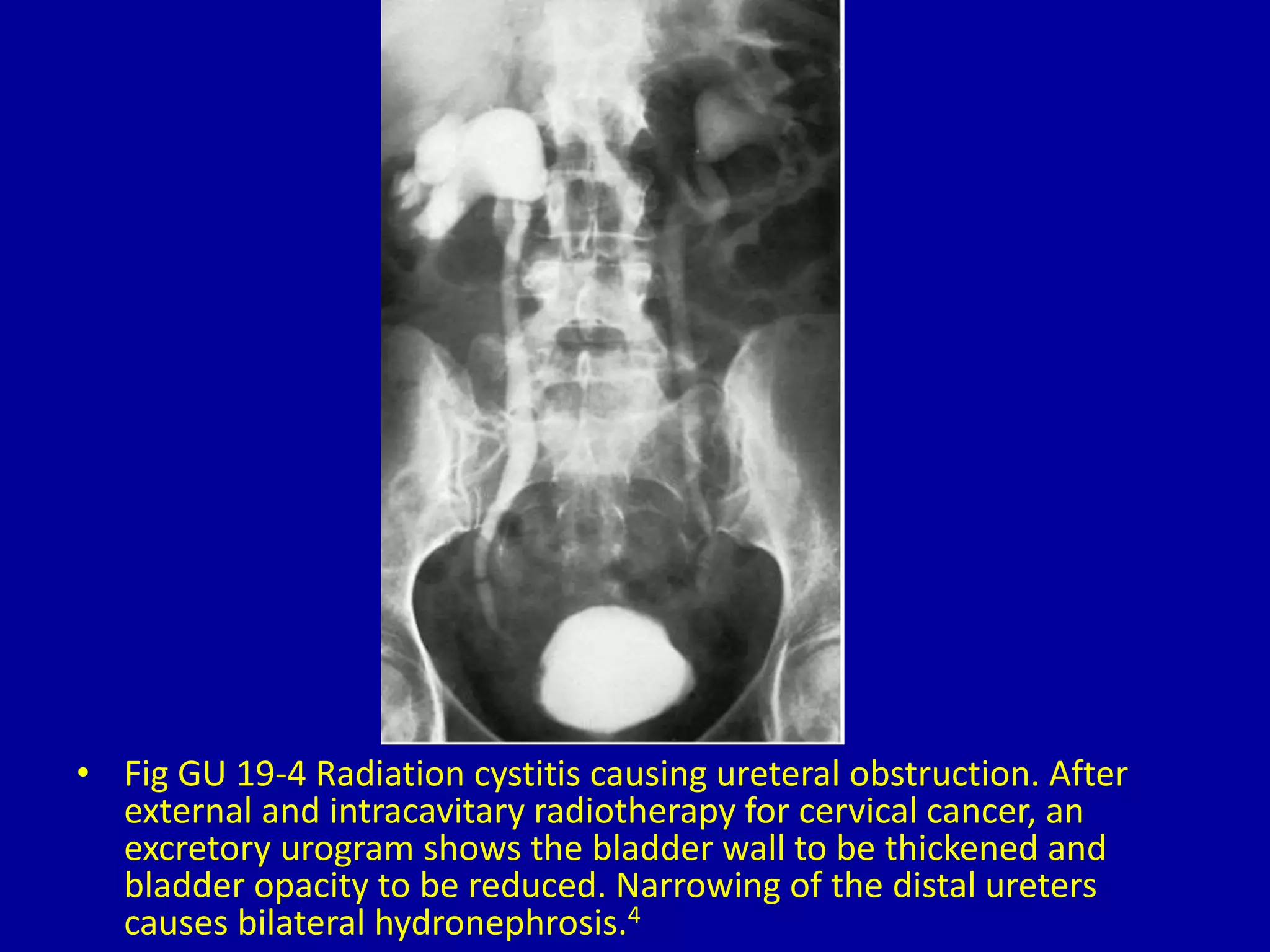
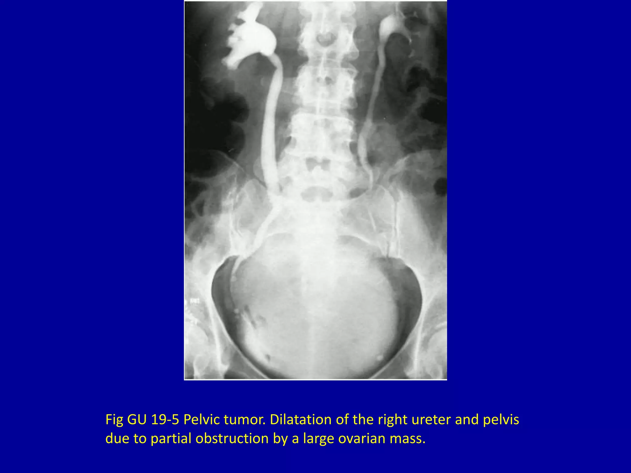
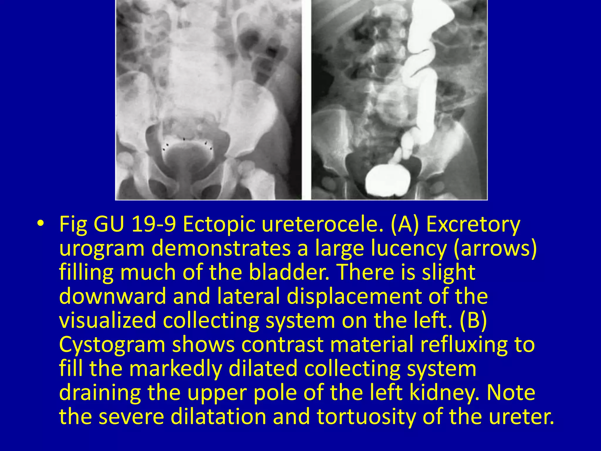
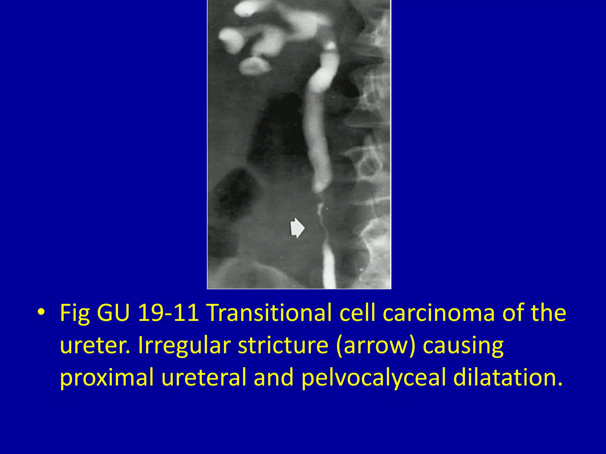
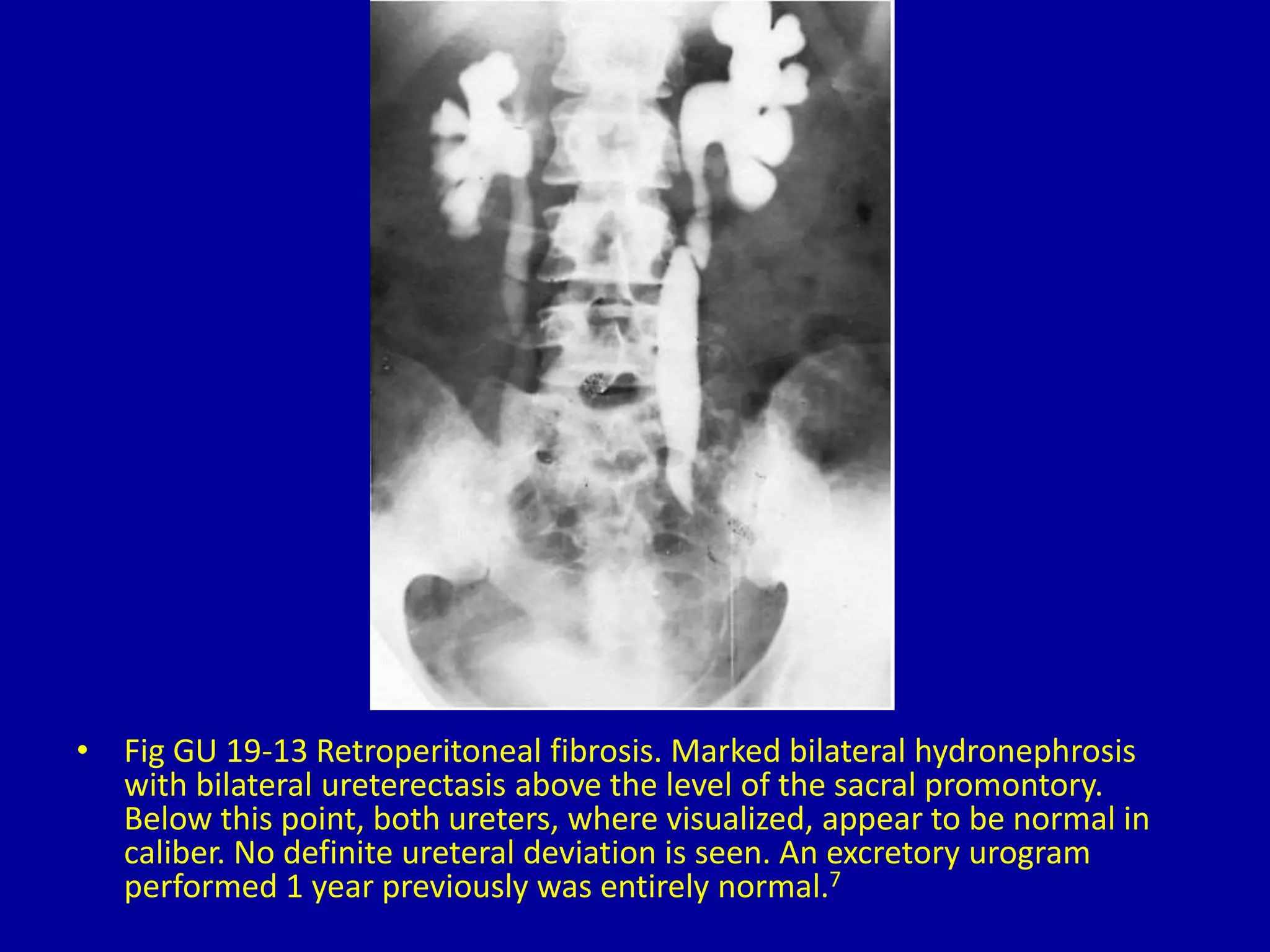
19Obstruction of the Ureter
CLINICAL IMAGAGING
AN ATLAS OF DIFFERENTIAL DAIGNOSIS
EISENBERG
DR. Muhammad Bin Zulfiqar
PGR-FCPS III SIMS/SHL
• Fig GU 19-1 Obstructing ureteral calculus.
Excretory urogram demonstrates a prolonged
nephrogram and marked dilatation of the
collecting system and pelvis proximal to the
obstructing stone (arrow).
• Fig GU 19-2 Congenital ureteropelvic junction
(UPJ) obstruction. Note the characteristic kink
or angulation at the UPJ (arrow).
Fig GU 19-3 Postsurgery stricture. Fibrotic narrowing of the
proximal ureter secondary to stone removal.
• Fig GU 19-4 Radiation cystitis causing ureteral obstruction. After
external and intracavitary radiotherapy for cervical cancer, an
excretory urogram shows the bladder wall to be thickened and
bladder opacity to be reduced. Narrowing of the distal ureters
causes bilateral hydronephrosis.4
Fig GU 19-5 Pelvic tumor. Dilatation of the right ureter and pelvis
due to partial obstruction by a large ovarian mass.
• Fig GU 19-6 Bladder cancer causing left ureteral
obstruction. Contrast material opacifies the distal left
ureter to the point of obstruction. The tumor involves
the ureteral orifice and produces a
“pseudoureterocele” appearance.4
• Fig GU 19-7 Severe interstitial cystitis. Bilateral ureteral
obstruction and marked contraction of the bladder
secondary to severe interstitial cystitis due to systemic
lupus erythematosus in a young woman on steroids. The
bladder capacity was reduced to approximately 1 ounce.4
• Fig GU 19-8 Simple ureterocele (arrows).
• Fig GU 19-9 Ectopic ureterocele. (A) Excretory
urogram demonstrates a large lucency (arrows)
filling much of the bladder. There is slight
downward and lateral displacement of the
visualized collecting system on the left. (B)
Cystogram shows contrast material refluxing to
fill the markedly dilated collecting system
draining the upper pole of the left kidney. Note
the severe dilatation and tortuosity of the ureter.
Fig GU 19-10 Retrocaval ureter. Note the medial swing of the
right ureter distal to the ureteropelvic junction.
• Fig GU 19-11 Transitional cell carcinoma of the
ureter. Irregular stricture (arrow) causing
proximal ureteral and pelvocalyceal dilatation.
• Fig GU 19-12 Hydronephrosis of pregnancy. Excretory urogram performed
3 days postpartum demonstrates bilateral large kidneys with dilatation of
the ureters and pelvocalyceal systems, especially on the right. The large
pelvic mass (arrows) indenting the superior surface of the bladder
represents the uterus, which is still causing extrinsic pressure on the
ureters.
• Fig GU 19-13 Retroperitoneal fibrosis. Marked bilateral hydronephrosis
with bilateral ureterectasis above the level of the sacral promontory.
Below this point, both ureters, where visualized, appear to be normal in
caliber. No definite ureteral deviation is seen. An excretory urogram
performed 1 year previously was entirely normal.7
• Fig GU 19-14 Obstructing valve (arrow) at the
ureteropelvic junction. Retrograde study shows smooth
infoldings below the valve representing fetal folds,
which usually regress as the child grows.19
19 obstruction of the ureter

19 obstruction of the ureter

  • 1.
  • 2.
    CLINICAL IMAGAGING AN ATLASOF DIFFERENTIAL DAIGNOSIS EISENBERG DR. Muhammad Bin Zulfiqar PGR-FCPS III SIMS/SHL
  • 3.
    • Fig GU19-1 Obstructing ureteral calculus. Excretory urogram demonstrates a prolonged nephrogram and marked dilatation of the collecting system and pelvis proximal to the obstructing stone (arrow).
  • 4.
    • Fig GU19-2 Congenital ureteropelvic junction (UPJ) obstruction. Note the characteristic kink or angulation at the UPJ (arrow).
  • 5.
    Fig GU 19-3Postsurgery stricture. Fibrotic narrowing of the proximal ureter secondary to stone removal.
  • 6.
    • Fig GU19-4 Radiation cystitis causing ureteral obstruction. After external and intracavitary radiotherapy for cervical cancer, an excretory urogram shows the bladder wall to be thickened and bladder opacity to be reduced. Narrowing of the distal ureters causes bilateral hydronephrosis.4
  • 7.
    Fig GU 19-5Pelvic tumor. Dilatation of the right ureter and pelvis due to partial obstruction by a large ovarian mass.
  • 8.
    • Fig GU19-6 Bladder cancer causing left ureteral obstruction. Contrast material opacifies the distal left ureter to the point of obstruction. The tumor involves the ureteral orifice and produces a “pseudoureterocele” appearance.4
  • 9.
    • Fig GU19-7 Severe interstitial cystitis. Bilateral ureteral obstruction and marked contraction of the bladder secondary to severe interstitial cystitis due to systemic lupus erythematosus in a young woman on steroids. The bladder capacity was reduced to approximately 1 ounce.4
  • 10.
    • Fig GU19-8 Simple ureterocele (arrows).
  • 11.
    • Fig GU19-9 Ectopic ureterocele. (A) Excretory urogram demonstrates a large lucency (arrows) filling much of the bladder. There is slight downward and lateral displacement of the visualized collecting system on the left. (B) Cystogram shows contrast material refluxing to fill the markedly dilated collecting system draining the upper pole of the left kidney. Note the severe dilatation and tortuosity of the ureter.
  • 12.
    Fig GU 19-10Retrocaval ureter. Note the medial swing of the right ureter distal to the ureteropelvic junction.
  • 13.
    • Fig GU19-11 Transitional cell carcinoma of the ureter. Irregular stricture (arrow) causing proximal ureteral and pelvocalyceal dilatation.
  • 14.
    • Fig GU19-12 Hydronephrosis of pregnancy. Excretory urogram performed 3 days postpartum demonstrates bilateral large kidneys with dilatation of the ureters and pelvocalyceal systems, especially on the right. The large pelvic mass (arrows) indenting the superior surface of the bladder represents the uterus, which is still causing extrinsic pressure on the ureters.
  • 15.
    • Fig GU19-13 Retroperitoneal fibrosis. Marked bilateral hydronephrosis with bilateral ureterectasis above the level of the sacral promontory. Below this point, both ureters, where visualized, appear to be normal in caliber. No definite ureteral deviation is seen. An excretory urogram performed 1 year previously was entirely normal.7
  • 16.
    • Fig GU19-14 Obstructing valve (arrow) at the ureteropelvic junction. Retrograde study shows smooth infoldings below the valve representing fetal folds, which usually regress as the child grows.19