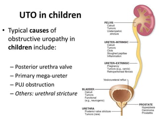
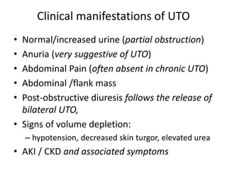
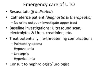
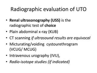
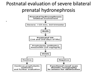
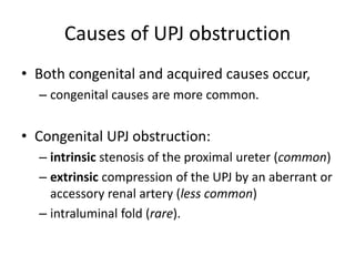
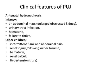
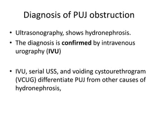
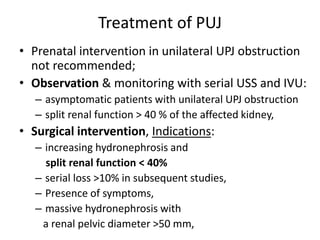
Obstructive uropathy in children
Dr. Abiodun MT
Outline
• Overview
• Selected causes in children
• Pathophysiology
• Clinical features
– Fetal/neonatal
– Older children
• Diagnosis/Differential diagnosis
• Management
• Outcome
OVERVIEW
• Obstructive uropathy or Urinary tract
obstruction (UTO) is a potentially reversible
urological disorder.
• Obstruction to urinary flow may be
– acute or chronic,
– partial or complete,
– unilateral or bilateral, and
– may occur at any site in the urinary tract,
especially at PUJ, Uretero-vesical jx & pelvic rim
UTO in children
• Typical causes of
obstructive uropathy in
children include:
– Posterior urethra valve
– Primary mega-ureter
– PUJ obstruction
– Others: urethral stricture
Clinical manifestations of UTO
• Normal/increased urine (partial obstruction)
• Anuria (very suggestive of UTO)
• Abdominal Pain (often absent in chronic UTO)
• Abdominal /flank mass
• Post-obstructive diuresis follows the release of
bilateral UTO,
• Signs of volume depletion:
– hypotension, decreased skin turgor, elevated urea
• AKI / CKD and associated symptoms
Emergency care of UTO
• Resuscitate (if indicated)
• Catheterize patient (diagnostic & therapeutic)
– No urine output = investigate upper tract
• Baseline investigations: Ultrasound scan,
electrolytes & Urea, creatinine, etc.
• Treat potentially life-threatening complications
– Pulmonary edema
– Hypovolemia
– Urosepsis
– Hyperkalemia
• Consult to nephrologist/ urologist
Radiographic evaluation of UTO
• Renal ultrasonography (USS) is the
radiographic test of choice
• Plain abdominal x-ray (KUB)
• CT scanning if ultrasound results are equivocal
• Micturating/voiding cystourethrogram
(VCUG/ MCUG)
• Intravenous urography (IVU),
• Radio-isotope studies (if indicated)
Postnatal evaluation of severe bilateral
prenatal hydronephrosis
.
Renal dysfunction in UTO
• Reduced glomerular filtration rate (GFR)
• Reduced renal blood flow (after initial rise)
• Impaired renal concentrating ability
• Impaired distal tubular function
– Nephrogenic diabetes insipidus
– Renal salt wasting
– Renal tubular acidosis (RTA)
– Impaired potassium concentration
• Post-obstructive diuresis
Post-obstructive diuresis
• Occurs in BUO or obstruction in a solitary kidney
• Physiologic: caused by retained urea, Na and H2O
• Pathologic: impaired concentrating ability or Na
reabsorption
• Iatrogenic: due to high-volume glucose containing
fluid replacement
• Common chronic obstruction, edema, CHF, uremia
• BUN, Creatinine returns to normal in 24-48 hours
• If persistent diuresis, and elevated creatinine
remains
– IVF (5% dextrose in 0.45% saline)infusion
– Na-wasting nephropathy
Obstructive nephropathy
• Damage to the renal parenchyma due to UTO
– Increased intratubular pressure
– renal vasoconstriction, reduced renal blood flow.
– Ischemia and an influx of inflammatory cells
– cytokines, proteases and oxygen free radicals
Prognosis of UTO
• Functional recovery ≤10 days following relief
of obstruction;
• Permanent kidney damage can occur:
– Tubulointerstitial fibrosis
– Tubular atrophy and apoptosis
– Interstitial inflammation
• Prognostic factors include:
– Severity &/or duration of obstruction
– Hypertension,
– Infection
– Preexisting renal disease
POSTERIOR URETHRAL VALVES (PUV)
• PUVs are obstructing membranous folds in
the posterior urethra
• Incidence: 1 in 5000 to 8000 pregnancies
• Most common cause of LUTO in male
newborn;
• Most common cause of CKD due to LUTO.
Pathogenesis of PUV
• Disruption of male urethral development
between 9 and 14 week of gestation:
– Persistence of the urogenital membrane with
abnormal canalization of the urethra.
– Overgrowth of urethrovaginal folds.
– Abnormal integration of the Wolffian duct into
the posterior urethra.
.
• .
An oblique membrane associated with the verumontanum, the congenital obstructive
posterior urethral membrane (COPUM), appears to be the cause of PUV
Classification of PUV
• Type 1 valve: The most common; a ridge from verumontanum
divides into two leaflets, attaches to anterior urethra
• Type 2 valve: from the verumontanum towards the internal
sphincter and bladder neck (?dissection artifact)
• Type 3 valve: a diaphragm distal to the verumontanum with a
central perforation (? Due to urethral catheterization).
New Classification:
• COPUM :congenital obstructive posterior urethral membrane,
related to verumontanum
• Cobb's collar: congenital urethral stricture caused by a
transverse membrane in the posterior urethra, not related to
verumontanum
Clinical Manifestations of PUV 1
• Prenatal: ultrasonographic findings:
– bilateral hydronephrosis,
– dilated bladder,
– dilated posterior urethra (keyhole sign),
– bladder wall thickened
• severe obstruction:
– oligohydramnios,
– urinary ascites
– perinephric urinoma,
– bladder diverticula /patent urachus,
– increased renal echogenicity/cortical cysts (renal dysplasia)
Clinical Manifestations of PUV 2
• Neonatal period:
– Respiratory distress due to lung hypoplasia (amniotic
fluid required for canalicular phase of lung devt 16
and 28 weeks GA)
– Abdominal distension (bladder or urinary ascites)
– Poor urinary stream or difficulty with voiding;
• Infancy
– Failure to thrive, Urosepsis,
– Poor urinary stream or straining while voiding;
• Older children
– Urinary tract infections,
– day time and nocturnal incontinence (enuresis),
– voiding dysfx (freq., straining, poor urinary stream)
Diagnosis
• Clinical history and examination findings
suggestive of LUTO;
• Diagnosis: Micturating cystourethrogram
(MCUG)
– hallmark is dilated, elongated posterior urethra
with a thin linear defect during the voiding phase;
• Confirmed diagnosis: cystoscopy.
.
• .
Voiding cystourethrogram of patient with PUV noted by white arrows with dilated
posterior urethra. Small capacity bladder with VUR (black arrows).
Courtesy of Nicholas Holmes, MD.
Radionuclide studies
• DMSA scan: Dimercaptosuccinic acid (DMSA)
detects focal renal parenchymal lesion and assess
the function of each kidney (static scan);
• Technetium 99-DTPA scan: Diethylenetriamine
pentaacetic acid (DTPA) scan assesses renal
excretory function (dynamic scans)
Laboratory studies
• Electrolytes, Urea & creatinine
• ESRD may occur early in infancy, and these
infants may require renal replacement
therapy, in addition to medical management
of the complications of renal failure.
Prenatal Management
• Prenatal surgery (vesicoamniotic shunt
placement):
– prevent lung hypoplasia
– reduce back pressure and nephron loss,
– improve neonatal survival.
– may reduce ESRD postnatally
• Fetal surgery for PUV is associated with a risk of
fetal and maternal morbidity without proven
benefit for long-term renal outcome.
Postnatal Management
• Stabilize the patient
– Correct electrolyte imbalance, treat respiratory distress,
– Treat urosepsis
– Monitor renal function
• Urinary tract drainage : a soft NG tube (size ≥8 French)
– NG tube has a large internal diameter, &inflated balloon of
the Foley catheter may occlude the ureteral orifices.
• Primary ablation during cystoscopy (preferred)
– Feasible in term and preterm infants.
• Vesicostomy (an alternative in very preterm infants )
Post-procedure Management
• Detect and treat bladder dysfunction:
– Evaluate with imaging/ urodynamic studies
– anticholinergic drugs lowers bladder pressures
– persistent hydronephrosis suggest abnormal
bladder function.
• Monitor renal function,
• Manage CKD, if present.
Outcome of PUV
• Chronic kidney disease (CKD)
• Despite early treatment, a significant
proportion of PUV patients develop ESRD
Differential diagnosis of PUV
• Agenesis or stricture/stenosis of the urethra
• Megalourethra
• Megacystis
• Micro-colon syndrome
• These causes are differentiated from PUV by
VCUG and cystoscopy.
PELVIC-URETERIC JUNCTION
OBSTRUCTION
• Pelvic-ureteric junction (PUJ) or ureteropelvic
junction (UPJ) obstruction is a partial or total
blockage of the flow of urine that occurs
where the ureter enters the kidney, and
results in hydronephrosis;
• Incidence of up to 1 in 500 fetuses;
• Most common cause of hydronephrosis in the
newborn
PUJ obstruction
.
Causes of UPJ obstruction
• Both congenital and acquired causes occur,
– congenital causes are more common.
• Congenital UPJ obstruction:
– intrinsic stenosis of the proximal ureter (common)
– extrinsic compression of the UPJ by an aberrant or
accessory renal artery (less common)
– intraluminal fold (rare).
Clinical features of PUJ
Antenatal hydronephrosis
Infancy:
• an abdominal mass (enlarged obstructed kidney),
• urinary tract infection,
• hematuria,
• failure to thrive.
Older children:
• intermittent flank and abdominal pain
• renal injury following minor trauma,
• hematuria,
• renal calculi,
• Hypertension (rare)
Diagnosis of PUJ obstruction
• Ultrasonography, shows hydronephrosis.
• The diagnosis is confirmed by intravenous
urography (IVU)
• IVU, serial USS, and voiding cystourethrogram
(VCUG) differentiate PUJ from other causes of
hydronephrosis,
Treatment of PUJ
• Prenatal intervention in unilateral UPJ obstruction
not recommended;
• Observation & monitoring with serial USS and IVU:
– asymptomatic patients with unilateral UPJ obstruction
– split renal function > 40 % of the affected kidney,
• Surgical intervention, Indications:
– increasing hydronephrosis and
split renal function < 40%
– serial loss >10% in subsequent studies,
– Presence of symptoms,
– massive hydronephrosis with
a renal pelvic diameter >50 mm,
Outcome of PUJ obstruction
• Stable or improved renal function, decreased
hydronephrosis, and lack of symptoms in most
patients following surgical correction
• Recurrent obstruction in 5%.
• Uncertain long-term outcome of patients
managed by observation alone
Differential diagnosis of PUJ
• Vesicoureteral reflux (VUR),
• Transient and functional hydronephrosis,
• Posterior urethral valves,
• Congenital megaureter,
• Ureterocele, and
• Multicystic dysplastic kidney
PRIMARY MEGAURETER
• Megaureter is a ureter that exceeds the upper
limits of normal size or , >7 mm in diameter in
children;
• Primary megaureter is due to a functional or
anatomical abnormality of the ureterovesical
junction;
• Secondary megaureter is due to bladder or
urethra disorders;
Incidence and Pathogenesis
• Incidence of primary megaureter is 0.36 per
1000 live births;
– Second most common cause of hydronephrosis in
the newborn (20% of cases )with an estimated;
• Pathogenesis: It is due to an abnormal or
delayed development of the muscle in the
distal ureter at 20 weeks gestation.
Clinical features and diagnosis
Clinical features of primary megaureter:
• Abdominal pain,
• Abdominal mass,
• UTI
• hematuria
• Uremia
Diagnosis: USS findings of both hydronephrosis and
a dilated ureter (antenatal / post-natal).
Investigation and treatment
MCUG: detects VUR
IVU: isotope clearance, delayed in urinary obstruction.
Treatments
• No prenatal intervention
• Non-surgical management
– In asymptomatic patients with nonrefluxing, non-
obstructed megaureters
– Monitoring with ultrasounds yearly
– Prophylactic antibiotics until the patient is toilet trained.
• Surgical intervention in symptomatic patients
– Relieve the obstruction
.
• .
Natural course of megaureter: Series of imaging studies that demonstrate
spontaneous resolution of a left megaureter without surgical intervention.
Courtesy of Dr. Laurence Baskin.
URETHRAL STRICTURE
• Stricture associated with urethral
catheterization occurs almost exclusively in
male patients
• Repeated urethral trauma from intermittent
catheterization can cause urethral stricture
formation, which in turn increases the
likelihood of traumatic catheterization.
• The incidence of urethral stricture increases
with duration of chronic catheterization;
CONCLUSION
• Obstructive uropathy is a major clinical
morbidity in children;
• Prompt diagnosis and management is
necessary to avoid kidney failure.
References
• Principles and practice of pediatric nephrology;
• LUTO. CME, ADC 2016;
• www.uptodate.com
• Urinary tract obstruction, ppt. Victor Federico
B. Acepcion, MD, FPUA
.
• Thank you

Obstructive uropathy FINAL presentation.pdf

  • 1.
    Obstructive uropathy inchildren Dr. Abiodun MT
  • 2.
    Outline • Overview • Selectedcauses in children • Pathophysiology • Clinical features – Fetal/neonatal – Older children • Diagnosis/Differential diagnosis • Management • Outcome
  • 3.
    OVERVIEW • Obstructive uropathyor Urinary tract obstruction (UTO) is a potentially reversible urological disorder. • Obstruction to urinary flow may be – acute or chronic, – partial or complete, – unilateral or bilateral, and – may occur at any site in the urinary tract, especially at PUJ, Uretero-vesical jx & pelvic rim
  • 4.
    UTO in children •Typical causes of obstructive uropathy in children include: – Posterior urethra valve – Primary mega-ureter – PUJ obstruction – Others: urethral stricture
  • 5.
    Clinical manifestations ofUTO • Normal/increased urine (partial obstruction) • Anuria (very suggestive of UTO) • Abdominal Pain (often absent in chronic UTO) • Abdominal /flank mass • Post-obstructive diuresis follows the release of bilateral UTO, • Signs of volume depletion: – hypotension, decreased skin turgor, elevated urea • AKI / CKD and associated symptoms
  • 6.
    Emergency care ofUTO • Resuscitate (if indicated) • Catheterize patient (diagnostic & therapeutic) – No urine output = investigate upper tract • Baseline investigations: Ultrasound scan, electrolytes & Urea, creatinine, etc. • Treat potentially life-threatening complications – Pulmonary edema – Hypovolemia – Urosepsis – Hyperkalemia • Consult to nephrologist/ urologist
  • 7.
    Radiographic evaluation ofUTO • Renal ultrasonography (USS) is the radiographic test of choice • Plain abdominal x-ray (KUB) • CT scanning if ultrasound results are equivocal • Micturating/voiding cystourethrogram (VCUG/ MCUG) • Intravenous urography (IVU), • Radio-isotope studies (if indicated)
  • 8.
    Postnatal evaluation ofsevere bilateral prenatal hydronephrosis .
  • 9.
    Renal dysfunction inUTO • Reduced glomerular filtration rate (GFR) • Reduced renal blood flow (after initial rise) • Impaired renal concentrating ability • Impaired distal tubular function – Nephrogenic diabetes insipidus – Renal salt wasting – Renal tubular acidosis (RTA) – Impaired potassium concentration • Post-obstructive diuresis
  • 10.
    Post-obstructive diuresis • Occursin BUO or obstruction in a solitary kidney • Physiologic: caused by retained urea, Na and H2O • Pathologic: impaired concentrating ability or Na reabsorption • Iatrogenic: due to high-volume glucose containing fluid replacement • Common chronic obstruction, edema, CHF, uremia • BUN, Creatinine returns to normal in 24-48 hours • If persistent diuresis, and elevated creatinine remains – IVF (5% dextrose in 0.45% saline)infusion – Na-wasting nephropathy
  • 11.
    Obstructive nephropathy • Damageto the renal parenchyma due to UTO – Increased intratubular pressure – renal vasoconstriction, reduced renal blood flow. – Ischemia and an influx of inflammatory cells – cytokines, proteases and oxygen free radicals
  • 12.
    Prognosis of UTO •Functional recovery ≤10 days following relief of obstruction; • Permanent kidney damage can occur: – Tubulointerstitial fibrosis – Tubular atrophy and apoptosis – Interstitial inflammation • Prognostic factors include: – Severity &/or duration of obstruction – Hypertension, – Infection – Preexisting renal disease
  • 13.
    POSTERIOR URETHRAL VALVES(PUV) • PUVs are obstructing membranous folds in the posterior urethra • Incidence: 1 in 5000 to 8000 pregnancies • Most common cause of LUTO in male newborn; • Most common cause of CKD due to LUTO.
  • 14.
    Pathogenesis of PUV •Disruption of male urethral development between 9 and 14 week of gestation: – Persistence of the urogenital membrane with abnormal canalization of the urethra. – Overgrowth of urethrovaginal folds. – Abnormal integration of the Wolffian duct into the posterior urethra.
  • 15.
    . • . An obliquemembrane associated with the verumontanum, the congenital obstructive posterior urethral membrane (COPUM), appears to be the cause of PUV
  • 16.
    Classification of PUV •Type 1 valve: The most common; a ridge from verumontanum divides into two leaflets, attaches to anterior urethra • Type 2 valve: from the verumontanum towards the internal sphincter and bladder neck (?dissection artifact) • Type 3 valve: a diaphragm distal to the verumontanum with a central perforation (? Due to urethral catheterization). New Classification: • COPUM :congenital obstructive posterior urethral membrane, related to verumontanum • Cobb's collar: congenital urethral stricture caused by a transverse membrane in the posterior urethra, not related to verumontanum
  • 17.
    Clinical Manifestations ofPUV 1 • Prenatal: ultrasonographic findings: – bilateral hydronephrosis, – dilated bladder, – dilated posterior urethra (keyhole sign), – bladder wall thickened • severe obstruction: – oligohydramnios, – urinary ascites – perinephric urinoma, – bladder diverticula /patent urachus, – increased renal echogenicity/cortical cysts (renal dysplasia)
  • 18.
    Clinical Manifestations ofPUV 2 • Neonatal period: – Respiratory distress due to lung hypoplasia (amniotic fluid required for canalicular phase of lung devt 16 and 28 weeks GA) – Abdominal distension (bladder or urinary ascites) – Poor urinary stream or difficulty with voiding; • Infancy – Failure to thrive, Urosepsis, – Poor urinary stream or straining while voiding; • Older children – Urinary tract infections, – day time and nocturnal incontinence (enuresis), – voiding dysfx (freq., straining, poor urinary stream)
  • 19.
    Diagnosis • Clinical historyand examination findings suggestive of LUTO; • Diagnosis: Micturating cystourethrogram (MCUG) – hallmark is dilated, elongated posterior urethra with a thin linear defect during the voiding phase; • Confirmed diagnosis: cystoscopy.
  • 20.
    . • . Voiding cystourethrogramof patient with PUV noted by white arrows with dilated posterior urethra. Small capacity bladder with VUR (black arrows). Courtesy of Nicholas Holmes, MD.
  • 21.
    Radionuclide studies • DMSAscan: Dimercaptosuccinic acid (DMSA) detects focal renal parenchymal lesion and assess the function of each kidney (static scan); • Technetium 99-DTPA scan: Diethylenetriamine pentaacetic acid (DTPA) scan assesses renal excretory function (dynamic scans)
  • 22.
    Laboratory studies • Electrolytes,Urea & creatinine • ESRD may occur early in infancy, and these infants may require renal replacement therapy, in addition to medical management of the complications of renal failure.
  • 23.
    Prenatal Management • Prenatalsurgery (vesicoamniotic shunt placement): – prevent lung hypoplasia – reduce back pressure and nephron loss, – improve neonatal survival. – may reduce ESRD postnatally • Fetal surgery for PUV is associated with a risk of fetal and maternal morbidity without proven benefit for long-term renal outcome.
  • 24.
    Postnatal Management • Stabilizethe patient – Correct electrolyte imbalance, treat respiratory distress, – Treat urosepsis – Monitor renal function • Urinary tract drainage : a soft NG tube (size ≥8 French) – NG tube has a large internal diameter, &inflated balloon of the Foley catheter may occlude the ureteral orifices. • Primary ablation during cystoscopy (preferred) – Feasible in term and preterm infants. • Vesicostomy (an alternative in very preterm infants )
  • 25.
    Post-procedure Management • Detectand treat bladder dysfunction: – Evaluate with imaging/ urodynamic studies – anticholinergic drugs lowers bladder pressures – persistent hydronephrosis suggest abnormal bladder function. • Monitor renal function, • Manage CKD, if present.
  • 26.
    Outcome of PUV •Chronic kidney disease (CKD) • Despite early treatment, a significant proportion of PUV patients develop ESRD
  • 27.
    Differential diagnosis ofPUV • Agenesis or stricture/stenosis of the urethra • Megalourethra • Megacystis • Micro-colon syndrome • These causes are differentiated from PUV by VCUG and cystoscopy.
  • 28.
    PELVIC-URETERIC JUNCTION OBSTRUCTION • Pelvic-uretericjunction (PUJ) or ureteropelvic junction (UPJ) obstruction is a partial or total blockage of the flow of urine that occurs where the ureter enters the kidney, and results in hydronephrosis; • Incidence of up to 1 in 500 fetuses; • Most common cause of hydronephrosis in the newborn
  • 29.
  • 30.
    Causes of UPJobstruction • Both congenital and acquired causes occur, – congenital causes are more common. • Congenital UPJ obstruction: – intrinsic stenosis of the proximal ureter (common) – extrinsic compression of the UPJ by an aberrant or accessory renal artery (less common) – intraluminal fold (rare).
  • 31.
    Clinical features ofPUJ Antenatal hydronephrosis Infancy: • an abdominal mass (enlarged obstructed kidney), • urinary tract infection, • hematuria, • failure to thrive. Older children: • intermittent flank and abdominal pain • renal injury following minor trauma, • hematuria, • renal calculi, • Hypertension (rare)
  • 32.
    Diagnosis of PUJobstruction • Ultrasonography, shows hydronephrosis. • The diagnosis is confirmed by intravenous urography (IVU) • IVU, serial USS, and voiding cystourethrogram (VCUG) differentiate PUJ from other causes of hydronephrosis,
  • 33.
    Treatment of PUJ •Prenatal intervention in unilateral UPJ obstruction not recommended; • Observation & monitoring with serial USS and IVU: – asymptomatic patients with unilateral UPJ obstruction – split renal function > 40 % of the affected kidney, • Surgical intervention, Indications: – increasing hydronephrosis and split renal function < 40% – serial loss >10% in subsequent studies, – Presence of symptoms, – massive hydronephrosis with a renal pelvic diameter >50 mm,
  • 34.
    Outcome of PUJobstruction • Stable or improved renal function, decreased hydronephrosis, and lack of symptoms in most patients following surgical correction • Recurrent obstruction in 5%. • Uncertain long-term outcome of patients managed by observation alone
  • 35.
    Differential diagnosis ofPUJ • Vesicoureteral reflux (VUR), • Transient and functional hydronephrosis, • Posterior urethral valves, • Congenital megaureter, • Ureterocele, and • Multicystic dysplastic kidney
  • 36.
    PRIMARY MEGAURETER • Megaureteris a ureter that exceeds the upper limits of normal size or , >7 mm in diameter in children; • Primary megaureter is due to a functional or anatomical abnormality of the ureterovesical junction; • Secondary megaureter is due to bladder or urethra disorders;
  • 37.
    Incidence and Pathogenesis •Incidence of primary megaureter is 0.36 per 1000 live births; – Second most common cause of hydronephrosis in the newborn (20% of cases )with an estimated; • Pathogenesis: It is due to an abnormal or delayed development of the muscle in the distal ureter at 20 weeks gestation.
  • 38.
    Clinical features anddiagnosis Clinical features of primary megaureter: • Abdominal pain, • Abdominal mass, • UTI • hematuria • Uremia Diagnosis: USS findings of both hydronephrosis and a dilated ureter (antenatal / post-natal).
  • 39.
    Investigation and treatment MCUG:detects VUR IVU: isotope clearance, delayed in urinary obstruction. Treatments • No prenatal intervention • Non-surgical management – In asymptomatic patients with nonrefluxing, non- obstructed megaureters – Monitoring with ultrasounds yearly – Prophylactic antibiotics until the patient is toilet trained. • Surgical intervention in symptomatic patients – Relieve the obstruction
  • 40.
    . • . Natural courseof megaureter: Series of imaging studies that demonstrate spontaneous resolution of a left megaureter without surgical intervention. Courtesy of Dr. Laurence Baskin.
  • 41.
    URETHRAL STRICTURE • Strictureassociated with urethral catheterization occurs almost exclusively in male patients • Repeated urethral trauma from intermittent catheterization can cause urethral stricture formation, which in turn increases the likelihood of traumatic catheterization. • The incidence of urethral stricture increases with duration of chronic catheterization;
  • 42.
    CONCLUSION • Obstructive uropathyis a major clinical morbidity in children; • Prompt diagnosis and management is necessary to avoid kidney failure.
  • 43.
    References • Principles andpractice of pediatric nephrology; • LUTO. CME, ADC 2016; • www.uptodate.com • Urinary tract obstruction, ppt. Victor Federico B. Acepcion, MD, FPUA
  • 44.