Downloaded 863 times


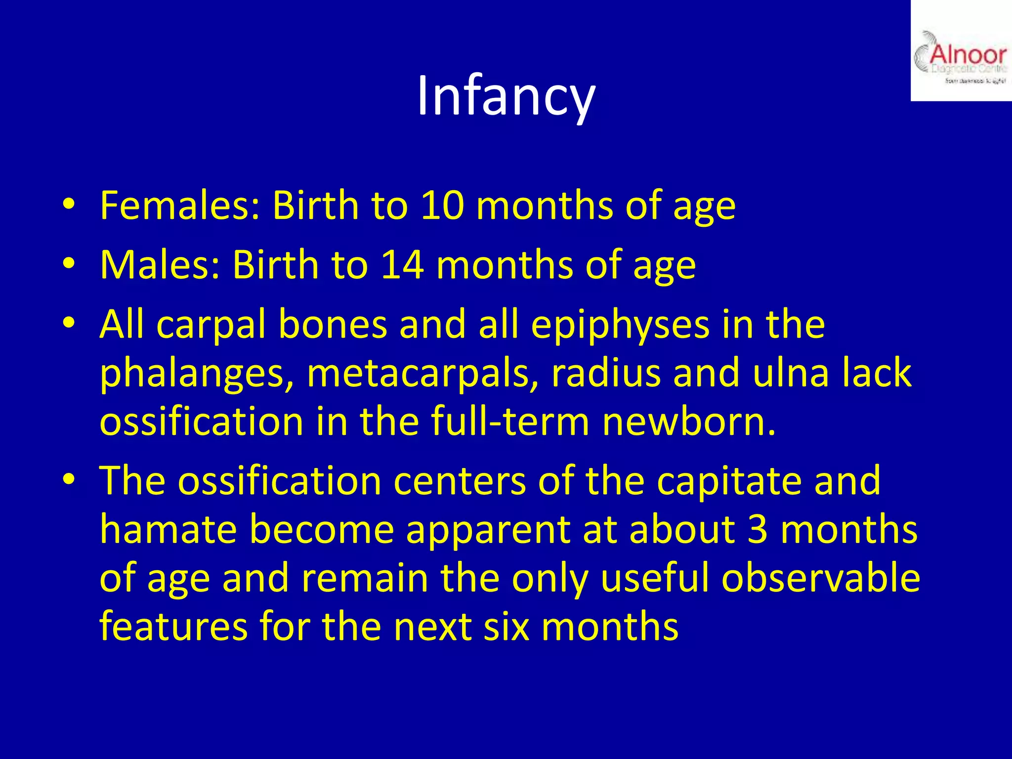


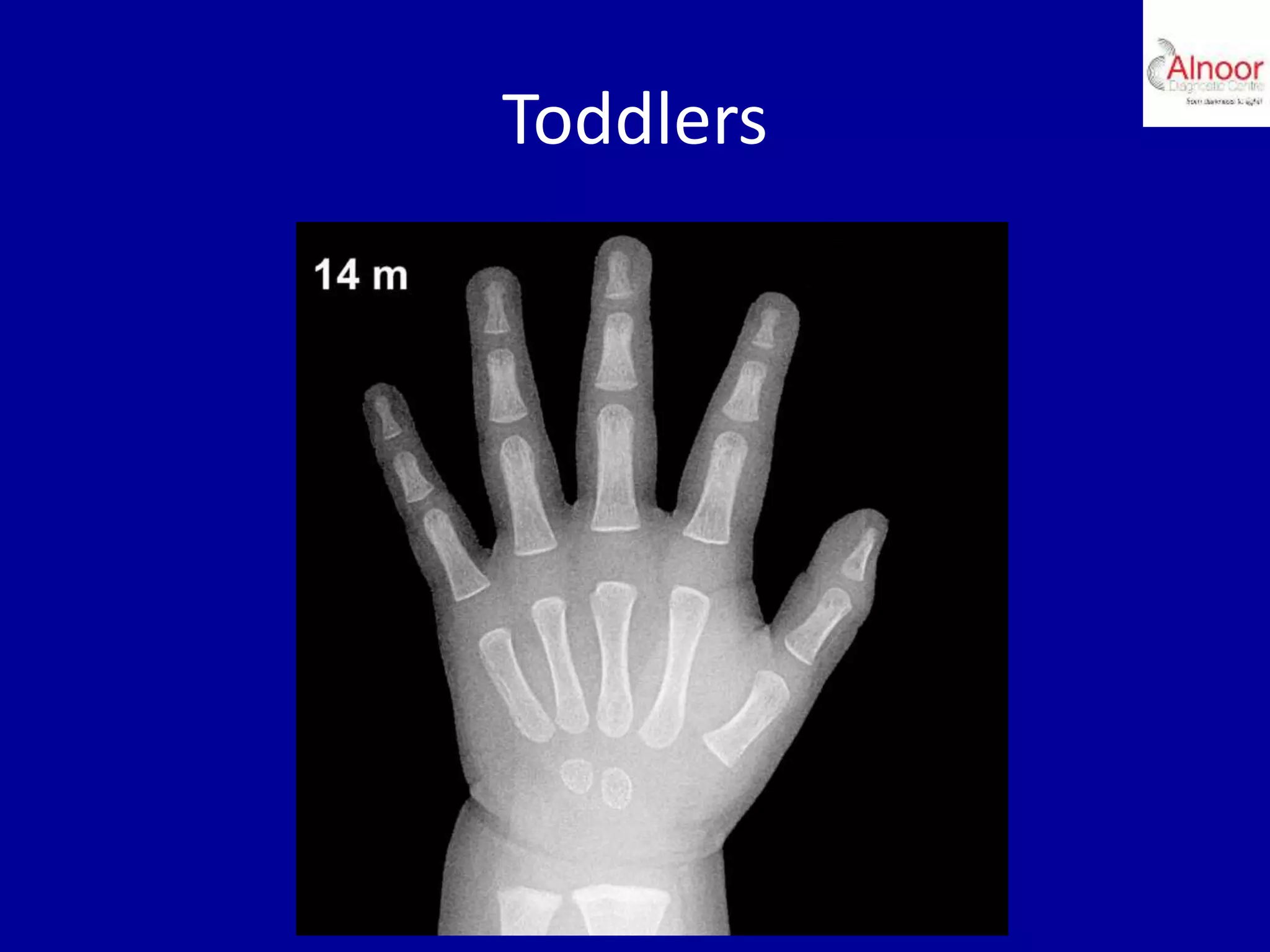
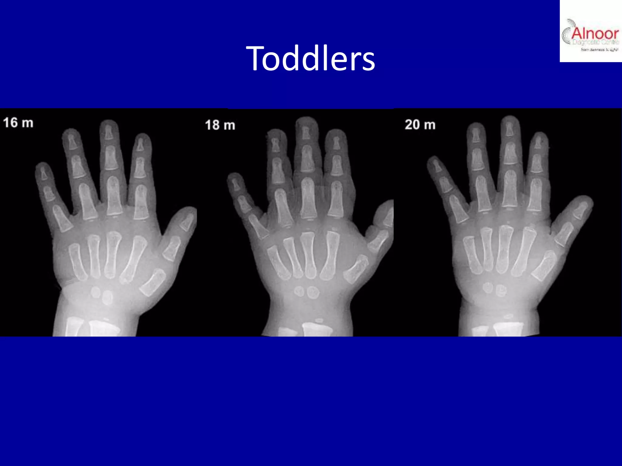


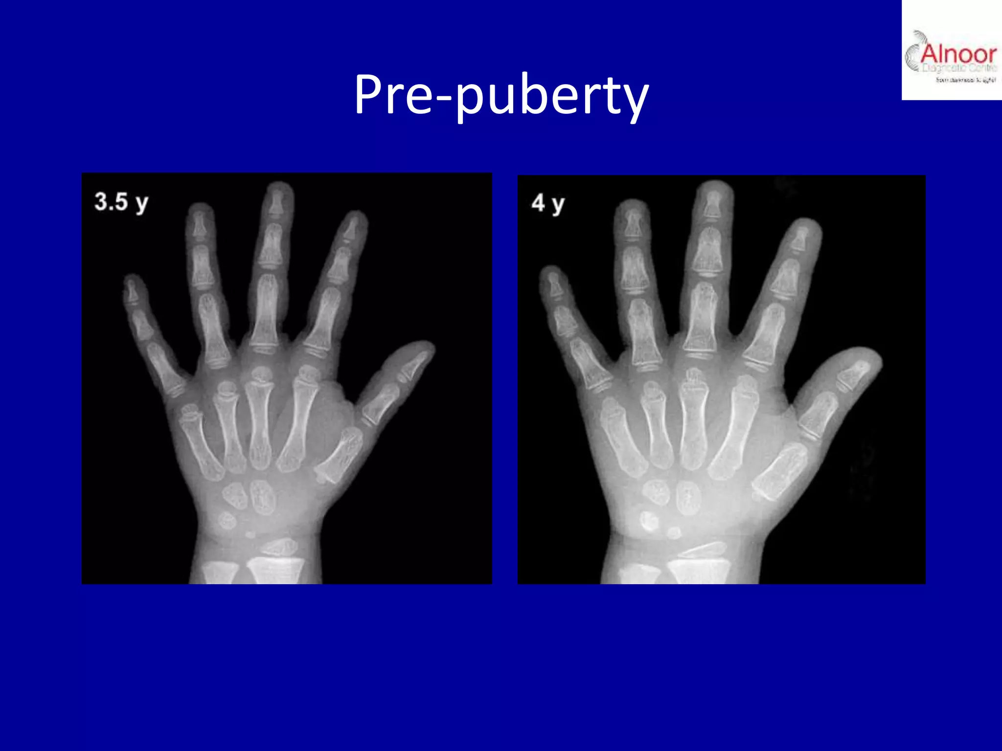
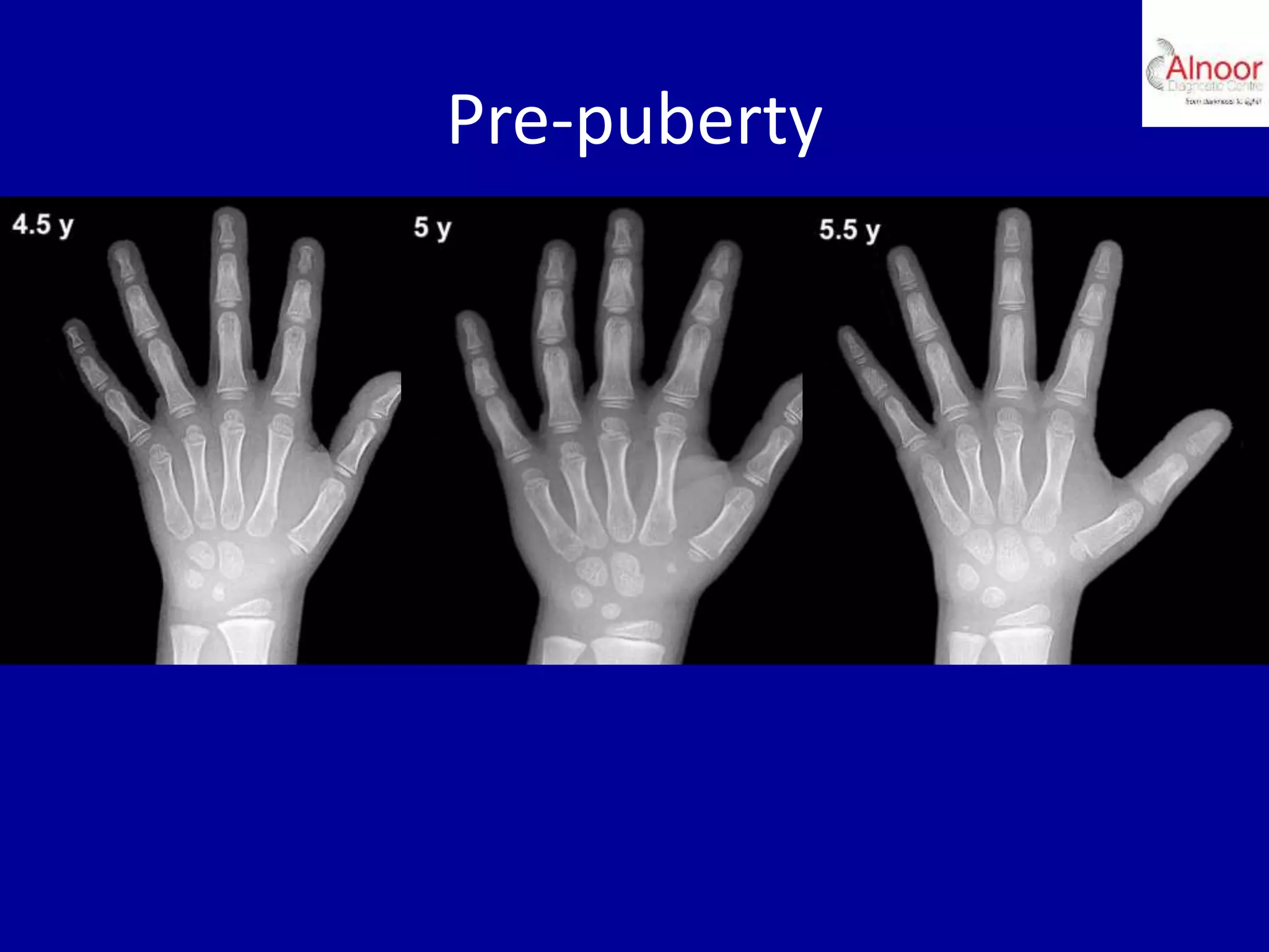


Bone age assessment is performed to evaluate growth in pediatric patients and diagnose endocrine disorders. It relies on visual evaluation of hand and wrist skeletal development compared to standard references. Key applications include diagnosing growth disorders and predicting final adult height. Skeletal development is divided into stages from infancy to post-puberty based on characteristics like appearance of ossification centers and epiphyseal fusion. Bone age is assessed by comparing a patient's hand and wrist radiograph to standardized images for their age and sex.


