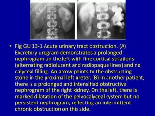
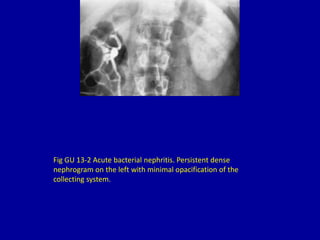
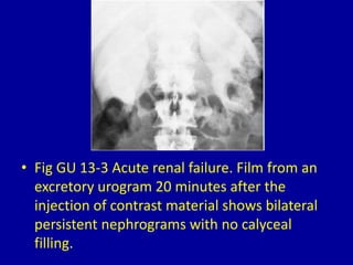
13Persistent or Increasingly
Dense Nephrogram
CLINICAL IMAGAGING
AN ATLAS OF DIFFERENTIAL DAIGNOSIS
EISENBERG
DR. Muhammad Bin Zulfiqar
PGR-FCPS III SIMS/SHL
• Fig GU 13-1 Acute urinary tract obstruction. (A)
Excretory urogram demonstrates a prolonged
nephrogram on the left with fine cortical striations
(alternating radiolucent and radiopaque lines) and no
calyceal filling. An arrow points to the obstructing
stone in the proximal left ureter. (B) In another patient,
there is a prolonged and intensified obstructive
nephrogram of the right kidney. On the left, there is
marked dilatation of the pelvocalyceal system but no
persistent nephrogram, reflecting an intermittent
chronic obstruction on this side.
Fig GU 13-2 Acute bacterial nephritis. Persistent dense
nephrogram on the left with minimal opacification of the
collecting system.
• Fig GU 13-3 Acute renal failure. Film from an
excretory urogram 20 minutes after the
injection of contrast material shows bilateral
persistent nephrograms with no calyceal
filling.
• Fig GU 13-4 Acute renal vein thrombosis. Film
of the right kidney taken 5 minutes after the
injection of contrast material shows a dense
nephrogram (arrows) and the absence of
calyceal filling.
13 persistent or increasingly dense nephrogram

13 persistent or increasingly dense nephrogram

  • 1.
  • 2.
    CLINICAL IMAGAGING AN ATLASOF DIFFERENTIAL DAIGNOSIS EISENBERG DR. Muhammad Bin Zulfiqar PGR-FCPS III SIMS/SHL
  • 3.
    • Fig GU13-1 Acute urinary tract obstruction. (A) Excretory urogram demonstrates a prolonged nephrogram on the left with fine cortical striations (alternating radiolucent and radiopaque lines) and no calyceal filling. An arrow points to the obstructing stone in the proximal left ureter. (B) In another patient, there is a prolonged and intensified obstructive nephrogram of the right kidney. On the left, there is marked dilatation of the pelvocalyceal system but no persistent nephrogram, reflecting an intermittent chronic obstruction on this side.
  • 4.
    Fig GU 13-2Acute bacterial nephritis. Persistent dense nephrogram on the left with minimal opacification of the collecting system.
  • 5.
    • Fig GU13-3 Acute renal failure. Film from an excretory urogram 20 minutes after the injection of contrast material shows bilateral persistent nephrograms with no calyceal filling.
  • 6.
    • Fig GU13-4 Acute renal vein thrombosis. Film of the right kidney taken 5 minutes after the injection of contrast material shows a dense nephrogram (arrows) and the absence of calyceal filling.