Downloaded 1,942 times


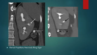


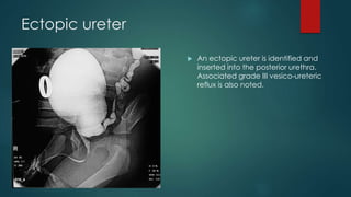


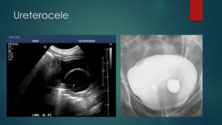


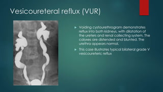


The document provides an overview of the anatomy and imaging techniques of the urinary tract. It describes the pelvicalyceal system including the minor and major calyces that drain into the renal pelvis. It notes the locations of the renal hilum and discusses the ureters, bladder, and urethra. Common congenital anomalies are described such as duplex collecting systems, ureteropelvic junction obstruction, megacalyces, and calyceal diverticula. Imaging modalities for evaluating the urinary tract include KUB, IVP, ultrasound, MRI, and urethrograms. Characteristic signs of conditions like papillary necrosis are also outlined.


