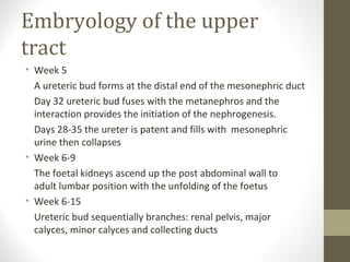
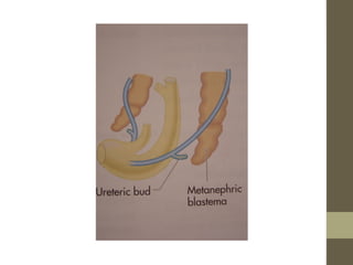
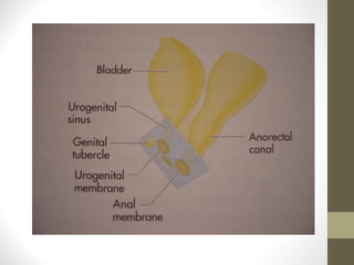
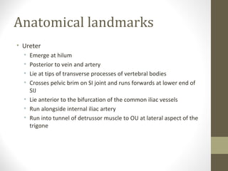
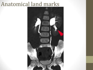
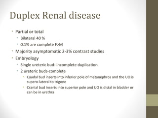
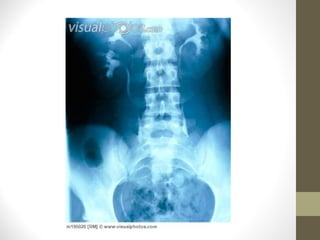
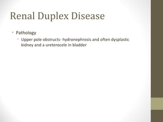
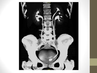
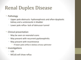
Urinary tract
anatomy and
relevant pathology
Miss Katie Chan – CT2 Urology
Miss Elizabeth Waine – consultant urologist
December 2013
Overview
• Embryology
• Anatomical landmarks
• Duplex systems
• PUJ Obstruction
• Renal Calculi
Basic Embryology
• Day 5/6 after fertilisation
32 cell embryo
Implants into endometrium
• Day 15/16 Embryo has proliferated and 2 cavities form with the
intra-embryonic disc between the 2 cavities
• Cells proliferate on the ectodermal side and form the mesoderm
which divides into 3 components
Paraxial , intermediate and lateral plate mesoderm.
Intermediate mesoderm becomes the genitourinary tract.
Embryology of the upper
tract
• Derived from caudal zone of the intermediate mesoderm (IM)
either side of the midline
• Week 4
Early-pronephros appears at the cervical portion of the
IM
Later-mesonephros forms but degenerates at week 11
Lateral to the mesonephros the mesonephric duct is forming
and advances caudally
The mesonephric duct canalises and functions briefly to
produce urine
Embryology of the upper
tract
• Week 5
A ureteric bud forms at the distal end of the mesonephric duct
Day 32 ureteric bud fuses with the metanephros and the
interaction provides the initiation of the nephrogenesis.
Days 28-35 the ureter is patent and fills with mesonephric
urine then collapses
• Week 6-9
The foetal kidneys ascend up the post abdominal wall to
adult lumbar position with the unfolding of the foetus
• Week 6-15
Ureteric bud sequentially branches: renal pelvis, major
calyces, minor calyces and collecting ducts
Embryology of the lower urinary tract
• The bladder forms from the anterior portion of the cloaca. The
posterior portion forms the rectum
• •Week 4-6 uro-rectal septum descends subdividing the cloaca and is
aided by the proliferation and in-growth of the folds of Rathke fully
divided at wk7
• –The area above the mesonephric ducts is the vesicourethral canal
• –The area below the mesonephric ducts is the urogenital sinus
• •Mesonephric ducts drain into urogenital sinus therefore mesoderm
becomes fused with endoderm. The ureters then evaginate from
the mesonephric duct and will enter the bladder at day37
• •The trigone is formed by the dilatation of the mesonephric ducts
terminus known as the common excretory duct. The overlying
mesoderm migrates to the midline and this is the primitive trigone
which is then covered by endoderm of the urogenital canal.
Anatomical land marks
• Left Kidney

• T11 to L2
• Relations
•
•
•
•

Spleen- supero-lateral
Adrenal- supero-medial
Tail of pancreas- Hilum
Splenic flexure- inferior and anterior

• Right kidney
• T12-L3
• Relations
•
•
•
•
•

Liver-superior
Adrenal – supero-medial
IVC
D2
Hepatic flexure
Anatomical landmarks
• Ureter
•
•
•
•

Emerge at hilum
Posterior to vein and artery
Lie at tips of transverse processes of vertebral bodies
Crosses pelvic brim on SI joint and runs forwards at lower end of
SIJ
• Lie anterior to the bifurcation of the common iliac vessels
• Run alongside internal iliac artery
• Run into tunnel of detrussor muscle to OU at lateral aspect of the
trigone
Anatomical land marks
Duplex Renal disease
• Partial or total
• Bilateral 40 %
• 0.1% are complete F>M

• Majority asymptomatic 2-3% contrast studies
• Embryology
• Single ureteric bud- incomplete duplication
• 2 ureteric buds-complete
• Caudal bud inserts into inferior pole of metanephros and the UO is
supero-lateral to trigone
• Cranial bud inserts into superior pole and UO is distal in bladder or
can be in urethra
Renal Duplex Disease
• Pathology
• Upper pole obstructs- hydronephrosis and often dysplastic
kidney and a ureterocele in bladder
Renal Duplex Disease
• Pathology
• Upper pole obstructs- hydronephrosis and often dysplastic
kidney and a ureterocele in bladder
• Lower pole reflux- lack of detrussor tunnel

• Clinical presentation
• May be seen on neonatal scans
• May present with recurrent pyelonephritis
• May present with incontinence
• If lower pole orifice is below urinary sphincter

• Investigations
• USS
• MCUG-will show reflux
PUJ Obstruction
• A pelvo-ureteric junction (PUJ) obstruction can
be thought of as a restriction to flow of urine,
from the renal pelvis to the ureter, which, if left
uncorrected, leads to progressive renal
deterioration
• Congenital obstructive uropathy
• Broad spectrum of severity
• Varied natural history
• Resolution of prenatal hydronephrosis
• New onset PUJ obst in previously normal kidneys.
PUJO
• Dilated renal pelvis with narrowed
tortuous segment of proximal ureter
• Pathology
• Intrinsic- defect in collagen fibres, circular
muscle fibres
• Extrinsic-Aberrant renal vessels (lower
pole)
PUJO-Presentation
• Prenatal• USS dilation apparent from 2nd trimester
• Renal pelvis diameter >15mm in 2nd
trimester
• 35-50% of all significant prenatal
uropathy
Incidental post natal finding
PUJO-clinical presentation
• Neonatal abdominal mass
• UTI- most common
• Loin Pain-after fluid load
• Adolescents after alcohol
• Late adults-diuretic meds

• Haematuria
• Hypertension
PUJO-investigations
• USS-assess renal pelvic diameter
• Good for surveillance

• Renogram
• Assess level of obstruction
• Indirect assessment of renal functionimportant for management decisions

• Contrast study
• Assess intrinsic obstruction
• Assess vasculature
PUJO-management
• Conservative
• Assymptomatic – esp elderly
• Infants No renal impairment & No infections-as it may resolve
• Adults- No renal impairment and no symptoms

• Surgical
• Pyeloplasty
• Excision of constriction
• Remove excess renal pelvis

• Endoscopic
• Incision
• Expansion balloons- essentially bursting ureteric constriction
Calculi
• Why do patients get pain from stones?
Renal Calculi
• Why do patients get pain from stones?
• At which positions do stones cause problems?
• Hint start at the top
Renal Calculi
• Why do patients get pain from stones?
• At which positions do stones cause problems?
•
•
•
•
•

Infundibulum
PUJ
Pelvic Brim
Over Iliac Vessels
VUJ
Questions

Urology presentation

  • 1.
    Urinary tract anatomy and relevantpathology Miss Katie Chan – CT2 Urology Miss Elizabeth Waine – consultant urologist December 2013
  • 2.
    Overview • Embryology • Anatomicallandmarks • Duplex systems • PUJ Obstruction • Renal Calculi
  • 3.
    Basic Embryology • Day5/6 after fertilisation 32 cell embryo Implants into endometrium • Day 15/16 Embryo has proliferated and 2 cavities form with the intra-embryonic disc between the 2 cavities • Cells proliferate on the ectodermal side and form the mesoderm which divides into 3 components Paraxial , intermediate and lateral plate mesoderm. Intermediate mesoderm becomes the genitourinary tract.
  • 5.
    Embryology of theupper tract • Derived from caudal zone of the intermediate mesoderm (IM) either side of the midline • Week 4 Early-pronephros appears at the cervical portion of the IM Later-mesonephros forms but degenerates at week 11 Lateral to the mesonephros the mesonephric duct is forming and advances caudally The mesonephric duct canalises and functions briefly to produce urine
  • 6.
    Embryology of theupper tract • Week 5 A ureteric bud forms at the distal end of the mesonephric duct Day 32 ureteric bud fuses with the metanephros and the interaction provides the initiation of the nephrogenesis. Days 28-35 the ureter is patent and fills with mesonephric urine then collapses • Week 6-9 The foetal kidneys ascend up the post abdominal wall to adult lumbar position with the unfolding of the foetus • Week 6-15 Ureteric bud sequentially branches: renal pelvis, major calyces, minor calyces and collecting ducts
  • 8.
    Embryology of thelower urinary tract • The bladder forms from the anterior portion of the cloaca. The posterior portion forms the rectum • •Week 4-6 uro-rectal septum descends subdividing the cloaca and is aided by the proliferation and in-growth of the folds of Rathke fully divided at wk7 • –The area above the mesonephric ducts is the vesicourethral canal • –The area below the mesonephric ducts is the urogenital sinus • •Mesonephric ducts drain into urogenital sinus therefore mesoderm becomes fused with endoderm. The ureters then evaginate from the mesonephric duct and will enter the bladder at day37 • •The trigone is formed by the dilatation of the mesonephric ducts terminus known as the common excretory duct. The overlying mesoderm migrates to the midline and this is the primitive trigone which is then covered by endoderm of the urogenital canal.
  • 10.
    Anatomical land marks •Left Kidney • T11 to L2 • Relations • • • • Spleen- supero-lateral Adrenal- supero-medial Tail of pancreas- Hilum Splenic flexure- inferior and anterior • Right kidney • T12-L3 • Relations • • • • • Liver-superior Adrenal – supero-medial IVC D2 Hepatic flexure
  • 11.
    Anatomical landmarks • Ureter • • • • Emergeat hilum Posterior to vein and artery Lie at tips of transverse processes of vertebral bodies Crosses pelvic brim on SI joint and runs forwards at lower end of SIJ • Lie anterior to the bifurcation of the common iliac vessels • Run alongside internal iliac artery • Run into tunnel of detrussor muscle to OU at lateral aspect of the trigone
  • 12.
  • 13.
    Duplex Renal disease •Partial or total • Bilateral 40 % • 0.1% are complete F>M • Majority asymptomatic 2-3% contrast studies • Embryology • Single ureteric bud- incomplete duplication • 2 ureteric buds-complete • Caudal bud inserts into inferior pole of metanephros and the UO is supero-lateral to trigone • Cranial bud inserts into superior pole and UO is distal in bladder or can be in urethra
  • 16.
    Renal Duplex Disease •Pathology • Upper pole obstructs- hydronephrosis and often dysplastic kidney and a ureterocele in bladder
  • 18.
    Renal Duplex Disease •Pathology • Upper pole obstructs- hydronephrosis and often dysplastic kidney and a ureterocele in bladder • Lower pole reflux- lack of detrussor tunnel • Clinical presentation • May be seen on neonatal scans • May present with recurrent pyelonephritis • May present with incontinence • If lower pole orifice is below urinary sphincter • Investigations • USS • MCUG-will show reflux
  • 19.
    PUJ Obstruction • Apelvo-ureteric junction (PUJ) obstruction can be thought of as a restriction to flow of urine, from the renal pelvis to the ureter, which, if left uncorrected, leads to progressive renal deterioration • Congenital obstructive uropathy • Broad spectrum of severity • Varied natural history • Resolution of prenatal hydronephrosis • New onset PUJ obst in previously normal kidneys.
  • 20.
    PUJO • Dilated renalpelvis with narrowed tortuous segment of proximal ureter • Pathology • Intrinsic- defect in collagen fibres, circular muscle fibres • Extrinsic-Aberrant renal vessels (lower pole)
  • 21.
    PUJO-Presentation • Prenatal• USSdilation apparent from 2nd trimester • Renal pelvis diameter >15mm in 2nd trimester • 35-50% of all significant prenatal uropathy Incidental post natal finding
  • 22.
    PUJO-clinical presentation • Neonatalabdominal mass • UTI- most common • Loin Pain-after fluid load • Adolescents after alcohol • Late adults-diuretic meds • Haematuria • Hypertension
  • 23.
    PUJO-investigations • USS-assess renalpelvic diameter • Good for surveillance • Renogram • Assess level of obstruction • Indirect assessment of renal functionimportant for management decisions • Contrast study • Assess intrinsic obstruction • Assess vasculature
  • 24.
    PUJO-management • Conservative • Assymptomatic– esp elderly • Infants No renal impairment & No infections-as it may resolve • Adults- No renal impairment and no symptoms • Surgical • Pyeloplasty • Excision of constriction • Remove excess renal pelvis • Endoscopic • Incision • Expansion balloons- essentially bursting ureteric constriction
  • 25.
    Calculi • Why dopatients get pain from stones?
  • 26.
    Renal Calculi • Whydo patients get pain from stones? • At which positions do stones cause problems? • Hint start at the top
  • 27.
    Renal Calculi • Whydo patients get pain from stones? • At which positions do stones cause problems? • • • • • Infundibulum PUJ Pelvic Brim Over Iliac Vessels VUJ
  • 28.

Editor's Notes