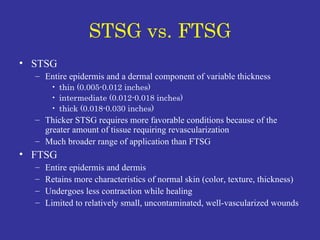
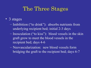
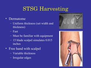
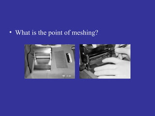
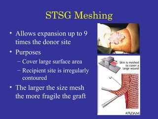
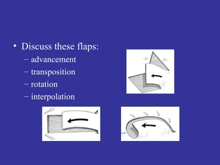
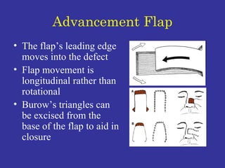
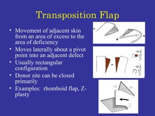
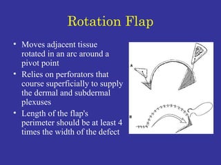
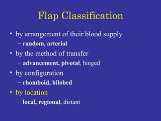
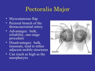
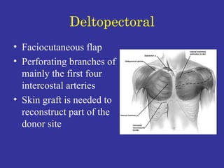
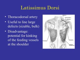
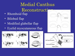
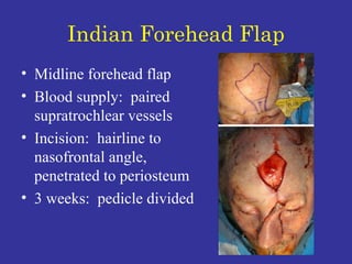
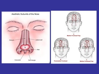
COCLIA 99: Grafts and Flaps in  Head & Neck Surgery The images and text in this presentation were borrowed from multiple resources available on the internet without permission.  This presentation is for education purposes only and should not be reproduced in any matter.
Skin Overview Epidermis stratified squamous epithelium no blood vessels:  receives nutrients by diffusion
Skin Overview Dermis 2 layers:  papillary & reticular reticular dermis:  larger blood vessels, epidermal appendages intradermal epithelial structures (sebaceous & sweat glands, hair follicles) are lined with epithelial cells with the potential for division and differentiation
STSG vs. FTSG STSG Entire epidermis and a dermal component of variable thickness thin (0.005-0.012 inches) intermediate (0.012-0.018 inches) thick (0.018-0.030 inches) Thicker STSG requires more favorable conditions because of the greater amount of tissue requiring revascularization Much broader range of application than FTSG FTSG Entire epidermis and dermis Retains more characteristics of normal skin (color, texture, thickness)  Undergoes less contraction while healing Limited to relatively small, uncontaminated, well-vascularized wounds
Describe the three stages of survival for a STSG…
The Three Stages 3 stages Imbibition (“to drink”):  absorbs nutrients from underlying recipient bed; initial 2-3 days Inosculation (“to kiss”):  blood vessels in the skin graft grow to meet the blood vessels in the recipient bed; days 4-6 Neovascularization:  new blood vessels form bridging the graft to the recipient bed; days 6-7
What factors are important in graft survival?
Survival Factors Good Nutrition and oxygenation delivery Removal of waste products Bad Mobility of graft Infection  Fluid collection beneath graft
How are STSG and FTSG harvested?
STSG Harvesting Dermatome Uniform thickness (set width and thickness) Fast Must be familiar with equipment 15 blade scalpel simulates 0.015 inches Free hand with scalpel Variable thickness Irregular edges
FTSG Harvesting Free hand with scalpel Enlarge by 3-5% Compensates for immediate primary contraction MUST  trim off all residual adipose tissue Relatively avascular
What is the point of meshing?
STSG Meshing Allows expansion up to 9 times the donor site Purposes Cover large surface area Recipient site is irregularly contoured The larger the size mesh the more fragile the graft
How is “pie-crusting” different from meshing?
Pie-Crusting Multiple stab wounds through the graft Made with scalpel or scissors Allows egress of wound fluid Does  not  expand surface area
How can you manage the donor graft site (STSG and FTSG)?
STSG Donor Site Options Semi-occlusive dressings (Op-Site, Tegaderm) Shown to be superior:  transparent, sterile, moist Semi-open dressings (Vaseline gauze, Xeroform) Might damage new fragile epithelial layer when removed No dressing Healing  begins within 24 hours of harvesting directly proportional to the number of epithelial appendages inversely proportional to the thickness of graft
FTSG Donor Site Usually closed primarily Can cover with STSG (rarely done)
Flap Classification by arrangement of their blood supply random, arterial by the method of transfer advancement, pivotal , hinged  by configuration rhomboid, bilobed by location  local, regional , distant
What is the difference between random and axial pattern flaps? Give examples of each type
Random Pattern Flaps Do not have named arterial or venous vessels Rely on blood flow through dermal and subdermal plexus Eventually connects with perforating vessels (neovascularization) Limited in length and width
Random Pattern Flaps Advancement flap Rotation advancement flap  Rhomboid flap
Axial Pattern Flaps Rely on blood supply from named direct cutaneous arteries and veins  Runs along longitudinal flap axis  Runs in subcutaneous tissue superficial to muscle Flap blood supply secure for at least length of blood vessels Can further lengthen flap with tacking on random pattern flap at distal end
Axial Pattern Flaps Nasolabial flap  angular Median or paramedian forehead flaps supratrochlear Lateral forehead flap superficial temporal
What is a Burow’s triangle?
Burow’s Triangle A triangle of skin can be excised from the base of a flap to aid in closure Effective for correcting dog ears
Flap Classification by arrangement of their blood supply random, arterial by the method of transfer advancement, pivotal , hinged  by configuration rhomboid, bilobed by location  local, regional , distant
Discuss these flaps: advancement transposition  rotation interpolation
Advancement Flap The flap’s leading edge moves into the defect Flap movement is longitudinal rather than rotational Burow’s triangles can be excised from the base of the flap to aid in closure
Transposition Flap Movement of adjacent skin from an area of excess to the area of deficiency Moves laterally about a pivot point into an adjacent defect Usually rectangular configuration Donor site can be closed primarily Examples:  rhomboid flap, Z-plasty
Rotation Flap Moves adjacent tissue rotated in an arc around a pivot point Relies on perforators that course superficially to supply the dermal and subdermal plexuses Length of the flap's perimeter should be at least 4 times the width of the defect
Interpolation Flaps Rotates about a pivot point into a nearby but not adjacent defect Usually linear configuration Pedicle passes above or below a skin bridge Base is located at some distance from the defect Flap is subsequently detached in a second surgical procedure
Flap Classification by arrangement of their blood supply random, arterial by the method of transfer advancement, pivotal , hinged  by configuration rhomboid, bilobed by location  local, regional , distant
Rhomboid Flap Pedicle width controls the amount of circulation within the dermal-subdermal plexus Closed with a choice of 4 different flaps Line of tension is greatest at donor site Point of greatest tension is at “C”
Rhomboid Flap
Bilobed Flap Each lobe of the flap is tethered to a cutaneous pedicle Two important variables: flap length, flap angle 1 st  lobe is designed to be equal to the width of the original defect 2 nd  lobe is constructed with an elliptical tip to facilitate side-to-side closure of the tertiary defect
 
 
What is the safe length-to-width ratio of a flap?
Length-to-Width Ratio 3:1 used as a rough guideline only Face is very vascular Random pattern pedicled flap blood supply originates from nearest cutaneous arterial perforator at the base Surviving length:  determined by the  perfusion pressure  of the feeding vessels and the intravascular resistance Increasing the width of a flap’s base does  not  increase the survivng length of the flap
Flap Classification by arrangement of their blood supply random, arterial by the method of transfer advancement, pivotal , hinged  by configuration rhomboid, bilobed by location   local, regional , distant
Describe some common regional flaps used in head and neck reconstruction Give the blood supply for each
Common Regional Flaps Pectoralis major Deltopectoral Latissimus dorsi
Pectoralis Major Myocutaneous flap Pectoral branch of the thoracoacromial artery  Advantages:  bulk, reliability, one-stage procedure Disadvantages:  bulk, insensate, tend to tether adjacent mobile structures Can reach as high as the nasopharynx
Deltopectoral Faciocutaneous flap Perforating branches of mainly the first four intercostal arteries  Skin graft is needed to reconstruct part of the donor site
Latissimus Dorsi Thoracodorsal artery  Useful to line large defects (sizable, bulk) Disadvantage:  potential for kinking of the feeding vessels at the shoulder
Nasolabial Fold Reconstruction Maintain nasolabial crease Do not distort lip or nasal alae Rhomboid flap Bilobed flap Advancement flap
Nasolabial Flap Medial cheek tissue located lateral to the nasolabial crease  Random blood supply from branches of the facial artery  Based either superiorly or inferiorly Superiorly based:  lower two thirds of the nose (nasal dorsum, alae, tip)  Inferiorly based:  upper lip, floor of nose, columella
Medial Canthus Reconstruction Rhomboid flap Bilobed flap Modified glabellar flap Eyelid myocutaneous flap
Indian Forehead Flap Midline forehead flap Blood supply:  paired supratrochlear vessels Incision:  hairline to nasofrontal angle, penetrated to periosteum 3 weeks:  pedicle divided
 

145d Coclia99 Grafts And Flaps

  • 1.
    COCLIA 99: Graftsand Flaps in Head & Neck Surgery The images and text in this presentation were borrowed from multiple resources available on the internet without permission. This presentation is for education purposes only and should not be reproduced in any matter.
  • 2.
    Skin Overview Epidermisstratified squamous epithelium no blood vessels: receives nutrients by diffusion
  • 3.
    Skin Overview Dermis2 layers: papillary & reticular reticular dermis: larger blood vessels, epidermal appendages intradermal epithelial structures (sebaceous & sweat glands, hair follicles) are lined with epithelial cells with the potential for division and differentiation
  • 4.
    STSG vs. FTSGSTSG Entire epidermis and a dermal component of variable thickness thin (0.005-0.012 inches) intermediate (0.012-0.018 inches) thick (0.018-0.030 inches) Thicker STSG requires more favorable conditions because of the greater amount of tissue requiring revascularization Much broader range of application than FTSG FTSG Entire epidermis and dermis Retains more characteristics of normal skin (color, texture, thickness) Undergoes less contraction while healing Limited to relatively small, uncontaminated, well-vascularized wounds
  • 5.
    Describe the threestages of survival for a STSG…
  • 6.
    The Three Stages3 stages Imbibition (“to drink”): absorbs nutrients from underlying recipient bed; initial 2-3 days Inosculation (“to kiss”): blood vessels in the skin graft grow to meet the blood vessels in the recipient bed; days 4-6 Neovascularization: new blood vessels form bridging the graft to the recipient bed; days 6-7
  • 7.
    What factors areimportant in graft survival?
  • 8.
    Survival Factors GoodNutrition and oxygenation delivery Removal of waste products Bad Mobility of graft Infection Fluid collection beneath graft
  • 9.
    How are STSGand FTSG harvested?
  • 10.
    STSG Harvesting DermatomeUniform thickness (set width and thickness) Fast Must be familiar with equipment 15 blade scalpel simulates 0.015 inches Free hand with scalpel Variable thickness Irregular edges
  • 11.
    FTSG Harvesting Freehand with scalpel Enlarge by 3-5% Compensates for immediate primary contraction MUST trim off all residual adipose tissue Relatively avascular
  • 12.
    What is thepoint of meshing?
  • 13.
    STSG Meshing Allowsexpansion up to 9 times the donor site Purposes Cover large surface area Recipient site is irregularly contoured The larger the size mesh the more fragile the graft
  • 14.
    How is “pie-crusting”different from meshing?
  • 15.
    Pie-Crusting Multiple stabwounds through the graft Made with scalpel or scissors Allows egress of wound fluid Does not expand surface area
  • 16.
    How can youmanage the donor graft site (STSG and FTSG)?
  • 17.
    STSG Donor SiteOptions Semi-occlusive dressings (Op-Site, Tegaderm) Shown to be superior: transparent, sterile, moist Semi-open dressings (Vaseline gauze, Xeroform) Might damage new fragile epithelial layer when removed No dressing Healing begins within 24 hours of harvesting directly proportional to the number of epithelial appendages inversely proportional to the thickness of graft
  • 18.
    FTSG Donor SiteUsually closed primarily Can cover with STSG (rarely done)
  • 19.
    Flap Classification byarrangement of their blood supply random, arterial by the method of transfer advancement, pivotal , hinged by configuration rhomboid, bilobed by location local, regional , distant
  • 20.
    What is thedifference between random and axial pattern flaps? Give examples of each type
  • 21.
    Random Pattern FlapsDo not have named arterial or venous vessels Rely on blood flow through dermal and subdermal plexus Eventually connects with perforating vessels (neovascularization) Limited in length and width
  • 22.
    Random Pattern FlapsAdvancement flap Rotation advancement flap Rhomboid flap
  • 23.
    Axial Pattern FlapsRely on blood supply from named direct cutaneous arteries and veins Runs along longitudinal flap axis Runs in subcutaneous tissue superficial to muscle Flap blood supply secure for at least length of blood vessels Can further lengthen flap with tacking on random pattern flap at distal end
  • 24.
    Axial Pattern FlapsNasolabial flap angular Median or paramedian forehead flaps supratrochlear Lateral forehead flap superficial temporal
  • 25.
    What is aBurow’s triangle?
  • 26.
    Burow’s Triangle Atriangle of skin can be excised from the base of a flap to aid in closure Effective for correcting dog ears
  • 27.
    Flap Classification byarrangement of their blood supply random, arterial by the method of transfer advancement, pivotal , hinged by configuration rhomboid, bilobed by location local, regional , distant
  • 28.
    Discuss these flaps:advancement transposition rotation interpolation
  • 29.
    Advancement Flap Theflap’s leading edge moves into the defect Flap movement is longitudinal rather than rotational Burow’s triangles can be excised from the base of the flap to aid in closure
  • 30.
    Transposition Flap Movementof adjacent skin from an area of excess to the area of deficiency Moves laterally about a pivot point into an adjacent defect Usually rectangular configuration Donor site can be closed primarily Examples: rhomboid flap, Z-plasty
  • 31.
    Rotation Flap Movesadjacent tissue rotated in an arc around a pivot point Relies on perforators that course superficially to supply the dermal and subdermal plexuses Length of the flap's perimeter should be at least 4 times the width of the defect
  • 32.
    Interpolation Flaps Rotatesabout a pivot point into a nearby but not adjacent defect Usually linear configuration Pedicle passes above or below a skin bridge Base is located at some distance from the defect Flap is subsequently detached in a second surgical procedure
  • 33.
    Flap Classification byarrangement of their blood supply random, arterial by the method of transfer advancement, pivotal , hinged by configuration rhomboid, bilobed by location local, regional , distant
  • 34.
    Rhomboid Flap Pediclewidth controls the amount of circulation within the dermal-subdermal plexus Closed with a choice of 4 different flaps Line of tension is greatest at donor site Point of greatest tension is at “C”
  • 35.
  • 36.
    Bilobed Flap Eachlobe of the flap is tethered to a cutaneous pedicle Two important variables: flap length, flap angle 1 st lobe is designed to be equal to the width of the original defect 2 nd lobe is constructed with an elliptical tip to facilitate side-to-side closure of the tertiary defect
  • 37.
  • 38.
  • 39.
    What is thesafe length-to-width ratio of a flap?
  • 40.
    Length-to-Width Ratio 3:1used as a rough guideline only Face is very vascular Random pattern pedicled flap blood supply originates from nearest cutaneous arterial perforator at the base Surviving length: determined by the perfusion pressure of the feeding vessels and the intravascular resistance Increasing the width of a flap’s base does not increase the survivng length of the flap
  • 41.
    Flap Classification byarrangement of their blood supply random, arterial by the method of transfer advancement, pivotal , hinged by configuration rhomboid, bilobed by location local, regional , distant
  • 42.
    Describe some commonregional flaps used in head and neck reconstruction Give the blood supply for each
  • 43.
    Common Regional FlapsPectoralis major Deltopectoral Latissimus dorsi
  • 44.
    Pectoralis Major Myocutaneousflap Pectoral branch of the thoracoacromial artery Advantages: bulk, reliability, one-stage procedure Disadvantages: bulk, insensate, tend to tether adjacent mobile structures Can reach as high as the nasopharynx
  • 45.
    Deltopectoral Faciocutaneous flapPerforating branches of mainly the first four intercostal arteries Skin graft is needed to reconstruct part of the donor site
  • 46.
    Latissimus Dorsi Thoracodorsalartery Useful to line large defects (sizable, bulk) Disadvantage: potential for kinking of the feeding vessels at the shoulder
  • 47.
    Nasolabial Fold ReconstructionMaintain nasolabial crease Do not distort lip or nasal alae Rhomboid flap Bilobed flap Advancement flap
  • 48.
    Nasolabial Flap Medialcheek tissue located lateral to the nasolabial crease Random blood supply from branches of the facial artery Based either superiorly or inferiorly Superiorly based: lower two thirds of the nose (nasal dorsum, alae, tip) Inferiorly based: upper lip, floor of nose, columella
  • 49.
    Medial Canthus ReconstructionRhomboid flap Bilobed flap Modified glabellar flap Eyelid myocutaneous flap
  • 50.
    Indian Forehead FlapMidline forehead flap Blood supply: paired supratrochlear vessels Incision: hairline to nasofrontal angle, penetrated to periosteum 3 weeks: pedicle divided
  • 51.