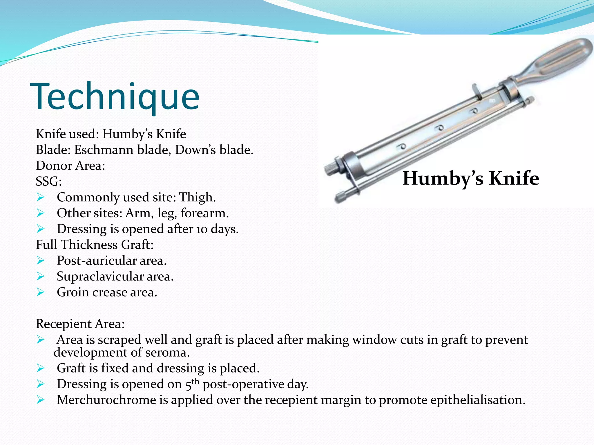
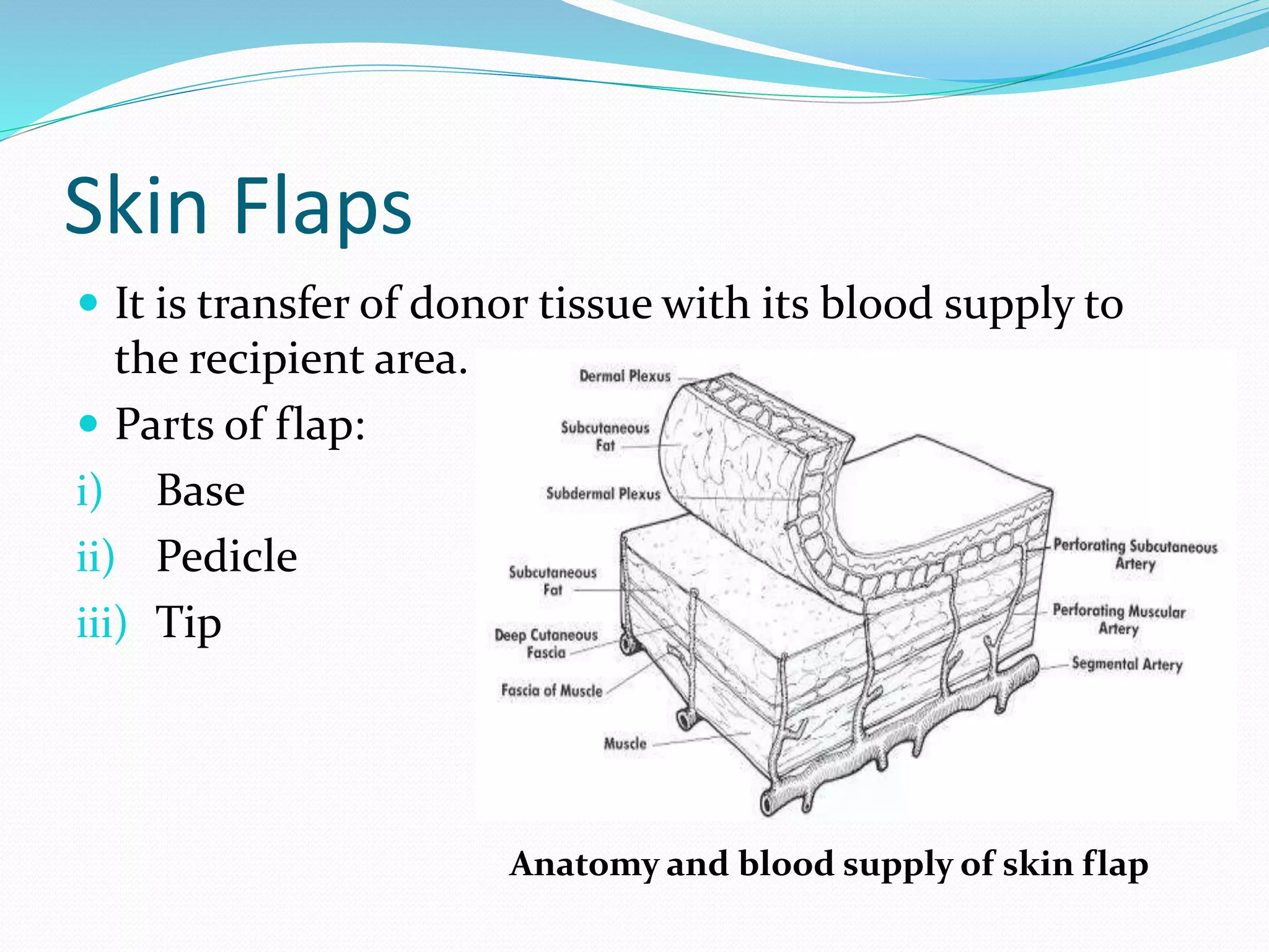
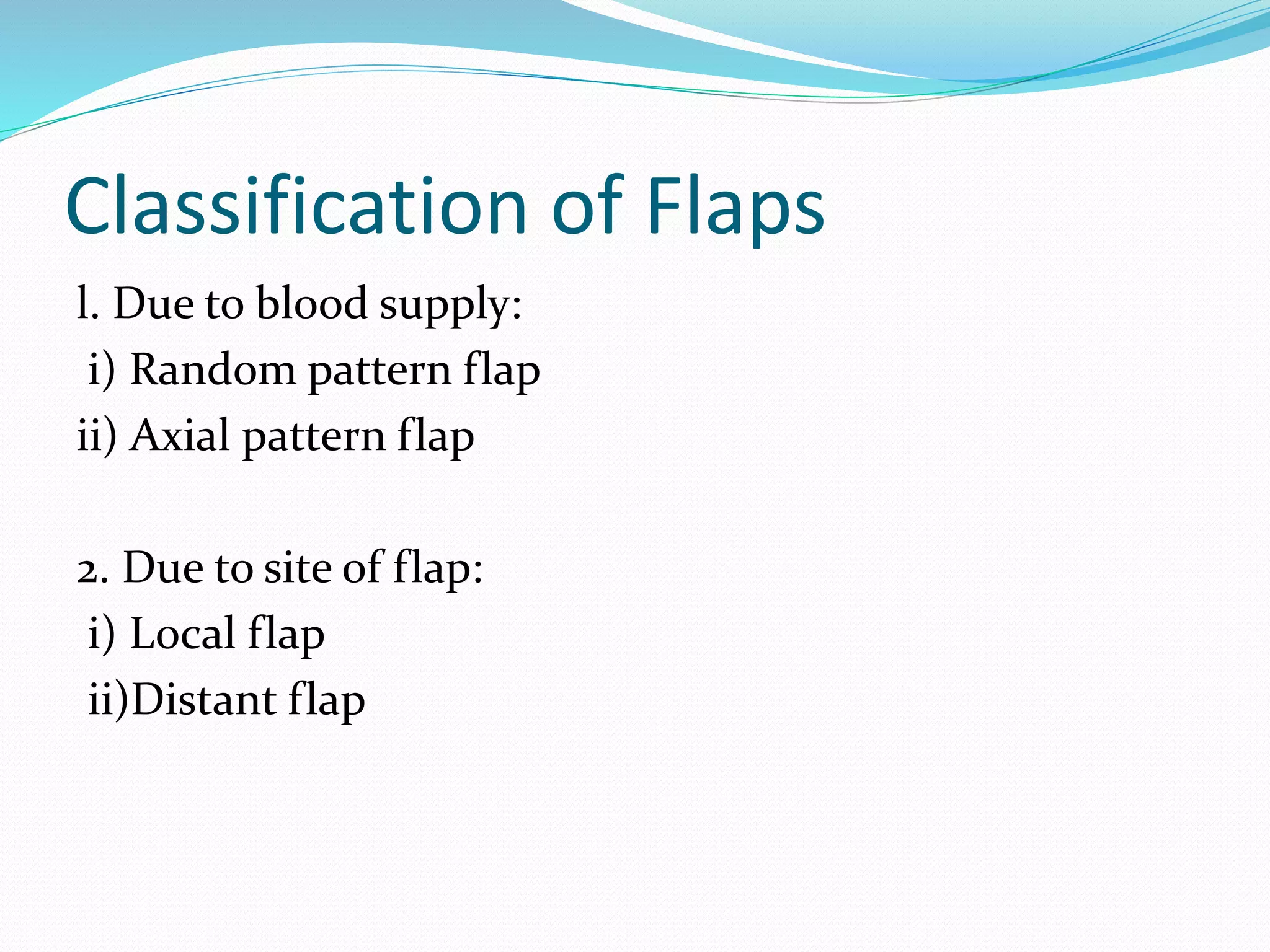
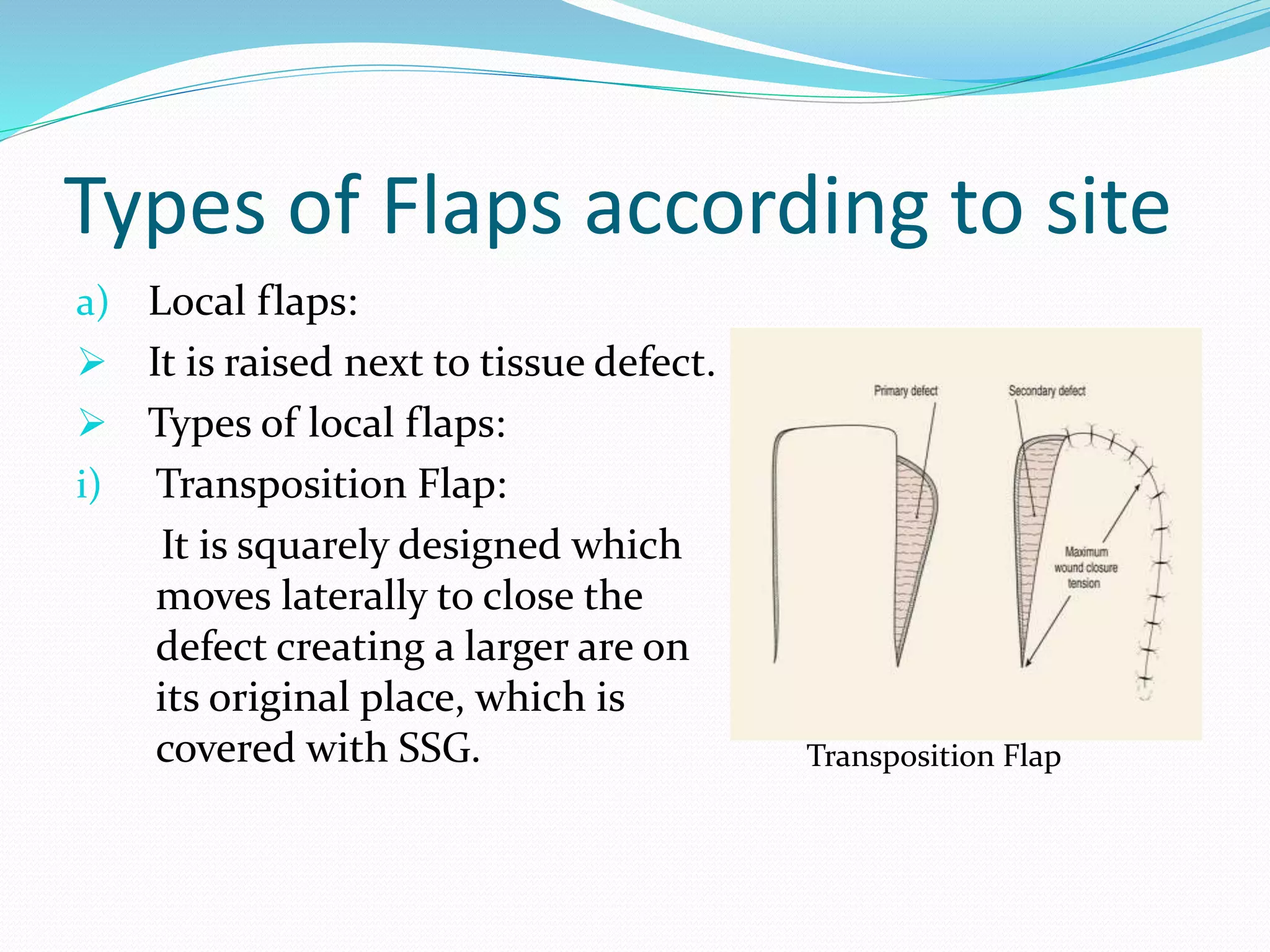
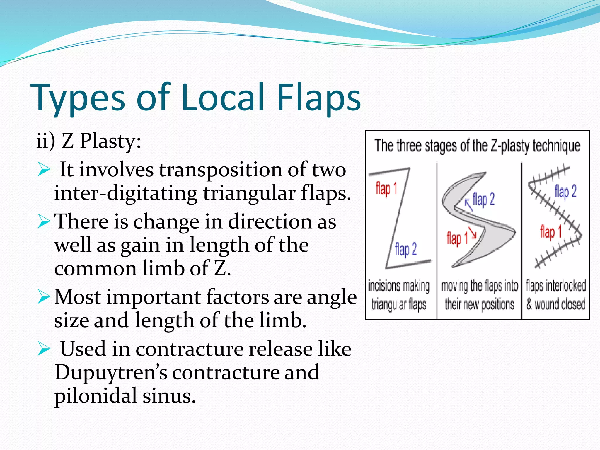
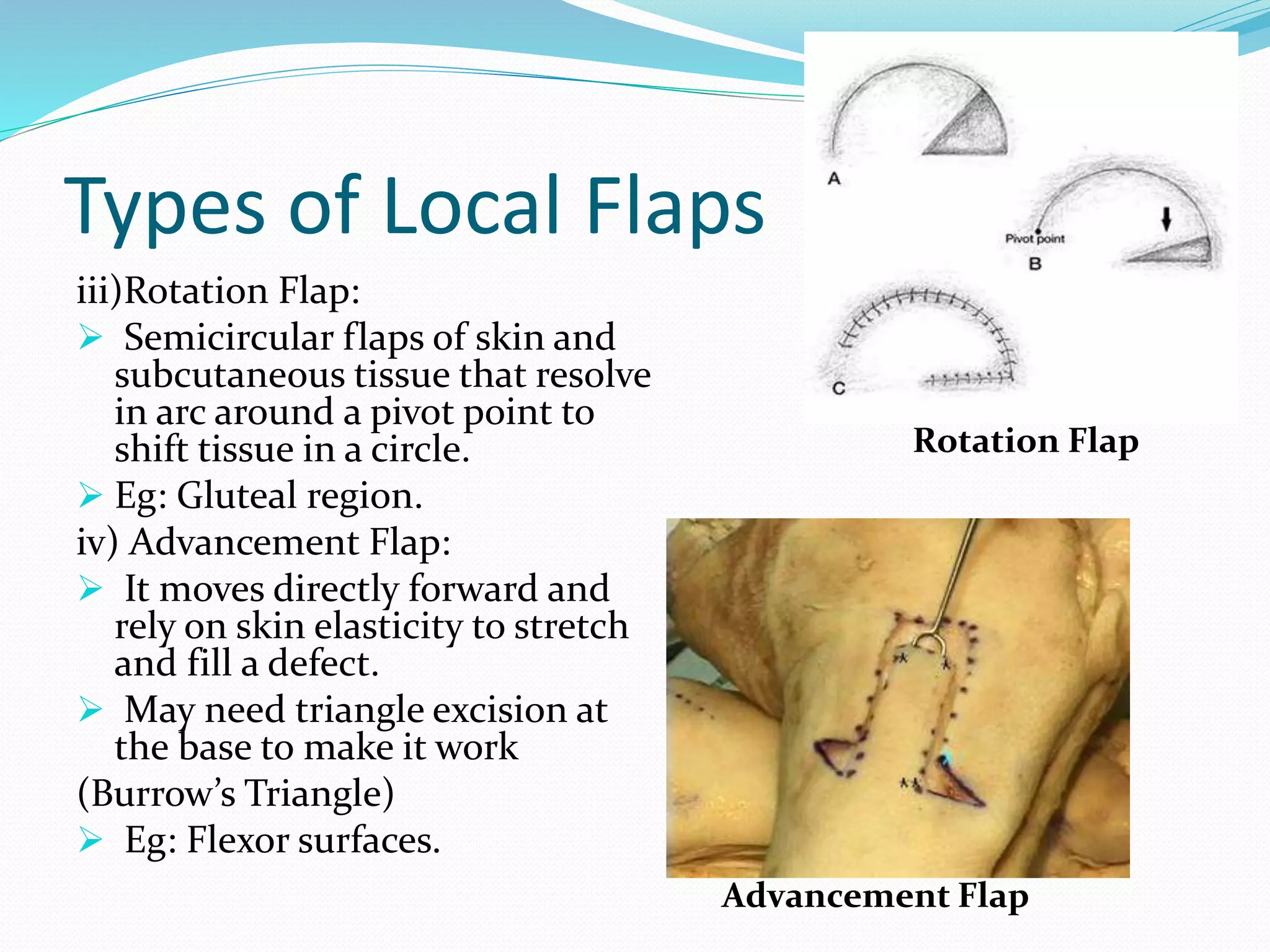
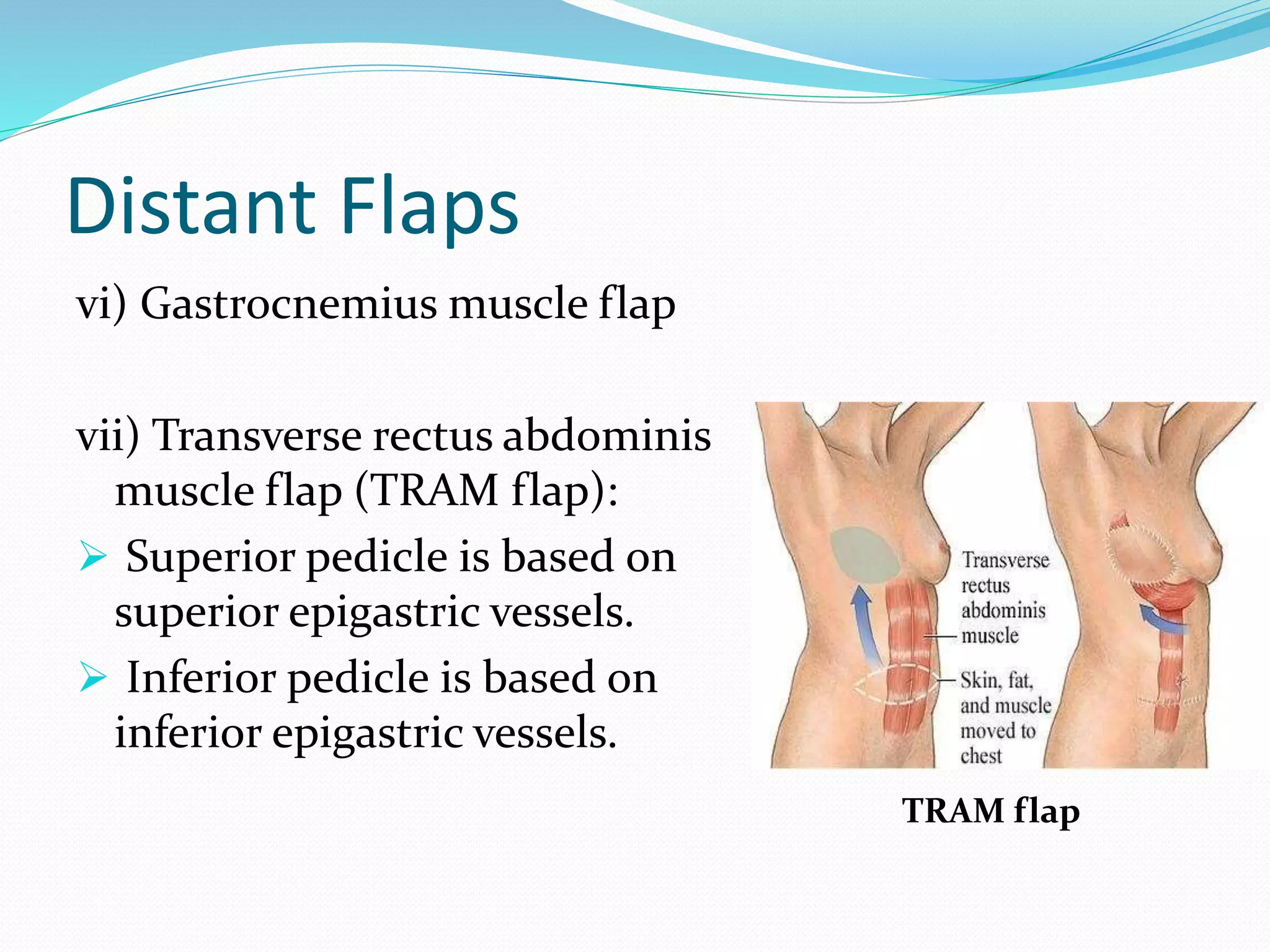
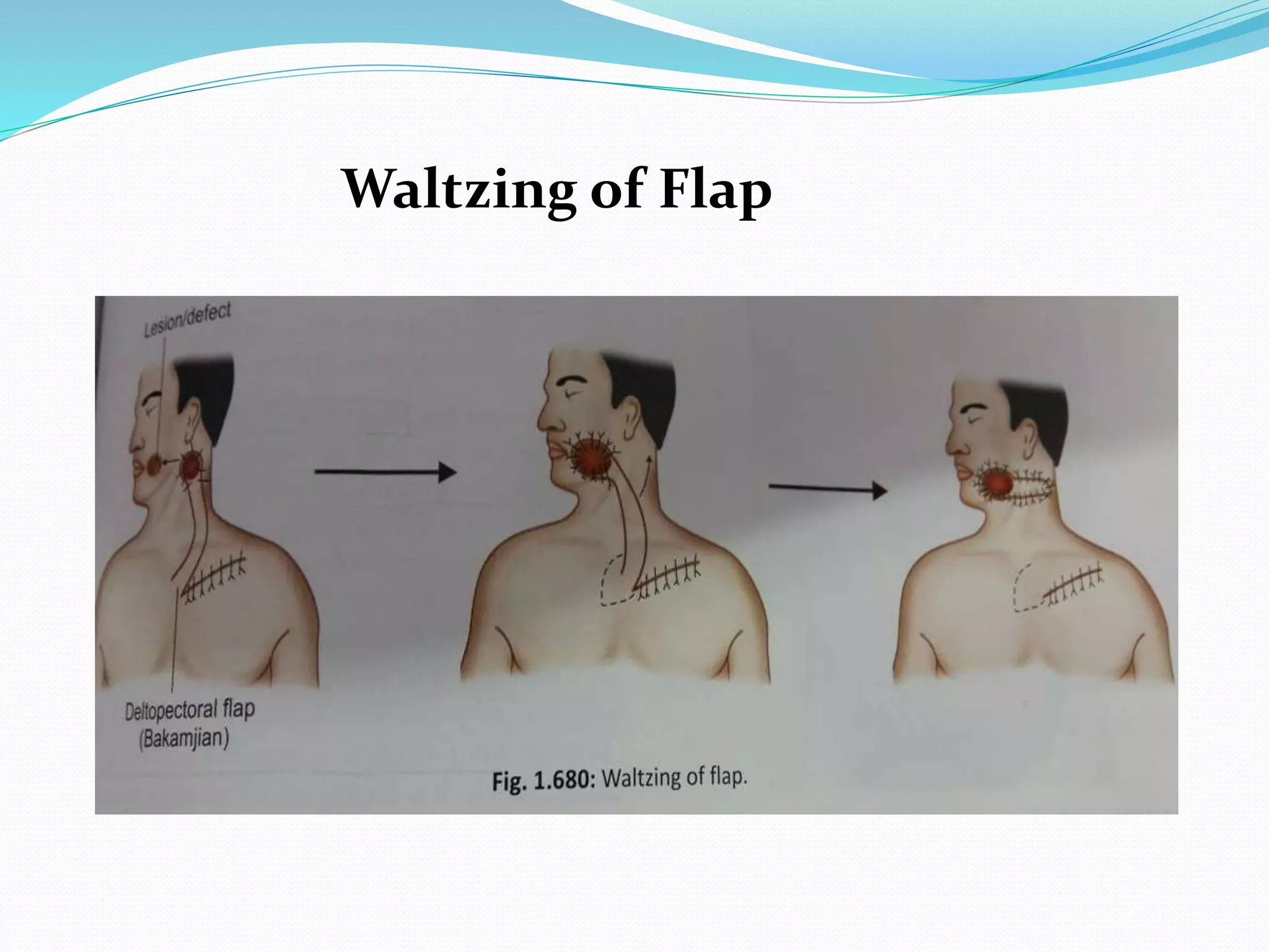
Dr. Ridhika Munjal
Skin
Skin is largest organ of human body and is composed of
two layers, i.e.,
i) Epidermis
ii) Dermis
Skin
 Epidermis:
• Stratified squamous
epithelium composed
primarily of
keratinocytes.
•No blood vessels.
•Relies on diffusion from
underlying tissues.
•Separated from the
dermis by a basement
membrane
Skin
 Dermis:
•Composed of two “sub-layers”:
(a)superficial papillary
(b)deep reticular.
•The dermis contains collagen, capillaries, elastic fibers,
fibroblasts, nerve endings, etc.
Skin Grafting:
Definitions:
 Graft:
It is transfer of tissue from one area to other without it’s blood supply
or nerve supply.
 Autograft:
It is tissue transfer from one location to another on the same patient.
 Isograft:
Tissue transfer between two genetically identical individuals, eg, twins.
 Allograft (Homograft):
Tissue transfer between two genetically different members, eg, kidney
transplant.
 Xenograft (Heterograft):
Tissue transfer from a donor of one species to a recipient of another
species.
Types Of Skin Graft
i) Partial Thickness Graft
ii) Full Thickness Graft
Partial Thickness Graft
 Also called as split thickness graft/ Thiersch graft.
 It is removal of full epidermis plus part of dermis from the donor area.
Advantages:
1. It is technically easier.
2. Graft take up is better.
3. Donor area heals on it’s own.
Disadvantages:
1. Infection
2. Contracture.
3. Loss of hair growth.
4. Seroma and haematoma formation will prevent graft take up.
5. Contraindicated in skin grafting over bone, tendon, cartilage and joint.
6. Can’t be done in group A beta haemolytic streptococcci infection.
Full Thickness Graft
 Also called as Wolfe graft.
 It includes both epidermis + full dermis.
Advantages:
1. Colour match is good.
2. No contracture.
3. Sensation and function of sebaceous gland, hair follicles
retained better.
Disadvantages:
1. Used only for small areas.
2. Wider donor area has to be covered with SSG .
Indications
1. Well granulated ulcer.
2. Clean wound.
3. After surgery to cover and close the defect created.
Stages Of Graft Intake
1. Stage of Plasmic Imbibition:
During 1st 48 hours nourishment of the graft occurs from
plasma exudate from host bed capillaries.
2. Inosculation of blood:
After 48 hours graft and host vessels form anastomosis.
3. Fibroblast Maturation:
 Capillary ingrowth completes the healing by fibroblast
maturation.
 The grafts are securely adhered to bed by 10-14 days.
Technique
Knife used: Humby’s Knife
Blade: Eschmann blade, Down’s blade.
Donor Area:
SSG:
 Commonly used site: Thigh.
 Other sites: Arm, leg, forearm.
 Dressing is opened after 10 days.
Full Thickness Graft:
 Post-auricular area.
 Supraclavicular area.
 Groin crease area.
Recepient Area:
 Area is scraped well and graft is placed after making window cuts in graft to prevent
development of seroma.
 Graft is fixed and dressing is placed.
 Dressing is opened on 5th post-operative day.
 Merchurochrome is applied over the recepient margin to promote epithelialisation.
Humby’s Knife
Technique Of SSG
Donor Site
Skin Flaps
 It is transfer of donor tissue with its blood supply to
the recipient area.
 Parts of flap:
i) Base
ii) Pedicle
iii) Tip
Anatomy and blood supply of skin flap
Indications
i) To cover wider and deeper defects.
ii) To cover bone, tendon and cartilage.
iii) If skin graft repeatedly fails.
Classification of Flaps
l. Due to blood supply:
i) Random pattern flap
ii) Axial pattern flap
2. Due to site of flap:
i) Local flap
ii)Distant flap
Flaps according to blood supply
i)Random Flaps:
These flaps consist of three sides of a rectangle,
bearing no specific relationship to where the blood
supply enters.
The length to breadth ratio is no more than 1.5:1.
ii) Axial Flaps:
These are much longer flaps, based on known blood
vessels.
Types of Flaps according to site
a) Local flaps:
 It is raised next to tissue defect.
 Types of local flaps:
i) Transposition Flap:
It is squarely designed which
moves laterally to close the
defect creating a larger are on
its original place, which is
covered with SSG. Transposition Flap
Types of Local Flaps
ii) Z Plasty:
 It involves transposition of two
inter-digitating triangular flaps.
There is change in direction as
well as gain in length of the
common limb of Z.
Most important factors are angle
size and length of the limb.
 Used in contracture release like
Dupuytren’s contracture and
pilonidal sinus.
Types of Local Flaps
iii)Rotation Flap:
 Semicircular flaps of skin and
subcutaneous tissue that resolve
in arc around a pivot point to
shift tissue in a circle.
 Eg: Gluteal region.
iv) Advancement Flap:
 It moves directly forward and
rely on skin elasticity to stretch
and fill a defect.
 May need triangle excision at
the base to make it work
(Burrow’s Triangle)
 Eg: Flexor surfaces.
Rotation Flap
Advancement Flap
Types Of Local Flaps
v) V-Y advancement flap:
 Advance skin on each side of a V- shaped
incision to close the wound a Y- shaped closure.
 Eg: Cut finger tip.
vi) Y-V advancement flap:
 Used to release multiple band scars over joints.
V-Y advancement Flap
Y-V advancement flap
Types of Local Flaps
vii) Bilobed Flap:
 It uses a flap to close a convex defect
and a second smaller flap to close the
donor site.
 Eg: nasal defects.
viii) Rhomboid Flap:
 It relies on the looseness of adjacent
skin to transfer a rhomboid shape flap
into a defect that has been converted
into similar rhomboid shape.
 Eg: cheek, temple, back and flat
surface defects.
Bilobed Flap
Rhomboid Flap
Local Flaps
Advantages :
 Best local cosmetic tissue match.
 Often a simple procedure.
 Local or regional anesthesia option.
Disadvantages :
 Possible local tissue shortage.
 Scarring may exacerbate the condition.
Types Of Flaps
(b) Distant Flaps:
 To repair defects in which local tissue is inadequate,
distant flaps can be moved on long pedicles that
contain blood supply.
 The pedicle may be buried beneath the skin to create
an island flap or left above the skin and formed into
tube.
Distant Flaps
Types of distant flaps:
i) Forehead flap:
 It is based on anterior branch
of superficial temporal artery.
ii) Deltopectoral flap (Bakamijan
Flap):
 It is based on three perforating
branches of internal mammary
artery.
Forehead flap
Deltopectoral Flap
Distant Flaps
iii) Groin flap:
 It is based on superficial circumflex iliac artery.
iv) Latissimus Dorsi muscle flap:
 It is based on thoracodorsal artery.
v) Pectoralis major flap:
It is based on pectoral branches of thoracoacromial
artery.
Groin Flap
Pectoralis major flap
Distant Flaps
vi) Gastrocnemius muscle flap
vii) Transverse rectus abdominis
muscle flap (TRAM flap):
 Superior pedicle is based on
superior epigastric vessels.
 Inferior pedicle is based on
inferior epigastric vessels.
TRAM flap
Distant Flaps
Advantages:
1. Good blood supply and good take up.
2. Gives bulk, texture and colour to the area.
Diasadvantages:
1. Long term hospitalization.
2. Infection.
3. Kinking, rotation and flap necrosis.
4. Staged procedure.
Saltatory Flap
 It is mobilizing the flaps in stages from distant donor
area towards recipient area.
 Waltzing:
It is a technique wherein flap is moved from donor
area and attached adjacent to recipient defect area.
Later, in 2nd stage, it is moved towards the defect
formally.
It reduces the tension on flap and increases
success rate.
Waltzing of Flap
THANK YOU!!!

Skin grafts and skin flaps

  • 1.
  • 2.
    Skin Skin is largestorgan of human body and is composed of two layers, i.e., i) Epidermis ii) Dermis
  • 3.
    Skin  Epidermis: • Stratifiedsquamous epithelium composed primarily of keratinocytes. •No blood vessels. •Relies on diffusion from underlying tissues. •Separated from the dermis by a basement membrane
  • 4.
    Skin  Dermis: •Composed oftwo “sub-layers”: (a)superficial papillary (b)deep reticular. •The dermis contains collagen, capillaries, elastic fibers, fibroblasts, nerve endings, etc.
  • 5.
    Skin Grafting: Definitions:  Graft: Itis transfer of tissue from one area to other without it’s blood supply or nerve supply.  Autograft: It is tissue transfer from one location to another on the same patient.  Isograft: Tissue transfer between two genetically identical individuals, eg, twins.  Allograft (Homograft): Tissue transfer between two genetically different members, eg, kidney transplant.  Xenograft (Heterograft): Tissue transfer from a donor of one species to a recipient of another species.
  • 6.
    Types Of SkinGraft i) Partial Thickness Graft ii) Full Thickness Graft
  • 7.
    Partial Thickness Graft Also called as split thickness graft/ Thiersch graft.  It is removal of full epidermis plus part of dermis from the donor area. Advantages: 1. It is technically easier. 2. Graft take up is better. 3. Donor area heals on it’s own. Disadvantages: 1. Infection 2. Contracture. 3. Loss of hair growth. 4. Seroma and haematoma formation will prevent graft take up. 5. Contraindicated in skin grafting over bone, tendon, cartilage and joint. 6. Can’t be done in group A beta haemolytic streptococcci infection.
  • 8.
    Full Thickness Graft Also called as Wolfe graft.  It includes both epidermis + full dermis. Advantages: 1. Colour match is good. 2. No contracture. 3. Sensation and function of sebaceous gland, hair follicles retained better. Disadvantages: 1. Used only for small areas. 2. Wider donor area has to be covered with SSG .
  • 9.
    Indications 1. Well granulatedulcer. 2. Clean wound. 3. After surgery to cover and close the defect created.
  • 10.
    Stages Of GraftIntake 1. Stage of Plasmic Imbibition: During 1st 48 hours nourishment of the graft occurs from plasma exudate from host bed capillaries. 2. Inosculation of blood: After 48 hours graft and host vessels form anastomosis. 3. Fibroblast Maturation:  Capillary ingrowth completes the healing by fibroblast maturation.  The grafts are securely adhered to bed by 10-14 days.
  • 11.
    Technique Knife used: Humby’sKnife Blade: Eschmann blade, Down’s blade. Donor Area: SSG:  Commonly used site: Thigh.  Other sites: Arm, leg, forearm.  Dressing is opened after 10 days. Full Thickness Graft:  Post-auricular area.  Supraclavicular area.  Groin crease area. Recepient Area:  Area is scraped well and graft is placed after making window cuts in graft to prevent development of seroma.  Graft is fixed and dressing is placed.  Dressing is opened on 5th post-operative day.  Merchurochrome is applied over the recepient margin to promote epithelialisation. Humby’s Knife
  • 12.
  • 13.
    Skin Flaps  Itis transfer of donor tissue with its blood supply to the recipient area.  Parts of flap: i) Base ii) Pedicle iii) Tip Anatomy and blood supply of skin flap
  • 14.
    Indications i) To coverwider and deeper defects. ii) To cover bone, tendon and cartilage. iii) If skin graft repeatedly fails.
  • 15.
    Classification of Flaps l.Due to blood supply: i) Random pattern flap ii) Axial pattern flap 2. Due to site of flap: i) Local flap ii)Distant flap
  • 16.
    Flaps according toblood supply i)Random Flaps: These flaps consist of three sides of a rectangle, bearing no specific relationship to where the blood supply enters. The length to breadth ratio is no more than 1.5:1. ii) Axial Flaps: These are much longer flaps, based on known blood vessels.
  • 17.
    Types of Flapsaccording to site a) Local flaps:  It is raised next to tissue defect.  Types of local flaps: i) Transposition Flap: It is squarely designed which moves laterally to close the defect creating a larger are on its original place, which is covered with SSG. Transposition Flap
  • 18.
    Types of LocalFlaps ii) Z Plasty:  It involves transposition of two inter-digitating triangular flaps. There is change in direction as well as gain in length of the common limb of Z. Most important factors are angle size and length of the limb.  Used in contracture release like Dupuytren’s contracture and pilonidal sinus.
  • 19.
    Types of LocalFlaps iii)Rotation Flap:  Semicircular flaps of skin and subcutaneous tissue that resolve in arc around a pivot point to shift tissue in a circle.  Eg: Gluteal region. iv) Advancement Flap:  It moves directly forward and rely on skin elasticity to stretch and fill a defect.  May need triangle excision at the base to make it work (Burrow’s Triangle)  Eg: Flexor surfaces. Rotation Flap Advancement Flap
  • 20.
    Types Of LocalFlaps v) V-Y advancement flap:  Advance skin on each side of a V- shaped incision to close the wound a Y- shaped closure.  Eg: Cut finger tip. vi) Y-V advancement flap:  Used to release multiple band scars over joints.
  • 21.
    V-Y advancement Flap Y-Vadvancement flap
  • 22.
    Types of LocalFlaps vii) Bilobed Flap:  It uses a flap to close a convex defect and a second smaller flap to close the donor site.  Eg: nasal defects. viii) Rhomboid Flap:  It relies on the looseness of adjacent skin to transfer a rhomboid shape flap into a defect that has been converted into similar rhomboid shape.  Eg: cheek, temple, back and flat surface defects. Bilobed Flap Rhomboid Flap
  • 23.
    Local Flaps Advantages : Best local cosmetic tissue match.  Often a simple procedure.  Local or regional anesthesia option. Disadvantages :  Possible local tissue shortage.  Scarring may exacerbate the condition.
  • 24.
    Types Of Flaps (b)Distant Flaps:  To repair defects in which local tissue is inadequate, distant flaps can be moved on long pedicles that contain blood supply.  The pedicle may be buried beneath the skin to create an island flap or left above the skin and formed into tube.
  • 25.
    Distant Flaps Types ofdistant flaps: i) Forehead flap:  It is based on anterior branch of superficial temporal artery. ii) Deltopectoral flap (Bakamijan Flap):  It is based on three perforating branches of internal mammary artery. Forehead flap Deltopectoral Flap
  • 26.
    Distant Flaps iii) Groinflap:  It is based on superficial circumflex iliac artery. iv) Latissimus Dorsi muscle flap:  It is based on thoracodorsal artery. v) Pectoralis major flap: It is based on pectoral branches of thoracoacromial artery.
  • 27.
  • 28.
    Distant Flaps vi) Gastrocnemiusmuscle flap vii) Transverse rectus abdominis muscle flap (TRAM flap):  Superior pedicle is based on superior epigastric vessels.  Inferior pedicle is based on inferior epigastric vessels. TRAM flap
  • 29.
    Distant Flaps Advantages: 1. Goodblood supply and good take up. 2. Gives bulk, texture and colour to the area. Diasadvantages: 1. Long term hospitalization. 2. Infection. 3. Kinking, rotation and flap necrosis. 4. Staged procedure.
  • 30.
    Saltatory Flap  Itis mobilizing the flaps in stages from distant donor area towards recipient area.  Waltzing: It is a technique wherein flap is moved from donor area and attached adjacent to recipient defect area. Later, in 2nd stage, it is moved towards the defect formally. It reduces the tension on flap and increases success rate.
  • 31.
  • 32.