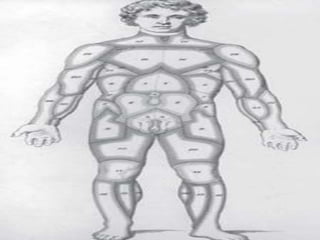
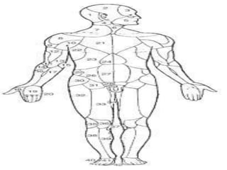
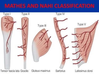
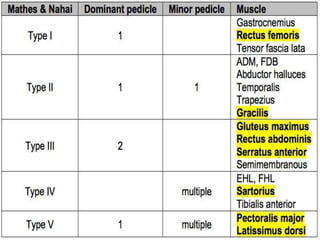
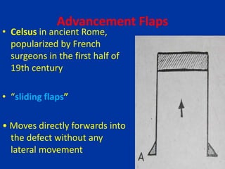
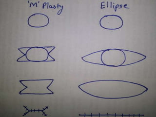
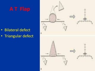
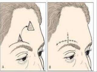
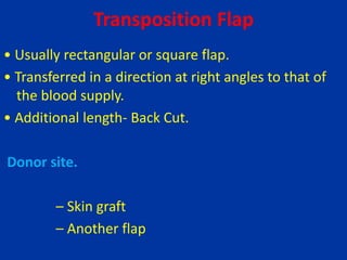
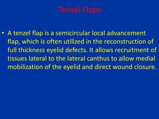
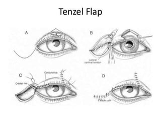
The document discusses various types of skin flaps used in reconstructive surgery. It defines a flap as a unit of tissue transferred from a donor site to a recipient site while maintaining its own blood supply. It then summarizes several types of local flaps used in reconstruction including advancement flaps, V-Y flaps, transposition flaps, and Z-plasties. The document also discusses regional flaps as well as modifications to flaps including flap delay and tissue expansion.
flaps in surgery slideshare
plastic surgery
cosmetic surgery
African experience
NIGERIAN SURGERY
HISTORY OF FLAPS
medicine
medical school
burrows triangle
rotational flaps
transpositional flaps
flaps in surgery slideshare
plastic surgery
cosmetic surgery
African experience
NIGERIAN SURGERY
HISTORY OF FLAPS
medicine
medical school
burrows triangle
rotational flaps
transpositional flaps
FLAPS IN ORAL AND MAXILLOFACIAL SURGERY (monday ppt).pptxaasthamoza
Method and type of flaps used for head and neck reconstruction. Comprising of local regional and free flaps. Indications and pitfalls in each type of flap .
Local & regional flaps /certified fixed orthodontic courses by Indian dental ...Indian dental academy
The Indian Dental Academy is the Leader in continuing dental education , training dentists in all aspects of dentistry and offering a wide range of dental certified courses in different formats.
Indian dental academy provides dental crown & Bridge,rotary endodontics,fixed orthodontics,
Dental implants courses.for details pls visit www.indiandentalacademy.com ,or call
0091-9248678078
Flap coverage in upper extremities in trauma VishalPatil483
SEMINAR PRESENTED BY DR VISHAL PATIL ,IN THE DEPT OF TRAUMA SURGERY AND CRITICAL CARE, AIIMS RISHIKESH
INCLUDES-INTRODUCTION-CLASSIFICATIONS OF FLAP-COMPLICATIONS RELATED TO FLAP COVERAGE- FLAP USED IN HAND AND UPPER EXTREMITY SOFT TISSUE RECONSTRUCTION WITH PICTURES OF IT
New Directions in Targeted Therapeutic Approaches for Older Adults With Mantl...i3 Health
i3 Health is pleased to make the speaker slides from this activity available for use as a non-accredited self-study or teaching resource.
This slide deck presented by Dr. Kami Maddocks, Professor-Clinical in the Division of Hematology and
Associate Division Director for Ambulatory Operations
The Ohio State University Comprehensive Cancer Center, will provide insight into new directions in targeted therapeutic approaches for older adults with mantle cell lymphoma.
STATEMENT OF NEED
Mantle cell lymphoma (MCL) is a rare, aggressive B-cell non-Hodgkin lymphoma (NHL) accounting for 5% to 7% of all lymphomas. Its prognosis ranges from indolent disease that does not require treatment for years to very aggressive disease, which is associated with poor survival (Silkenstedt et al, 2021). Typically, MCL is diagnosed at advanced stage and in older patients who cannot tolerate intensive therapy (NCCN, 2022). Although recent advances have slightly increased remission rates, recurrence and relapse remain very common, leading to a median overall survival between 3 and 6 years (LLS, 2021). Though there are several effective options, progress is still needed towards establishing an accepted frontline approach for MCL (Castellino et al, 2022). Treatment selection and management of MCL are complicated by the heterogeneity of prognosis, advanced age and comorbidities of patients, and lack of an established standard approach for treatment, making it vital that clinicians be familiar with the latest research and advances in this area. In this activity chaired by Michael Wang, MD, Professor in the Department of Lymphoma & Myeloma at MD Anderson Cancer Center, expert faculty will discuss prognostic factors informing treatment, the promising results of recent trials in new therapeutic approaches, and the implications of treatment resistance in therapeutic selection for MCL.
Target Audience
Hematology/oncology fellows, attending faculty, and other health care professionals involved in the treatment of patients with mantle cell lymphoma (MCL).
Learning Objectives
1.) Identify clinical and biological prognostic factors that can guide treatment decision making for older adults with MCL
2.) Evaluate emerging data on targeted therapeutic approaches for treatment-naive and relapsed/refractory MCL and their applicability to older adults
3.) Assess mechanisms of resistance to targeted therapies for MCL and their implications for treatment selection
Ozempic: Preoperative Management of Patients on GLP-1 Receptor Agonists Saeid Safari
Preoperative Management of Patients on GLP-1 Receptor Agonists like Ozempic and Semiglutide
ASA GUIDELINE
NYSORA Guideline
2 Case Reports of Gastric Ultrasound
Pulmonary Thromboembolism - etilogy, types, medical- Surgical and nursing man...VarunMahajani
Disruption of blood supply to lung alveoli due to blockage of one or more pulmonary blood vessels is called as Pulmonary thromboembolism. In this presentation we will discuss its causes, types and its management in depth.
ARTIFICIAL INTELLIGENCE IN HEALTHCARE.pdfAnujkumaranit
Artificial intelligence (AI) refers to the simulation of human intelligence processes by machines, especially computer systems. It encompasses tasks such as learning, reasoning, problem-solving, perception, and language understanding. AI technologies are revolutionizing various fields, from healthcare to finance, by enabling machines to perform tasks that typically require human intelligence.
Ethanol (CH3CH2OH), or beverage alcohol, is a two-carbon alcohol
that is rapidly distributed in the body and brain. Ethanol alters many
neurochemical systems and has rewarding and addictive properties. It
is the oldest recreational drug and likely contributes to more morbidity,
mortality, and public health costs than all illicit drugs combined. The
5th edition of the Diagnostic and Statistical Manual of Mental Disorders
(DSM-5) integrates alcohol abuse and alcohol dependence into a single
disorder called alcohol use disorder (AUD), with mild, moderate,
and severe subclassifications (American Psychiatric Association, 2013).
In the DSM-5, all types of substance abuse and dependence have been
combined into a single substance use disorder (SUD) on a continuum
from mild to severe. A diagnosis of AUD requires that at least two of
the 11 DSM-5 behaviors be present within a 12-month period (mild
AUD: 2–3 criteria; moderate AUD: 4–5 criteria; severe AUD: 6–11 criteria).
The four main behavioral effects of AUD are impaired control over
drinking, negative social consequences, risky use, and altered physiological
effects (tolerance, withdrawal). This chapter presents an overview
of the prevalence and harmful consequences of AUD in the U.S.,
the systemic nature of the disease, neurocircuitry and stages of AUD,
comorbidities, fetal alcohol spectrum disorders, genetic risk factors, and
pharmacotherapies for AUD.
Title: Sense of Taste
Presenter: Dr. Faiza, Assistant Professor of Physiology
Qualifications:
MBBS (Best Graduate, AIMC Lahore)
FCPS Physiology
ICMT, CHPE, DHPE (STMU)
MPH (GC University, Faisalabad)
MBA (Virtual University of Pakistan)
Learning Objectives:
Describe the structure and function of taste buds.
Describe the relationship between the taste threshold and taste index of common substances.
Explain the chemical basis and signal transduction of taste perception for each type of primary taste sensation.
Recognize different abnormalities of taste perception and their causes.
Key Topics:
Significance of Taste Sensation:
Differentiation between pleasant and harmful food
Influence on behavior
Selection of food based on metabolic needs
Receptors of Taste:
Taste buds on the tongue
Influence of sense of smell, texture of food, and pain stimulation (e.g., by pepper)
Primary and Secondary Taste Sensations:
Primary taste sensations: Sweet, Sour, Salty, Bitter, Umami
Chemical basis and signal transduction mechanisms for each taste
Taste Threshold and Index:
Taste threshold values for Sweet (sucrose), Salty (NaCl), Sour (HCl), and Bitter (Quinine)
Taste index relationship: Inversely proportional to taste threshold
Taste Blindness:
Inability to taste certain substances, particularly thiourea compounds
Example: Phenylthiocarbamide
Structure and Function of Taste Buds:
Composition: Epithelial cells, Sustentacular/Supporting cells, Taste cells, Basal cells
Features: Taste pores, Taste hairs/microvilli, and Taste nerve fibers
Location of Taste Buds:
Found in papillae of the tongue (Fungiform, Circumvallate, Foliate)
Also present on the palate, tonsillar pillars, epiglottis, and proximal esophagus
Mechanism of Taste Stimulation:
Interaction of taste substances with receptors on microvilli
Signal transduction pathways for Umami, Sweet, Bitter, Sour, and Salty tastes
Taste Sensitivity and Adaptation:
Decrease in sensitivity with age
Rapid adaptation of taste sensation
Role of Saliva in Taste:
Dissolution of tastants to reach receptors
Washing away the stimulus
Taste Preferences and Aversions:
Mechanisms behind taste preference and aversion
Influence of receptors and neural pathways
Impact of Sensory Nerve Damage:
Degeneration of taste buds if the sensory nerve fiber is cut
Abnormalities of Taste Detection:
Conditions: Ageusia, Hypogeusia, Dysgeusia (parageusia)
Causes: Nerve damage, neurological disorders, infections, poor oral hygiene, adverse drug effects, deficiencies, aging, tobacco use, altered neurotransmitter levels
Neurotransmitters and Taste Threshold:
Effects of serotonin (5-HT) and norepinephrine (NE) on taste sensitivity
Supertasters:
25% of the population with heightened sensitivity to taste, especially bitterness
Increased number of fungiform papillae
These simplified slides by Dr. Sidra Arshad present an overview of the non-respiratory functions of the respiratory tract.
Learning objectives:
1. Enlist the non-respiratory functions of the respiratory tract
2. Briefly explain how these functions are carried out
3. Discuss the significance of dead space
4. Differentiate between minute ventilation and alveolar ventilation
5. Describe the cough and sneeze reflexes
Study Resources:
1. Chapter 39, Guyton and Hall Textbook of Medical Physiology, 14th edition
2. Chapter 34, Ganong’s Review of Medical Physiology, 26th edition
3. Chapter 17, Human Physiology by Lauralee Sherwood, 9th edition
4. Non-respiratory functions of the lungs https://academic.oup.com/bjaed/article/13/3/98/278874
Anti ulcer drugs and their Advance pharmacology ||
Anti-ulcer drugs are medications used to prevent and treat ulcers in the stomach and upper part of the small intestine (duodenal ulcers). These ulcers are often caused by an imbalance between stomach acid and the mucosal lining, which protects the stomach lining.
||Scope: Overview of various classes of anti-ulcer drugs, their mechanisms of action, indications, side effects, and clinical considerations.
3. Definition
A flap is a unit of tissue that is transferred from
donor site to recipient site while maintaining its
own blood supply.
Term “Flap”
Originated from the 16th century Dutch word
“FLAPPE” which means “anything that hung broad
and loose, fastened only by one side
4. Timeline
• 600 BC Sushruta Samhita Pedicle flaps in the face
and forehead for nasal reconstruction
• 1597 Tagliacozzi Nasal reconstruction by tubed
pedicle flap from arm; described “delay” of pedicle
flap
• 1896 Tansini Latissimus dorsi musculocutaneous
flap for breast reconstruction (post- mastectomy)
• 1920 Gillies Tubed pedicle flap
• 1946 Stark Muscle flaps for osteomyelitis
6. • 1972 Orticochea Musculocutaneous flaps
• 1977 Mc Craw et al Musculocutaneous territories
• 1981 Mathes and Nahai Classification of muscle
flaps based on vascular anatomy
• 1981 Ponten described fasciocutaneous flap
8. Carl Manchot (1889 )
• Performed the first examination of the vascular
supply of the human integument.
• 40 cutaneous territories
• His work “Die Hautarterien des menschlichen
Körpers “ [The Cutaneous Arteries of the Human
Body] was initially published in German and later
translated to English by Milton.
9.
10. Spalteholz (1893)
• Published paper on the origin, course and
distribution of the cutaneous perforators in adult
and neonatal cadavers.
• He performed arterial injections of gelatin and
various pigments. The soft tissues were fixed in
alcohol and subtracted in xylol and the resulting
vascular network was embedded in Canada
Balsam.
11. Salmon (1930)
• French anatomist and surgeon charted more than
80 cutaneous territories encompassing the entire
body.
• Salmon dissected 15 human cadavers and took
radiographs of integument which enabled him to
demonstrate much smaller vessel than Manchot
12.
13. • The blood reaching the skin originates from deep
vessels.
• DANIEL AND WILLIAMS(1973) defined that the deep
vessels supplying skin are fundamentally two type
of arteries i.e. either musculocutaneous or direct
cutaneous arteries
• Originally described as Direct cutaneous arteries ,
are now called as septocutaneous arteries.
14. Musculo cutaneous
• Origin: Major vessel
supplying muscle
• Travel perpendicularly
through underlying muscle
into the overlying
cutaneous circulation
• Prevalent supply of skin
covering the broad, flat
muscles of the torso.
• Example: latissimus dorsi
flap, rectus abdominis flap
Septo cutaneous
• Origin: segmental or
muscular artery
• Arise originally from either
segmental or
musculocutaneous vessels,
pass directly within
intermuscular fascial
septae
• common between the
longer, thinner muscles of
the extremitie
• Example :Radial forearm
flap, Dorsalis pedis flap
16. BASED ON LOCATION OF DONOR SITE
LOCAL FLAP
Flap transferred from an area adjacent to the defect.
This is the least complex type of flap and includes
advancement flaps, rotation flaps, and transposition
flaps.
17. REGIONAL FLAP
Regional flaps are not immediately adjacent to the
defect
Instead, the freed tissue "island" is moved over or
underneath normal tissue to reach the defect to be
filled, with the blood supply still connected to the
donor site via a pedicle
Examples PMMC, DP flaps for head and neck defects,
TRAM for breast reconstruction
18. DISTANT FLAP
Flap transferred from an noncontiguous anatomic
site.
The distant flap can be used as a distant pedicled or
free flap
Flapfailure is a severe complication.
19. Based on blood supply
Random .blood supply is not derived from a
recognized artery ,rather, from many little unnamed
vessels.eg local cutaneous flaps
Axial flap, blood supply derived from a recognized
artery or group of arteries eg most muscle flaps
20. Random flaps
Based on the rich sub -
dermal vascular plexus of
the skin
Most of the local flap are
random flaps
Maximum length : breadth
ratio of 1 : 1 in the lower
extremity. length :
breadth ratio of up to 3 :
1 in the face.
Axial flaps
Derive their blood supply from
a direct cutaneous artery or
named blood vessel
Examples : Nasolabial flap
(angular artery) Forehead
flap (supra trochlear artery).
The surviving length of an axial
pattern flap is entirely
related to the length of the
included artery.
23. CLASSIFICATION OF LOCAL FLAPS
• The composition of the defect to be reconstructed
should dictate the correct composition of the flap
used for reconstruction.
It includes :
• 1.Cutaneous
• 2.Fasciocutaneous
• 3.Musculocutaneous
24. Advancement Flaps
• Celsus in ancient Rome,
popularized by French
surgeons in the first half of
19th century
• “sliding flaps”
• Moves directly forwards into
the defect without any
lateral movement
25. Advancement Flaps
• Execution is facilitated by presence of excess
Skin
• More feasible in elderly or when skin elasticity is
more like in very young
• Usually rectangular, perpendicular to the lines of
minimal tension
• Uses – forehead , brow
26. Procedures devised to facilitate advancement
•Excision of Burrow’s
triangle
•Counterincision at the flap
base
•Triangular design
•Curvilinear design
•Z-plasty at the base
29. V-Y Advancement Flap
Design
•Advancement should be
directed over the shortest
diameter of the defect
•The size of the V base should
match the size of the largest
diameter of the lesion
•The V must be long enough
to allow tension free suture
of the Y
30. V-Y Advancement Flap
•Advancement flap involves movement in two planes
vertical and horizontal
•Pivot point on vertical plane which actually acts as a
pivot plane
•Pivot plane is the base of the flap at which the flap is
attached to the body
31. V-Y Advancement Flap
• α angle is determined by
– Location of defect
– Elasticity of the surrounding tissues
– Recommended to range between 20°-40°
• For leg defects, small angle is recommended as
there is less elasticity
• Gluteal region- large angle is planned
34. W Plasty or Zigzag plasty
• Used to break up a
single linear scar
• For scars that do not
require lengthening
• It redistributes tension
along the length of the
scar
35. M-Plasty
• A useful technique to
preserve healthy tissue in
scar revision
• lessen the chance of standing
cone (ie, dog-ear) deformity
• The M-plasty, by creating 2
separate 30° angles instead of
one
36.
37. A T Flap
• Bilateral defect
• Triangular defect
38.
39. Transposition Flap
• Usually rectangular or square flap.
• Transferred in a direction at right angles to that of
the blood supply.
• Additional length- Back Cut.
Donor site.
– Skin graft
– Another flap
40. Transposition Flap- DESIGN
Recipient defect is triangulated
– Right angle triangle
– Hypotenuse- near border of the flap
– The right angle assumes a position opposite the flap
– In scalp defects, apex should direct towards the
periphery of the scalp
45. Dufourmentel Flap
• Designed by a French Surgeon, Claude
Dufourmental in 1962
• The defect is tailored in the shape of a rhombus
• The short diagonal (BD) and one of the adjacent
side (CD) are extended
50. Triple Rhombic flap
• Circular defect conceptualized as hexagon.
• Sides of hexagon are equal to radius (r) of circle.
• First side of flap created by direct extension equal
in length to radius at alternative corners.
• Second side of flap designed parallel to adjacent
side of hexagon.
51.
52. Bilobed Flap
• Consists of two lobes of skin and subcutaneous
tissue based on a common pedicle
Design
– Primary flap is smaller than the defect
– Secondary flap is more triangular in shape
•
53. Optimal angle between the two flaps is 90°, can vary between
45° and 180°; greater the angle, larger the resultant dog-ear
54. Zitelli's modification (1989)
• Primary flap oriented 45°
• And the secondary 90°
• Convert the defect to a "tear drop" shape
• Use a caliper with one tip placed at the apex of the
wound, to mark out two semicircles
55.
56. • Outer semicircle defines the necessary length of the
two lobes
• Inner semicircle bisects the center of the original
wound and continues across the donor skin, defines
the limit of the common pedicle of the two lobes
60. Z Plasty
• Involves transposition of two interdigitating
triangular flaps
• Effects
– Gain in length along the direction of the common
limb of the Z
– Direction of the common limb is changed
• Uses
– Prevention and treatment of contracted scars
– Scar revision
61. •
In 1856, Denonvilliers first described the Z-plasty
technique as a surgical treatment for lower lid
ectropion.
• The first reference to this technique in American
literature was in 1913, by McCurdy, as treatment for
contracture at the oral commissure.
62. • Limberg, in 1929, provided a more detailed
geometric description.
• Numerical data showing optimal angles and length
relationships of Z-plasty limbs are credited to Davis
(1946).
63. • Release of contracture
– The central limb is placed along the line of
contracture- contractural diagonal
– 60° angle taken on each side and limbs of Z drawn,
all equal in size
– Longer diagonal is the transverse diagonal
64.
65.
66.
67. Mechanism of lengthening by Z plasty
– Length of contractural diagonal less than transverse
diagonal before release
– Contractural diagonal lengthens at the expense of
transverse diagonal
– Thus need for transverse skin laxity for contracture
lengthening
– Variables in construction of Z Plasty
• Angle size
• Limb length
69. Angle size
– Increasing the angle beyond 60° will increase
lengthening but also cause increased amount of
transverse shortening.
– Tension produced in the surrounding tissues tend
to be so great that the flaps can not readily be
brought in to their transposed position
70. Limb length
– With almost fixed angle, length provides the major
variable
– Amount of tissue available determines the limb
length
– More is the amount of tissue, larger the length and
vice versa
71. Multiple Z Plasty
• Way of reducing the amount of transverse
shortening without significantly reducing the
amount of lengthening
• Also distributes the lateral tension over various
limbs of multiple Z s
• In place of one large Z plasty, a series of multiple
small Z-plasties are constructed
72.
73.
74.
75. Planning of Z-plasty for contracture
release
• Narrow contractures with lax surrounding skin eg.
Bowstring contracture
• Draw an equilateral triangle on each side of the
contracture and select the more suitable of the two
sets of limbs
– Better blood supply; avoid a flap with scar at base
– Resultant scar falling into a cosmetically favorable
line
76.
77. Use in scar revision
Straight line scar
– Break the continuity of a straight line scar, thus
rendering them more conspicuous
• Bridle scar- scar crossing a hollow
• Curving scar
80. • management of long-standing flexion
contractures of the proximal interphalangeal
joint ...
•
81. Rotation –advancement Flap
• Semicircular flap which rotates around a pivot point
• Located along tension lines
• Flap designed quite large than the defect to ensure
primary closure of the donor site
• skin graft or another flap are alternatives for the
donor site
82. • Tissue can move into anadjacent defect in 2
directions.
• It can advance in a straight line (ie, advancement
flap), or the tissue can rotate into the defect (ie,
rotation flap).
• The distinction between the two is not always clear,
and one type of motion blends into the other .
• Furthermore, a single flap can have both straight
(advancement) movement and rotational (rotation)
movement
83. •Triangulation of the defect, Isoceles triangle made
•Apex towards flap pedicle angle <30° to avoid
buckling of the skin
•PIVOT POINT D- on a projection of line AC, atleast
CD>2AC
•E is located midway between A ,An arc is drawn from
B to D
•CBD constitutes the flap
84.
85. • Conventional rotation flap are successful only in
places where lax skin is present or a back cut is
needed.
• Flap template employs tissue just adjacent to the
triangulated defect thus ensures coverage of the
defect and closure of the donor site
89. • A Mustarde; cheek flap is a large
rotation flap with its leading edge at the
lateral canthus extending laterally and arching
like a Tenzel flap
90. Flag Flap
"Flag flap." Skin moved over distance to palmar
surface or to neighboring digit.
"Flagstaff" contains the pedicle consisting of dorsal
vein, dorsal branch of digital artery, and dorsal
branch of digital nerve
92. Indication
• Soft-tissue defects on the dorsal aspect of fingers
between the metacarpophalangeal and the distal
interphalangeal joint as well as on the tip of the
thumb.
93. Dorsoulnar thumb flap
• Pedicled flap based on the dorsoulnar artery, a
branch off the princeps pollicis artery after it
branches fr Dorsoulnar flap
• Harvested from inner side of thumb
metacarpophalangeal area reaches distal area of
thumb on the radial artery.
• Artery with a distal pedicle.
94.
95. CROSS-FINGER FLAPS
• CROSS-FINGER FLAPS useful for covering a defect of
the skin and other soft tissues on the volar surface
of the finger when tendons and neurovascular
structures are exposed and a small amount of
subcutaneous fat is needed.
96. • They also are useful for some amputations of the
thumb.
• Best avoided in patients over 50 years of age, in
hands with arthritic changes or a tendency to
finger stiffness for some other reason, or if local
infection is present
98. Tenzel Flaps
• A tenzel flap is a semicircular local advancement
flap, which is often utilized in the reconstruction of
full thickness eyelid defects. It allows recruitment of
tissues lateral to the lateral canthus to allow medial
mobilization of the eyelid and direct wound closure.
99. • indicated in the reconstruction of moderate upper
or lower full thickness eyelid defects involving
approximately 1/3 to 1/2 of the eyelid
• most commonly employed in eyelid reconstruction
following resection of a neoplasm at the eyelid
margin or in cases of trauma with full thickness
tissue loss.
100.
101. FLAP MODIFICATION
• Modifications and refinements in both technique
and design of flaps have been used for the optimal
results in reconstructive surgery. Important
modifications are :
• 1. Flap delay.
• 2. Tissue expansion.
102. DELAY PHENOMENON
It can be defined as “ preliminary surgical intervention
wherein a portion of the vascular supply to a flap is
divided before definitive elevation and transfer of
the flap
103. Delay procedure has been used for
several hundred years.
• 16th century : Tagliacozzi delayed his upper arm
flaps
• It was not until the early 1900s that the concept
was recognized.
• 1921 :Blair introduced the term “DELAYED
TRANSFER “
• 1965 : Milton using the pig model, investigated the
effectiveness of four different methods of delaying
a flap .
104. MECHANISM
• Increased axiality of blood flow: Removal of blood
flow from periphery of a random flap promotes
development of axial flow.
• Opening of choke vessels.
• Tolerance to ischemia :adaptive metabolic changes
• Sympathectomy vasodilation theory : leading to
vasodilation
105. FLAP DELAY
1.STANDARD DELAY :
• (A) with an incision at the periphery of the
cutaneous territory.
• (B) partial flap elevation.
106. 2. STRATEGIC DELAY
• Involves division of selected pedicles to the flap to
enhance perfusion through the remaining pedicle
or pedicles .
107. TISSUE EXPANSION
• 1957 : Neumann is credited with the first modern
report of this technique.
• 1976 : Radovan further described the use of this
technique for breast reconstruction.
108. Advantages
1. Reconstruction with tissue of a similar colour and
texture to that of the donor defect.
2. Reconstruction with sensate skin containing skin
appendages.
3. Limited donor-site deformity.
109. • History
Peripheral vascular disease/Coronary artery disease
Collagen vascular disease
Diabetes mellitis
Prior radiotherapy
• Social habits
cigarettes?
• Medications
ASA, anticoagulants
• Cause of defect
recurrence?