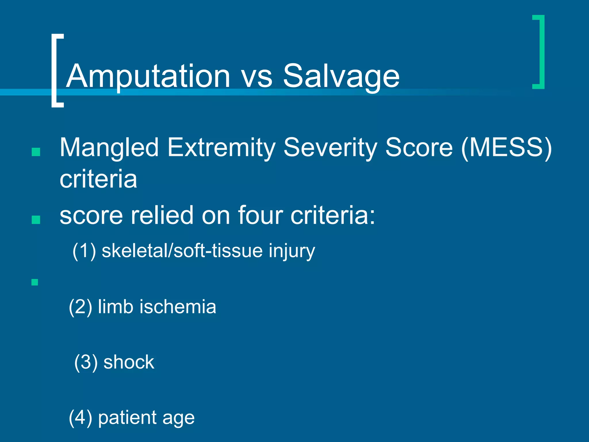
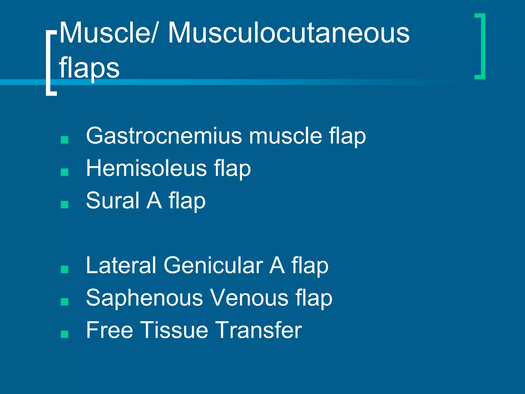
SlideShare a Scribd company logo
Dr Raghav Shrotriya
Department of Plastic Surgery
KEM Hospital, Mumbai
Reconstruction of Upper and
Middle 1/3rd Leg Defects
Heads
■ Introduction
■ Patient evaluation
■ Criteria for amputation
■ Reconstructive Ladder
■ Algorithm for management
■ Options for wound cover
Introduction
■ Most major limb injuries were historically treated with
amputations and the patient was destined to die of infection
■ Pierre Joseph coined the term- “Debridement”.
■ Ollier introduced concept of immobilisation and developed
plaster cast
■ Till the second world war, amputation was still the primary
treatment
■ Refinements and improved knowledge of plastic surgery has
since then helped limb salvage
Types of wounds in the upper
third of the leg
■ Loss of skin/ skin and subcutaneous
tissue with no exposure of bone or
tendon.
■ Loss of skin and subcut with exposure
of periosteum.
■ Loss of skin and soft tissue with
exposure of bone/tendon/nerves
■ Loss of skin and soft tissue with bone
defect
Gustilo and Anderson
Classification
Amputation vs Salvage
■ Mangled Extremity Severity Score (MESS)
criteria
■ score relied on four criteria:
(1) skeletal/soft-tissue injury
■
(2) limb ischemia
(3) shock
(4) patient age
Risk factors for amputation
■ Gustilo IIIC tibial injuries
■ sciatic or tibial nerve injury
■ prolonged ischemia (>4–6 hours)/muscle
necrosis
■ crush or destructive soft-tissue injury
■ significant wound contamination
■ multiple/severely comminuted fractures;
segmental bone loss
■ old age or severe comorbidity
■ apparent futility of revascularization or failed
revascularization.
Goal of Reconstruction
➢ To restore or maintain function
➢ Coverage of defects and open wounds of
leg to give patients a healed wound
➢ To let them resume their life, ambulate, and
go back to work while preventing
amputation
■ The focus in recent times has been –
ideal tissue selection to achieve
optimal functional and cosmetic results
as well as reduced donor site morbidity
Anatomy
Anterior TA, EDL,
EHL, PT
Deep
peroneal
nerve
Ant. tibial
artery
Dorsal Flex,
ext. toes &
invert
lateral PL, PB Superficial
peroneal n.
Peroneal
artery
Flex, eversion
Superficial
posterior
Gast, Plant,
Soleus
Posterior
tibial N.
posterior
Tibial artery
Plant. flex
Deep
posterior
Popletius,
flexors ,
tibialis post.
Posterior
Tibial N.
posterior
Tibial artery
Plantar.
Flexion,
inversion
compartment muscles Nerve artery function
Indications for reconstruction
■ Trauma
■ Osteomyelitis
■ Post cellulitis debridement wound
■ Exposed hard wares
■ Tumors
Patient Evaluation
■ Primary Survey – A B C of trauma
■ Triage
■ Secondary Survey- limb trauma
Assessment of injury
■ History or mechanism of injury
■ Vascular status of the extremity
■ Size of the skin wound
■ Muscle crush or loss
■ Periosteal stripping or bone necrosis
■ Fracture pattern, fragmentation, bone loss
■ Contamination
■ Compartment syndrome
■ Coexisting co-morbid conditions
The Reconstructive Ladder
Concept of Reconstructive
Elevator
Primary management
■ Hemodynamic stability is ensured
■ Vascular status is assessed
■ All the devitalised area and dead bone is
debrided
■ If wound is suitable for cover, it should be
covered with a muscle or fasciocutaneous
flap
■ If there is a lot of contamination and
osteomyelitis is a distinct possibility, then
antibiotic impregnated beads are packed into
the dead space
Primary management
■ X rays to evaluate any fractures or
osteomyelitis
■ Presence of any implants
■ Orientation and position of external
fixators if any
■ Doppler examination if any suspected
arterial trauma
■ Further dressings are done till the
wound is healthy
■ In case of severe contamination,
hyperbaric oxygen therapy which
causes release of nascent oxygen in
the wound thereby killing microbes can
be commenced
Skin and subcutaneous tissue loss
only with no exposure of vital
structures
■ These wounds are usually simple and can
be covered with STSG once they are
ready
■ VAC therapy can be applied to hasten the
granulation and also to reduce the size of
the wound
■ Such wounds are predominantly seen on
the posterior aspect of the leg
Loss of skin and subcut with
exposure of paratenon/ periosteum
■ Such wounds occur on the anterior aspect
of the leg and are not very common
■ If the exposed paratenon or periosteum is
viable and unbreached, interim cover can
be given with STSG
■ However, STSG take may not be very
sound and a muscle flap cover may be
required subsequently
Value of autologous tissue
■ The skin harvested from the degloved or
amputated part can be utilized as biologic
dressings to permanent skin grafts.
■ The leg length can be preserved using soft tissue
distal from the zone of injury as fillet pedicled or
free flaps.
■ Amputated bones can be banked or used as a
flap to reconstruct the leg.
Primary Limb Amputation
■ Absolute indications:
○ anatomically complete disruption of the posterior
tibial nerve in adults
○ crush injuries with warm ischemia time greater than
6 h.
■ Relative indications:
○ serious associated polytrauma,
○ severe ipsilateral foot trauma
○ anticipated protracted course to obtain soft tissue
coverage and tibial reconstruction.
Soft tissue coverage timing
Timing Failure rate Infection rate Bone healing
time
Hospital time
<72 hours 1% 2% 6.8 months 27 days
72 hours-3
weeks
12% 18% 12.3 months 130 days
> 3 weeks 10% 6% 29 months 256 days
How timing of free flap coverage affects outcome in treatment of open fractures, from
Godina
Timing of Reconstruction
■ Early intervention:Lowest complication
rate and highest success rate
associated with closure within the first
72 hrs
■ Minimizes the risk for increasing
bacterial colonization and inflammation
leading to complications.
Factors affecting wound cover timing
■ General condition of the patient and
vascular status of extremity
■ Condition of the wound
■ Bacterial status of the wound
■ Type of fracture, different types of tissues
involved in the injury
■ Presence of exposed structures
Direct Closure
■ Direct closure is simplest and often most effective
means of achieving viable coverage
■ May need to “recruit” more skin to achieve a tension
free closure
■ Decreasing wound tension can be
accomplished by:
○ Relaxing skin incisions
○ Application of negative pressure wound therapy
Soft Tissue Management: Split
Thickness Skin Grafts
■ Exposed muscle or soft tissue
■ Bone or tendon with healthy periosteum /
paratenon
■ IIIA fractures, without exposure of
vessels, nerves or bone
❖ Inadequate for IIIB and IIIC injuries
Indications for Flap Coverage
■ Skin graft cannot be used
○ Exposed cartilage, tendon (without paratenon),
bone, open joints, metal implants
■ Flap coverage is preferable
○ Secondary reconstruction anticipated, flexor joint
surfaces, exposed nerves and vessels, durablitiy
required, multiple tissues required, dead space
present
Flaps used for coverage of
defects over upper and middle
1/3rd leg
■ Transposition flaps
■ Perforator based flaps
■ Local and regional flaps
■ Microvascular free flaps
■ Cross leg flaps
■ Management of bone defects
Local flap – upper and middle third
■ Skin flaps: transposition
■ Fasciocutaneous flap
■ Medial gastrocnemius musculoctaneous
flap
■ Soleus
■ Tibialis anterior
■ Flexor digitorum longus
■ Flexor hallucis longus
■ Extensor digitorum longus
■ Extensor hallucis longus
Sliding Transposition Flap
■ Lateral movement of standard transposition
flap or “bipedicle flap”: transverse tension
across pedicle and embarrassment of its
blood supply
■ Use of sliding transposition flap obviates
transverse tension by insetting distal
oblique edge of flap in to contralateral side
of defect-draping long axis of flap loosely
across convex surface
Flap Design
■ Suitable donor site selected medial or
lateral to defect
■ From lower end of defect , oblique line
is drawn which is the distal edge of
flap (equal in length to contralateral
side of defect)
■ From end of this line , lateral edge of
flap drawn towards the base
■ Width of base should be adequate to
sustain circulation
■ Length to width ratio of 3:2 is usually
adequate
Perforator based flaps
■ Medial and lateral superior genicular arteries supply
the region around the knee, and are a source of
local perforator flaps in knee reconstruction
■ Musculocutaneous perforators from the femoral
artery, as well as several septocutaneous branches
including the descending genicular artery, medial/
lateral superior genicular arteries, medial/lateral
inferior genicular arteries, anterior tibial recurrent
artery, and popliteal artery cutaneous branch
■ The saphenous artery is a superficial branch of
the descending genicular artery, arising medially
with the saphenous nerve and vein
■ Sural arteries (medial/lateral) arise from the
popliteal trunk
■ PTA perforators (4–5 on average) arise in the
intermuscular septum between the soleus and
flexor digitorum longus.
■ ATA perforators that occur in two rows – one
from the tibialis anterior muscle or between the
TA and EHL and the second from the
anteromedial septum between the peroneus
tertius and peroneus brevis
■ The peroneal artery perforator flap is based on
perforators that emerge distally in the
posterolateral septum between the peroneus
longus and soleus muscles
Propeller Flap Concept
■ Local island fasciocutaneous flap
based on a single dissected perforator
which can rotate up to 180
■ Better contour by avoiding dogear and
bulk
Muscle/ Musculocutaneous
flaps
■ Gastrocnemius muscle flap
■ Hemisoleus flap
■ Sural A flap
■ Lateral Genicular A flap
■ Saphenous Venous flap
■ Free Tissue Transfer
Gastrocnemius flaps
Anatomy : Superficial muscle
■ 2 heads arise separately
from lower femur , femoral
condyles and posterior
knee capsule
■ Achilles tendon
■ Action : plantarflexion of
foot and flexion of knee
joint
Vascular Anatomy:
■ Type I
■ Medial sural artery
■ Lateral sural artery
■ L=6cm
■ D=2mm
Operative Technique
■ Incision : Posterior midline or
longitudinal between muscle and
defect
■ Sural N. and Saphenous V. : 2 key
landmarks in midline preserved :
help to locate natural cleavage
between 2 muscle bellies
■ Muscle fascia incised and blunt
dissection used to create plane
between it and underlying soleus
■ Muscle transected distally with a
cuff of tendon which can then be
brought anterior to the tibia through
a subcutaneous tunnel and
insetted
Sural Artery Flap
Saphenous Venous Flap
■ First described by Dr.Thatte and Dr. Wagh
■ Unipedicled venous flap based on the long
saphenous vein
■ Survival of the flap is attributed to perivenous
or perineural capillary network
■ Flap can be used to cover defects of :
➢ Upper third of the leg including posterior surface
➢ Popliteal fossa
Flap Design
■ Rectangular fasciocutaneous
island,designed as a 1:3 proportioned
rectangle with LSV in middle
■ Base of flap : midpoint of leg
■ Lowermost limit : above lower 3rd of leg
■ Medial extension : stops around area
of subcutaneous portion of tibia-3-5cm
from LSV
■ Equal breadth available on
posteromedial surface
■ Base of island consists of LSV with its
adventitia and surrounding areolar
tissue
■ Narrow base : flap mobility in arc from
0 to 170 degrees
Saphenous venous flap
Soleus
■ Muscle or
myocutaneous flap
■ Can be carried 5 cm
proximal to its
insertion
■ Important in venous
pumping mechanism
■ Long term effects on
venous flow not
documented
Anatomy
■ Deep to gastrocnemius muscle
■ Origin: posterior head and body of fibula
■ Insertion: calcaneus through achilles tendon
■ Motor nerve: posterior tibial and medial popliteal nerves
■ Size : 8 x 28 cm
Vascular anatomy
■ Type II 8 x 28 cm
■ Dominant pedicle:
■ Popliteal artery
■ Posterior tibial
artery (Proximal 2
branches)
■ Peroneal artery
(proximal 2
branches )
■ Minor pedicle
3 or 4 segmental
branches of posterior
tibial artery
Arc of rotation
■ Distally based flap
■ Based on minor
pedicles
■ Reverse transposition
■ Covers lower 1/3 of
tibia
Soft Tissue Coverage for the
Middle 1/3 Tibia
▶ Soleus flap
▶ Narrower muscle belly
compared to gastrocs
and a somewhat less
robust vascular supply
▶ Less tolerant of tension
compared to gastrocs
flap so harvesting and
mobilization of muscle
belly can be technically
demanding
Distally based Gracilis flap
Microvascular free tissue transfer
■ Revolutionized the treatment of high-energy
lower extremity injuries
■ Provides excellent vascularised tissue and
most helpful in cases of osteomyelitis
■ Adequate tissue for large defects
■ Well vascularized tissue controls local
bacterial innoculum
■ Delivers oxygen and antibiotic to tissues
■ Promotes soft tissue healing
■ Anterolateral thigh flap and gracilis for
smaller defects are favoured.
Considerations
■ Length of pedicle
■ Contour
■ Easy of harvest without leaving a noticeable
donor deficit (lattisimus dorsi muscle, gracilis
muscle, anterolateral thigh flap)
■ The type of tissue deficiency and volume
■ Donor-site morbidity
■ Recipient-site requirements
■ Vascular pedicle length
■ The anticipated aesthetic result
■ Free Anterolateral Thigh Flap
■ Free Gracilis Muscle Flap
Free ALT flap
Free Gracilis Muscle Flap
Bone defect- options
■ Tibial bone gaps up to 4 cm are ideal for
autogenous cancellous grafting
■ Defects ≥6 cm warrant consideration of
either Ilizarov bone transport or vascularized
bone transfer
■ Bone gaps ≥12 cm :
○ indication for free or pedicled vascularized bone
flaps
Distraction osteogenesis (Ilizarov)
■ Used for bone defects upto 10 cm (ideally upto
6cm)
■ Fracture ends debrided, cortical osteotomy
done away from zone of trauma and distractor
rings placed
■ Distraction begins after 7 days and proceeds at
1 mm per day till defect covered. Frame kept for
1 year
■ Provides strong and anatomically correct bone.
The soft tissue defect can be covered with a
flap
Distraction osteogenesis (Ilizarov)
Free vascularised bone grafts
■ Free fibula flap (segmental and
nutrient supply from peroneal artery)
■ iliac crest vascularized bone flap (deep
circumflex iliac artery)
■ the vascularized scapular flap
(circumflex scapular artery).
Vascularised free fibula graft
■ Used for defects 6cm and above
■ Based on peroneal artery, which supplies
nutrient artery to the fibula
■ Fibula has an excellent capacity for
remoulding and hypertrophy
■ Can be raised along with a soft tissue and
skin pedicle
■ An average of 5 months for partial weight
bearing and 15 months for full weight
bearing required
Cross leg flaps
■ Usually the last resort
■ Axial blood supply by posterior descending
subfascial cutaneous branch of the
popliteal artery
■ Indications:
○ Major lower extremity injury with axial vessels damage
○ History of previous trauma and thrombosis
○ Bone tumour resection with chemotherapy &/or radiotherapy.
■ Contraindications of free flap
○ A single vessel run off, scarcity of local tissue, damaged
recipient vessels and poor general condition.
○ No access to microsurgery
■ Disadvantage: Requires immobilization
of both extremities for extended
periods of time
Compound Flaps
■ Consists of multiple tissue components
linked together in a manner that allows
their simultaneous transfer.
■ These separate components can be
maneuvered and placed in a three-
dimensional manner to achieve an ideal
one-stage reconstruction
Hallock’s classification
Supermicrosurgery
■ Microsurgical anastomosis of vessels, with
a diameter <0.8 mm.
■ Lymphaticovenous shunting to treat
lymphedema
■ relatively a new concept for lower extremity
reconstruction.
■ perforator-to-perforator anastomoses
approach
■ allow an increase in the selection of recipient
pedicles.
■ less time is consumed to secure the recipient
vessel
■ to elevate the flap by taking just a short segment
of the perforator pedicle
■ minimizes any risk for major vessel injury or can
utilize collateral circulation without apparent flow
of major vessels while having acceptable flap
survival
Summary
■ Upper and middle third leg defects are common
problems post traumatic injury
■ Early cover gives best long term results
■ Gastrocnemius flap is the first choice for cover
■ Bone defects are managed either by cancellous
grafts, free fibula flaps or Ilizarow’s technique
■ Osteomyelitis remains the most important
complication
Thank You

More Related Content

What's hot

Nose reconstruction
Nose reconstructionNose reconstruction
Nose reconstruction
zenebe teklu
 
Basic Principles Of Local Flap In Plastic Surgery
Basic Principles Of Local Flap In Plastic SurgeryBasic Principles Of Local Flap In Plastic Surgery
Basic Principles Of Local Flap In Plastic Surgery
Shamendra Sahu
 
Nose reconstruction
Nose reconstructionNose reconstruction
Nose reconstruction
Dr. Suiyibangbe
 
Flaps and grafts in plastic surgery
Flaps and grafts in plastic surgeryFlaps and grafts in plastic surgery
Flaps and grafts in plastic surgery
Sintayehu Asrat
 
Distally-Based Sural Flap
Distally-Based Sural Flap Distally-Based Sural Flap
Distally-Based Sural Flap
Nattakul Yamprasert
 
scope of Pedicled flaps in oral and maxillofacial surgery
scope of Pedicled flaps in oral and maxillofacial surgeryscope of Pedicled flaps in oral and maxillofacial surgery
scope of Pedicled flaps in oral and maxillofacial surgeryAnil Narayanam
 
lip reconstruction
 lip reconstruction lip reconstruction
lip reconstruction
Sumer Yadav
 
Algorithm to approach the lower extremity defect and to select appropriate fl...
Algorithm to approach the lower extremity defect and to select appropriate fl...Algorithm to approach the lower extremity defect and to select appropriate fl...
Algorithm to approach the lower extremity defect and to select appropriate fl...
Binh Phuoc
 
Superficial circumflex iliac artery perforator flap
Superficial circumflex iliac artery perforator flapSuperficial circumflex iliac artery perforator flap
Superficial circumflex iliac artery perforator flap
Dr. Junaid Khurshid
 
Flap-Delay-Phenomena.pptx
Flap-Delay-Phenomena.pptxFlap-Delay-Phenomena.pptx
Flap-Delay-Phenomena.pptx
kiranpoudel12
 
The decline of free flap surgery in lower limb reconstruction
The decline of free flap surgery in lower limb reconstruction The decline of free flap surgery in lower limb reconstruction
The decline of free flap surgery in lower limb reconstruction
Vaikunthan Rajaratnam
 
Flaps and its classification
Flaps and its classificationFlaps and its classification
Flaps and its classification
Dr. Kiran Pandey
 
Free Functional Muscle Transfer
Free Functional Muscle TransferFree Functional Muscle Transfer
Free Functional Muscle Transfer
Dr.Avinash Rao Gundavarapu
 
Perforator flaps
Perforator flapsPerforator flaps
Perforator flaps
Dr.Avinash Rao Gundavarapu
 
Flaps Basics and Important Leg Flaps and Trauma to Leg management
Flaps Basics and Important Leg Flaps and Trauma to Leg managementFlaps Basics and Important Leg Flaps and Trauma to Leg management
Flaps Basics and Important Leg Flaps and Trauma to Leg management
Dr. Hardik Dodia
 
Pollicization
PollicizationPollicization
Types of flaps
Types of flaps Types of flaps
Types of flaps
Yeswanth Mohan
 

What's hot (20)

Perforasome
PerforasomePerforasome
Perforasome
 
Nose reconstruction
Nose reconstructionNose reconstruction
Nose reconstruction
 
Basic Principles Of Local Flap In Plastic Surgery
Basic Principles Of Local Flap In Plastic SurgeryBasic Principles Of Local Flap In Plastic Surgery
Basic Principles Of Local Flap In Plastic Surgery
 
Nose reconstruction
Nose reconstructionNose reconstruction
Nose reconstruction
 
Flaps and grafts in plastic surgery
Flaps and grafts in plastic surgeryFlaps and grafts in plastic surgery
Flaps and grafts in plastic surgery
 
Distally-Based Sural Flap
Distally-Based Sural Flap Distally-Based Sural Flap
Distally-Based Sural Flap
 
Lower limb-flaps
Lower limb-flapsLower limb-flaps
Lower limb-flaps
 
scope of Pedicled flaps in oral and maxillofacial surgery
scope of Pedicled flaps in oral and maxillofacial surgeryscope of Pedicled flaps in oral and maxillofacial surgery
scope of Pedicled flaps in oral and maxillofacial surgery
 
lip reconstruction
 lip reconstruction lip reconstruction
lip reconstruction
 
Algorithm to approach the lower extremity defect and to select appropriate fl...
Algorithm to approach the lower extremity defect and to select appropriate fl...Algorithm to approach the lower extremity defect and to select appropriate fl...
Algorithm to approach the lower extremity defect and to select appropriate fl...
 
Superficial circumflex iliac artery perforator flap
Superficial circumflex iliac artery perforator flapSuperficial circumflex iliac artery perforator flap
Superficial circumflex iliac artery perforator flap
 
Flaps in plastic surgery
Flaps in plastic surgeryFlaps in plastic surgery
Flaps in plastic surgery
 
Flap-Delay-Phenomena.pptx
Flap-Delay-Phenomena.pptxFlap-Delay-Phenomena.pptx
Flap-Delay-Phenomena.pptx
 
The decline of free flap surgery in lower limb reconstruction
The decline of free flap surgery in lower limb reconstruction The decline of free flap surgery in lower limb reconstruction
The decline of free flap surgery in lower limb reconstruction
 
Flaps and its classification
Flaps and its classificationFlaps and its classification
Flaps and its classification
 
Free Functional Muscle Transfer
Free Functional Muscle TransferFree Functional Muscle Transfer
Free Functional Muscle Transfer
 
Perforator flaps
Perforator flapsPerforator flaps
Perforator flaps
 
Flaps Basics and Important Leg Flaps and Trauma to Leg management
Flaps Basics and Important Leg Flaps and Trauma to Leg managementFlaps Basics and Important Leg Flaps and Trauma to Leg management
Flaps Basics and Important Leg Flaps and Trauma to Leg management
 
Pollicization
PollicizationPollicization
Pollicization
 
Types of flaps
Types of flaps Types of flaps
Types of flaps
 

Similar to Uper n middle third leg defects

amputation
amputationamputation
amputation
Nikhil Drolia
 
Mangled extremity
Mangled extremityMangled extremity
Mangled extremity
Rohit Vikas
 
CONSERVATIVE MANAGEMENT OF FRACTURE
CONSERVATIVE MANAGEMENT OF FRACTURECONSERVATIVE MANAGEMENT OF FRACTURE
CONSERVATIVE MANAGEMENT OF FRACTURE
Naveed Jumani
 
Free LD flap for scalp reconstruction DR VIPIN V NAIR
Free LD flap for scalp  reconstruction DR VIPIN V NAIRFree LD flap for scalp  reconstruction DR VIPIN V NAIR
Free LD flap for scalp reconstruction DR VIPIN V NAIR
PGIMER Chandigarh
 
PMMC FLAP
PMMC FLAPPMMC FLAP
PMMC FLAP
Syed Mohammed
 
Plastic and reconstructive surgery presentation.pptx
Plastic and reconstructive surgery presentation.pptxPlastic and reconstructive surgery presentation.pptx
Plastic and reconstructive surgery presentation.pptx
All India Institute of Medical Sciences, Bhopal
 
G15 soft tissue coverage
G15 soft tissue coverageG15 soft tissue coverage
G15 soft tissue coverage
Claudiu Cucu
 
Amputation stump
Amputation stumpAmputation stump
Amputation stump
PratikDhabalia
 
Mandibular reconstruction
Mandibular reconstructionMandibular reconstruction
Mandibular reconstruction
Akashah Ambar
 
Amputation and disarticulation
Amputation and disarticulationAmputation and disarticulation
Amputation and disarticulation
Yash Oza
 
Mangled extremity and it’s management
Mangled    extremity          and   it’s managementMangled    extremity          and   it’s management
Mangled extremity and it’s management
Shorifuddin Ahmed
 
Acetabulum ant approaches
Acetabulum ant approachesAcetabulum ant approaches
Acetabulum ant approaches
Parthasarathy Suyambu
 
Fractures.pptx
Fractures.pptxFractures.pptx
Fractures.pptx
JaneBwalya1
 
amputation prof dr seif.ppt
amputation prof dr seif.pptamputation prof dr seif.ppt
amputation prof dr seif.ppt
abdullah520979
 
Principles Of Amputation
Principles Of  AmputationPrinciples Of  Amputation
Principles Of Amputation
Joe Antony
 
amputations.pptx
amputations.pptxamputations.pptx
amputations.pptx
MubashirHussan2
 
Hand injury
Hand injuryHand injury
Hand injury
Salihi Abdulmalik
 
Flaps (2).pptx
Flaps (2).pptxFlaps (2).pptx
Flaps (2).pptx
RajSwaroob3
 
amputations pg.pptx
amputations pg.pptxamputations pg.pptx
amputations pg.pptx
Arya RaveendranNair
 
Pedicled flaps in head and neck surgery
Pedicled flaps in head and neck surgeryPedicled flaps in head and neck surgery
Pedicled flaps in head and neck surgery
Ram Raju
 

Similar to Uper n middle third leg defects (20)

amputation
amputationamputation
amputation
 
Mangled extremity
Mangled extremityMangled extremity
Mangled extremity
 
CONSERVATIVE MANAGEMENT OF FRACTURE
CONSERVATIVE MANAGEMENT OF FRACTURECONSERVATIVE MANAGEMENT OF FRACTURE
CONSERVATIVE MANAGEMENT OF FRACTURE
 
Free LD flap for scalp reconstruction DR VIPIN V NAIR
Free LD flap for scalp  reconstruction DR VIPIN V NAIRFree LD flap for scalp  reconstruction DR VIPIN V NAIR
Free LD flap for scalp reconstruction DR VIPIN V NAIR
 
PMMC FLAP
PMMC FLAPPMMC FLAP
PMMC FLAP
 
Plastic and reconstructive surgery presentation.pptx
Plastic and reconstructive surgery presentation.pptxPlastic and reconstructive surgery presentation.pptx
Plastic and reconstructive surgery presentation.pptx
 
G15 soft tissue coverage
G15 soft tissue coverageG15 soft tissue coverage
G15 soft tissue coverage
 
Amputation stump
Amputation stumpAmputation stump
Amputation stump
 
Mandibular reconstruction
Mandibular reconstructionMandibular reconstruction
Mandibular reconstruction
 
Amputation and disarticulation
Amputation and disarticulationAmputation and disarticulation
Amputation and disarticulation
 
Mangled extremity and it’s management
Mangled    extremity          and   it’s managementMangled    extremity          and   it’s management
Mangled extremity and it’s management
 
Acetabulum ant approaches
Acetabulum ant approachesAcetabulum ant approaches
Acetabulum ant approaches
 
Fractures.pptx
Fractures.pptxFractures.pptx
Fractures.pptx
 
amputation prof dr seif.ppt
amputation prof dr seif.pptamputation prof dr seif.ppt
amputation prof dr seif.ppt
 
Principles Of Amputation
Principles Of  AmputationPrinciples Of  Amputation
Principles Of Amputation
 
amputations.pptx
amputations.pptxamputations.pptx
amputations.pptx
 
Hand injury
Hand injuryHand injury
Hand injury
 
Flaps (2).pptx
Flaps (2).pptxFlaps (2).pptx
Flaps (2).pptx
 
amputations pg.pptx
amputations pg.pptxamputations pg.pptx
amputations pg.pptx
 
Pedicled flaps in head and neck surgery
Pedicled flaps in head and neck surgeryPedicled flaps in head and neck surgery
Pedicled flaps in head and neck surgery
 

More from Raghav Shrotriya

Trophic ulcers
Trophic ulcersTrophic ulcers
Trophic ulcers
Raghav Shrotriya
 
Rehabilitation of Upper Limb Amputee
Rehabilitation of Upper Limb AmputeeRehabilitation of Upper Limb Amputee
Rehabilitation of Upper Limb Amputee
Raghav Shrotriya
 
Obstetric brachial plexus injury...ppt
Obstetric brachial plexus injury...pptObstetric brachial plexus injury...ppt
Obstetric brachial plexus injury...ppt
Raghav Shrotriya
 
Biogeometry of flaps.ppt
Biogeometry of flaps.pptBiogeometry of flaps.ppt
Biogeometry of flaps.ppt
Raghav Shrotriya
 
Paralysis of facial nerve
Paralysis of facial nerveParalysis of facial nerve
Paralysis of facial nerve
Raghav Shrotriya
 
Skin substitutes
Skin substitutesSkin substitutes
Skin substitutes
Raghav Shrotriya
 

More from Raghav Shrotriya (6)

Trophic ulcers
Trophic ulcersTrophic ulcers
Trophic ulcers
 
Rehabilitation of Upper Limb Amputee
Rehabilitation of Upper Limb AmputeeRehabilitation of Upper Limb Amputee
Rehabilitation of Upper Limb Amputee
 
Obstetric brachial plexus injury...ppt
Obstetric brachial plexus injury...pptObstetric brachial plexus injury...ppt
Obstetric brachial plexus injury...ppt
 
Biogeometry of flaps.ppt
Biogeometry of flaps.pptBiogeometry of flaps.ppt
Biogeometry of flaps.ppt
 
Paralysis of facial nerve
Paralysis of facial nerveParalysis of facial nerve
Paralysis of facial nerve
 
Skin substitutes
Skin substitutesSkin substitutes
Skin substitutes
 

Recently uploaded

Are There Any Natural Remedies To Treat Syphilis.pdf
Are There Any Natural Remedies To Treat Syphilis.pdfAre There Any Natural Remedies To Treat Syphilis.pdf
Are There Any Natural Remedies To Treat Syphilis.pdf
Little Cross Family Clinic
 
Knee anatomy and clinical tests 2024.pdf
Knee anatomy and clinical tests 2024.pdfKnee anatomy and clinical tests 2024.pdf
Knee anatomy and clinical tests 2024.pdf
vimalpl1234
 
Maxilla, Mandible & Hyoid Bone & Clinical Correlations by Dr. RIG.pptx
Maxilla, Mandible & Hyoid Bone & Clinical Correlations by Dr. RIG.pptxMaxilla, Mandible & Hyoid Bone & Clinical Correlations by Dr. RIG.pptx
Maxilla, Mandible & Hyoid Bone & Clinical Correlations by Dr. RIG.pptx
Dr. Rabia Inam Gandapore
 
BENIGN PROSTATIC HYPERPLASIA.BPH. BPHpdf
BENIGN PROSTATIC HYPERPLASIA.BPH. BPHpdfBENIGN PROSTATIC HYPERPLASIA.BPH. BPHpdf
BENIGN PROSTATIC HYPERPLASIA.BPH. BPHpdf
DR SETH JOTHAM
 
Tom Selleck Health: A Comprehensive Look at the Iconic Actor’s Wellness Journey
Tom Selleck Health: A Comprehensive Look at the Iconic Actor’s Wellness JourneyTom Selleck Health: A Comprehensive Look at the Iconic Actor’s Wellness Journey
Tom Selleck Health: A Comprehensive Look at the Iconic Actor’s Wellness Journey
greendigital
 
Alcohol_Dr. Jeenal Mistry MD Pharmacology.pdf
Alcohol_Dr. Jeenal Mistry MD Pharmacology.pdfAlcohol_Dr. Jeenal Mistry MD Pharmacology.pdf
Alcohol_Dr. Jeenal Mistry MD Pharmacology.pdf
Dr Jeenal Mistry
 
ARTHROLOGY PPT NCISM SYLLABUS AYURVEDA STUDENTS
ARTHROLOGY PPT NCISM SYLLABUS AYURVEDA STUDENTSARTHROLOGY PPT NCISM SYLLABUS AYURVEDA STUDENTS
ARTHROLOGY PPT NCISM SYLLABUS AYURVEDA STUDENTS
Dr. Vinay Pareek
 
Non-respiratory Functions of the Lungs.pdf
Non-respiratory Functions of the Lungs.pdfNon-respiratory Functions of the Lungs.pdf
Non-respiratory Functions of the Lungs.pdf
MedicoseAcademics
 
Novas diretrizes da OMS para os cuidados perinatais de mais qualidade
Novas diretrizes da OMS para os cuidados perinatais de mais qualidadeNovas diretrizes da OMS para os cuidados perinatais de mais qualidade
Novas diretrizes da OMS para os cuidados perinatais de mais qualidade
Prof. Marcus Renato de Carvalho
 
basicmodesofventilation2022-220313203758.pdf
basicmodesofventilation2022-220313203758.pdfbasicmodesofventilation2022-220313203758.pdf
basicmodesofventilation2022-220313203758.pdf
aljamhori teaching hospital
 
Pharynx and Clinical Correlations BY Dr.Rabia Inam Gandapore.pptx
Pharynx and Clinical Correlations BY Dr.Rabia Inam Gandapore.pptxPharynx and Clinical Correlations BY Dr.Rabia Inam Gandapore.pptx
Pharynx and Clinical Correlations BY Dr.Rabia Inam Gandapore.pptx
Dr. Rabia Inam Gandapore
 
ANATOMY AND PHYSIOLOGY OF URINARY SYSTEM.pptx
ANATOMY AND PHYSIOLOGY OF URINARY SYSTEM.pptxANATOMY AND PHYSIOLOGY OF URINARY SYSTEM.pptx
ANATOMY AND PHYSIOLOGY OF URINARY SYSTEM.pptx
Swetaba Besh
 
HOT NEW PRODUCT! BIG SALES FAST SHIPPING NOW FROM CHINA!! EU KU DB BK substit...
HOT NEW PRODUCT! BIG SALES FAST SHIPPING NOW FROM CHINA!! EU KU DB BK substit...HOT NEW PRODUCT! BIG SALES FAST SHIPPING NOW FROM CHINA!! EU KU DB BK substit...
HOT NEW PRODUCT! BIG SALES FAST SHIPPING NOW FROM CHINA!! EU KU DB BK substit...
GL Anaacs
 
The Normal Electrocardiogram - Part I of II
The Normal Electrocardiogram - Part I of IIThe Normal Electrocardiogram - Part I of II
The Normal Electrocardiogram - Part I of II
MedicoseAcademics
 
ACUTE SCROTUM.....pdf. ACUTE SCROTAL CONDITIOND
ACUTE SCROTUM.....pdf. ACUTE SCROTAL CONDITIONDACUTE SCROTUM.....pdf. ACUTE SCROTAL CONDITIOND
ACUTE SCROTUM.....pdf. ACUTE SCROTAL CONDITIOND
DR SETH JOTHAM
 
Cervical & Brachial Plexus By Dr. RIG.pptx
Cervical & Brachial Plexus By Dr. RIG.pptxCervical & Brachial Plexus By Dr. RIG.pptx
Cervical & Brachial Plexus By Dr. RIG.pptx
Dr. Rabia Inam Gandapore
 
Surat @ℂall @Girls ꧁❤8527049040❤꧂@ℂall @Girls Service Vip Top Model Safe
Surat @ℂall @Girls ꧁❤8527049040❤꧂@ℂall @Girls Service Vip Top Model SafeSurat @ℂall @Girls ꧁❤8527049040❤꧂@ℂall @Girls Service Vip Top Model Safe
Surat @ℂall @Girls ꧁❤8527049040❤꧂@ℂall @Girls Service Vip Top Model Safe
Savita Shen $i11
 
New Directions in Targeted Therapeutic Approaches for Older Adults With Mantl...
New Directions in Targeted Therapeutic Approaches for Older Adults With Mantl...New Directions in Targeted Therapeutic Approaches for Older Adults With Mantl...
New Directions in Targeted Therapeutic Approaches for Older Adults With Mantl...
i3 Health
 
Ozempic: Preoperative Management of Patients on GLP-1 Receptor Agonists
Ozempic: Preoperative Management of Patients on GLP-1 Receptor Agonists  Ozempic: Preoperative Management of Patients on GLP-1 Receptor Agonists
Ozempic: Preoperative Management of Patients on GLP-1 Receptor Agonists
Saeid Safari
 
Triangles of Neck and Clinical Correlation by Dr. RIG.pptx
Triangles of Neck and Clinical Correlation by Dr. RIG.pptxTriangles of Neck and Clinical Correlation by Dr. RIG.pptx
Triangles of Neck and Clinical Correlation by Dr. RIG.pptx
Dr. Rabia Inam Gandapore
 

Recently uploaded (20)

Are There Any Natural Remedies To Treat Syphilis.pdf
Are There Any Natural Remedies To Treat Syphilis.pdfAre There Any Natural Remedies To Treat Syphilis.pdf
Are There Any Natural Remedies To Treat Syphilis.pdf
 
Knee anatomy and clinical tests 2024.pdf
Knee anatomy and clinical tests 2024.pdfKnee anatomy and clinical tests 2024.pdf
Knee anatomy and clinical tests 2024.pdf
 
Maxilla, Mandible & Hyoid Bone & Clinical Correlations by Dr. RIG.pptx
Maxilla, Mandible & Hyoid Bone & Clinical Correlations by Dr. RIG.pptxMaxilla, Mandible & Hyoid Bone & Clinical Correlations by Dr. RIG.pptx
Maxilla, Mandible & Hyoid Bone & Clinical Correlations by Dr. RIG.pptx
 
BENIGN PROSTATIC HYPERPLASIA.BPH. BPHpdf
BENIGN PROSTATIC HYPERPLASIA.BPH. BPHpdfBENIGN PROSTATIC HYPERPLASIA.BPH. BPHpdf
BENIGN PROSTATIC HYPERPLASIA.BPH. BPHpdf
 
Tom Selleck Health: A Comprehensive Look at the Iconic Actor’s Wellness Journey
Tom Selleck Health: A Comprehensive Look at the Iconic Actor’s Wellness JourneyTom Selleck Health: A Comprehensive Look at the Iconic Actor’s Wellness Journey
Tom Selleck Health: A Comprehensive Look at the Iconic Actor’s Wellness Journey
 
Alcohol_Dr. Jeenal Mistry MD Pharmacology.pdf
Alcohol_Dr. Jeenal Mistry MD Pharmacology.pdfAlcohol_Dr. Jeenal Mistry MD Pharmacology.pdf
Alcohol_Dr. Jeenal Mistry MD Pharmacology.pdf
 
ARTHROLOGY PPT NCISM SYLLABUS AYURVEDA STUDENTS
ARTHROLOGY PPT NCISM SYLLABUS AYURVEDA STUDENTSARTHROLOGY PPT NCISM SYLLABUS AYURVEDA STUDENTS
ARTHROLOGY PPT NCISM SYLLABUS AYURVEDA STUDENTS
 
Non-respiratory Functions of the Lungs.pdf
Non-respiratory Functions of the Lungs.pdfNon-respiratory Functions of the Lungs.pdf
Non-respiratory Functions of the Lungs.pdf
 
Novas diretrizes da OMS para os cuidados perinatais de mais qualidade
Novas diretrizes da OMS para os cuidados perinatais de mais qualidadeNovas diretrizes da OMS para os cuidados perinatais de mais qualidade
Novas diretrizes da OMS para os cuidados perinatais de mais qualidade
 
basicmodesofventilation2022-220313203758.pdf
basicmodesofventilation2022-220313203758.pdfbasicmodesofventilation2022-220313203758.pdf
basicmodesofventilation2022-220313203758.pdf
 
Pharynx and Clinical Correlations BY Dr.Rabia Inam Gandapore.pptx
Pharynx and Clinical Correlations BY Dr.Rabia Inam Gandapore.pptxPharynx and Clinical Correlations BY Dr.Rabia Inam Gandapore.pptx
Pharynx and Clinical Correlations BY Dr.Rabia Inam Gandapore.pptx
 
ANATOMY AND PHYSIOLOGY OF URINARY SYSTEM.pptx
ANATOMY AND PHYSIOLOGY OF URINARY SYSTEM.pptxANATOMY AND PHYSIOLOGY OF URINARY SYSTEM.pptx
ANATOMY AND PHYSIOLOGY OF URINARY SYSTEM.pptx
 
HOT NEW PRODUCT! BIG SALES FAST SHIPPING NOW FROM CHINA!! EU KU DB BK substit...
HOT NEW PRODUCT! BIG SALES FAST SHIPPING NOW FROM CHINA!! EU KU DB BK substit...HOT NEW PRODUCT! BIG SALES FAST SHIPPING NOW FROM CHINA!! EU KU DB BK substit...
HOT NEW PRODUCT! BIG SALES FAST SHIPPING NOW FROM CHINA!! EU KU DB BK substit...
 
The Normal Electrocardiogram - Part I of II
The Normal Electrocardiogram - Part I of IIThe Normal Electrocardiogram - Part I of II
The Normal Electrocardiogram - Part I of II
 
ACUTE SCROTUM.....pdf. ACUTE SCROTAL CONDITIOND
ACUTE SCROTUM.....pdf. ACUTE SCROTAL CONDITIONDACUTE SCROTUM.....pdf. ACUTE SCROTAL CONDITIOND
ACUTE SCROTUM.....pdf. ACUTE SCROTAL CONDITIOND
 
Cervical & Brachial Plexus By Dr. RIG.pptx
Cervical & Brachial Plexus By Dr. RIG.pptxCervical & Brachial Plexus By Dr. RIG.pptx
Cervical & Brachial Plexus By Dr. RIG.pptx
 
Surat @ℂall @Girls ꧁❤8527049040❤꧂@ℂall @Girls Service Vip Top Model Safe
Surat @ℂall @Girls ꧁❤8527049040❤꧂@ℂall @Girls Service Vip Top Model SafeSurat @ℂall @Girls ꧁❤8527049040❤꧂@ℂall @Girls Service Vip Top Model Safe
Surat @ℂall @Girls ꧁❤8527049040❤꧂@ℂall @Girls Service Vip Top Model Safe
 
New Directions in Targeted Therapeutic Approaches for Older Adults With Mantl...
New Directions in Targeted Therapeutic Approaches for Older Adults With Mantl...New Directions in Targeted Therapeutic Approaches for Older Adults With Mantl...
New Directions in Targeted Therapeutic Approaches for Older Adults With Mantl...
 
Ozempic: Preoperative Management of Patients on GLP-1 Receptor Agonists
Ozempic: Preoperative Management of Patients on GLP-1 Receptor Agonists  Ozempic: Preoperative Management of Patients on GLP-1 Receptor Agonists
Ozempic: Preoperative Management of Patients on GLP-1 Receptor Agonists
 
Triangles of Neck and Clinical Correlation by Dr. RIG.pptx
Triangles of Neck and Clinical Correlation by Dr. RIG.pptxTriangles of Neck and Clinical Correlation by Dr. RIG.pptx
Triangles of Neck and Clinical Correlation by Dr. RIG.pptx
 

Uper n middle third leg defects

  • 1. Dr Raghav Shrotriya Department of Plastic Surgery KEM Hospital, Mumbai Reconstruction of Upper and Middle 1/3rd Leg Defects
  • 2. Heads ■ Introduction ■ Patient evaluation ■ Criteria for amputation ■ Reconstructive Ladder ■ Algorithm for management ■ Options for wound cover
  • 3. Introduction ■ Most major limb injuries were historically treated with amputations and the patient was destined to die of infection ■ Pierre Joseph coined the term- “Debridement”. ■ Ollier introduced concept of immobilisation and developed plaster cast ■ Till the second world war, amputation was still the primary treatment ■ Refinements and improved knowledge of plastic surgery has since then helped limb salvage
  • 4. Types of wounds in the upper third of the leg ■ Loss of skin/ skin and subcutaneous tissue with no exposure of bone or tendon. ■ Loss of skin and subcut with exposure of periosteum. ■ Loss of skin and soft tissue with exposure of bone/tendon/nerves ■ Loss of skin and soft tissue with bone defect
  • 6. Amputation vs Salvage ■ Mangled Extremity Severity Score (MESS) criteria ■ score relied on four criteria: (1) skeletal/soft-tissue injury ■ (2) limb ischemia (3) shock (4) patient age
  • 7.
  • 8. Risk factors for amputation ■ Gustilo IIIC tibial injuries ■ sciatic or tibial nerve injury ■ prolonged ischemia (>4–6 hours)/muscle necrosis ■ crush or destructive soft-tissue injury
  • 9. ■ significant wound contamination ■ multiple/severely comminuted fractures; segmental bone loss ■ old age or severe comorbidity ■ apparent futility of revascularization or failed revascularization.
  • 10. Goal of Reconstruction ➢ To restore or maintain function ➢ Coverage of defects and open wounds of leg to give patients a healed wound ➢ To let them resume their life, ambulate, and go back to work while preventing amputation
  • 11. ■ The focus in recent times has been – ideal tissue selection to achieve optimal functional and cosmetic results as well as reduced donor site morbidity
  • 13.
  • 14. Anterior TA, EDL, EHL, PT Deep peroneal nerve Ant. tibial artery Dorsal Flex, ext. toes & invert lateral PL, PB Superficial peroneal n. Peroneal artery Flex, eversion Superficial posterior Gast, Plant, Soleus Posterior tibial N. posterior Tibial artery Plant. flex Deep posterior Popletius, flexors , tibialis post. Posterior Tibial N. posterior Tibial artery Plantar. Flexion, inversion compartment muscles Nerve artery function
  • 15. Indications for reconstruction ■ Trauma ■ Osteomyelitis ■ Post cellulitis debridement wound ■ Exposed hard wares ■ Tumors
  • 16. Patient Evaluation ■ Primary Survey – A B C of trauma ■ Triage ■ Secondary Survey- limb trauma
  • 17. Assessment of injury ■ History or mechanism of injury ■ Vascular status of the extremity ■ Size of the skin wound ■ Muscle crush or loss ■ Periosteal stripping or bone necrosis ■ Fracture pattern, fragmentation, bone loss ■ Contamination ■ Compartment syndrome ■ Coexisting co-morbid conditions
  • 20. Primary management ■ Hemodynamic stability is ensured ■ Vascular status is assessed ■ All the devitalised area and dead bone is debrided ■ If wound is suitable for cover, it should be covered with a muscle or fasciocutaneous flap ■ If there is a lot of contamination and osteomyelitis is a distinct possibility, then antibiotic impregnated beads are packed into the dead space
  • 21. Primary management ■ X rays to evaluate any fractures or osteomyelitis ■ Presence of any implants ■ Orientation and position of external fixators if any ■ Doppler examination if any suspected arterial trauma
  • 22. ■ Further dressings are done till the wound is healthy ■ In case of severe contamination, hyperbaric oxygen therapy which causes release of nascent oxygen in the wound thereby killing microbes can be commenced
  • 23.
  • 24. Skin and subcutaneous tissue loss only with no exposure of vital structures ■ These wounds are usually simple and can be covered with STSG once they are ready ■ VAC therapy can be applied to hasten the granulation and also to reduce the size of the wound ■ Such wounds are predominantly seen on the posterior aspect of the leg
  • 25. Loss of skin and subcut with exposure of paratenon/ periosteum ■ Such wounds occur on the anterior aspect of the leg and are not very common ■ If the exposed paratenon or periosteum is viable and unbreached, interim cover can be given with STSG ■ However, STSG take may not be very sound and a muscle flap cover may be required subsequently
  • 26. Value of autologous tissue ■ The skin harvested from the degloved or amputated part can be utilized as biologic dressings to permanent skin grafts. ■ The leg length can be preserved using soft tissue distal from the zone of injury as fillet pedicled or free flaps. ■ Amputated bones can be banked or used as a flap to reconstruct the leg.
  • 27. Primary Limb Amputation ■ Absolute indications: ○ anatomically complete disruption of the posterior tibial nerve in adults ○ crush injuries with warm ischemia time greater than 6 h. ■ Relative indications: ○ serious associated polytrauma, ○ severe ipsilateral foot trauma ○ anticipated protracted course to obtain soft tissue coverage and tibial reconstruction.
  • 28. Soft tissue coverage timing Timing Failure rate Infection rate Bone healing time Hospital time <72 hours 1% 2% 6.8 months 27 days 72 hours-3 weeks 12% 18% 12.3 months 130 days > 3 weeks 10% 6% 29 months 256 days How timing of free flap coverage affects outcome in treatment of open fractures, from Godina
  • 29. Timing of Reconstruction ■ Early intervention:Lowest complication rate and highest success rate associated with closure within the first 72 hrs ■ Minimizes the risk for increasing bacterial colonization and inflammation leading to complications.
  • 30. Factors affecting wound cover timing ■ General condition of the patient and vascular status of extremity ■ Condition of the wound ■ Bacterial status of the wound ■ Type of fracture, different types of tissues involved in the injury ■ Presence of exposed structures
  • 31. Direct Closure ■ Direct closure is simplest and often most effective means of achieving viable coverage ■ May need to “recruit” more skin to achieve a tension free closure ■ Decreasing wound tension can be accomplished by: ○ Relaxing skin incisions ○ Application of negative pressure wound therapy
  • 32. Soft Tissue Management: Split Thickness Skin Grafts ■ Exposed muscle or soft tissue ■ Bone or tendon with healthy periosteum / paratenon ■ IIIA fractures, without exposure of vessels, nerves or bone ❖ Inadequate for IIIB and IIIC injuries
  • 33. Indications for Flap Coverage ■ Skin graft cannot be used ○ Exposed cartilage, tendon (without paratenon), bone, open joints, metal implants ■ Flap coverage is preferable ○ Secondary reconstruction anticipated, flexor joint surfaces, exposed nerves and vessels, durablitiy required, multiple tissues required, dead space present
  • 34. Flaps used for coverage of defects over upper and middle 1/3rd leg ■ Transposition flaps ■ Perforator based flaps ■ Local and regional flaps ■ Microvascular free flaps ■ Cross leg flaps ■ Management of bone defects
  • 35. Local flap – upper and middle third ■ Skin flaps: transposition ■ Fasciocutaneous flap ■ Medial gastrocnemius musculoctaneous flap ■ Soleus ■ Tibialis anterior ■ Flexor digitorum longus ■ Flexor hallucis longus ■ Extensor digitorum longus ■ Extensor hallucis longus
  • 36. Sliding Transposition Flap ■ Lateral movement of standard transposition flap or “bipedicle flap”: transverse tension across pedicle and embarrassment of its blood supply ■ Use of sliding transposition flap obviates transverse tension by insetting distal oblique edge of flap in to contralateral side of defect-draping long axis of flap loosely across convex surface
  • 37. Flap Design ■ Suitable donor site selected medial or lateral to defect ■ From lower end of defect , oblique line is drawn which is the distal edge of flap (equal in length to contralateral side of defect)
  • 38. ■ From end of this line , lateral edge of flap drawn towards the base ■ Width of base should be adequate to sustain circulation ■ Length to width ratio of 3:2 is usually adequate
  • 39.
  • 40. Perforator based flaps ■ Medial and lateral superior genicular arteries supply the region around the knee, and are a source of local perforator flaps in knee reconstruction ■ Musculocutaneous perforators from the femoral artery, as well as several septocutaneous branches including the descending genicular artery, medial/ lateral superior genicular arteries, medial/lateral inferior genicular arteries, anterior tibial recurrent artery, and popliteal artery cutaneous branch
  • 41. ■ The saphenous artery is a superficial branch of the descending genicular artery, arising medially with the saphenous nerve and vein ■ Sural arteries (medial/lateral) arise from the popliteal trunk ■ PTA perforators (4–5 on average) arise in the intermuscular septum between the soleus and flexor digitorum longus.
  • 42. ■ ATA perforators that occur in two rows – one from the tibialis anterior muscle or between the TA and EHL and the second from the anteromedial septum between the peroneus tertius and peroneus brevis ■ The peroneal artery perforator flap is based on perforators that emerge distally in the posterolateral septum between the peroneus longus and soleus muscles
  • 43.
  • 44.
  • 45.
  • 46.
  • 47. Propeller Flap Concept ■ Local island fasciocutaneous flap based on a single dissected perforator which can rotate up to 180 ■ Better contour by avoiding dogear and bulk
  • 48.
  • 49. Muscle/ Musculocutaneous flaps ■ Gastrocnemius muscle flap ■ Hemisoleus flap ■ Sural A flap ■ Lateral Genicular A flap ■ Saphenous Venous flap ■ Free Tissue Transfer
  • 50. Gastrocnemius flaps Anatomy : Superficial muscle ■ 2 heads arise separately from lower femur , femoral condyles and posterior knee capsule ■ Achilles tendon ■ Action : plantarflexion of foot and flexion of knee joint Vascular Anatomy: ■ Type I ■ Medial sural artery ■ Lateral sural artery ■ L=6cm ■ D=2mm
  • 51. Operative Technique ■ Incision : Posterior midline or longitudinal between muscle and defect ■ Sural N. and Saphenous V. : 2 key landmarks in midline preserved : help to locate natural cleavage between 2 muscle bellies ■ Muscle fascia incised and blunt dissection used to create plane between it and underlying soleus ■ Muscle transected distally with a cuff of tendon which can then be brought anterior to the tibia through a subcutaneous tunnel and insetted
  • 53. Saphenous Venous Flap ■ First described by Dr.Thatte and Dr. Wagh ■ Unipedicled venous flap based on the long saphenous vein ■ Survival of the flap is attributed to perivenous or perineural capillary network ■ Flap can be used to cover defects of : ➢ Upper third of the leg including posterior surface ➢ Popliteal fossa
  • 54. Flap Design ■ Rectangular fasciocutaneous island,designed as a 1:3 proportioned rectangle with LSV in middle ■ Base of flap : midpoint of leg ■ Lowermost limit : above lower 3rd of leg ■ Medial extension : stops around area of subcutaneous portion of tibia-3-5cm from LSV ■ Equal breadth available on posteromedial surface ■ Base of island consists of LSV with its adventitia and surrounding areolar tissue ■ Narrow base : flap mobility in arc from 0 to 170 degrees
  • 56. Soleus ■ Muscle or myocutaneous flap ■ Can be carried 5 cm proximal to its insertion ■ Important in venous pumping mechanism ■ Long term effects on venous flow not documented
  • 57. Anatomy ■ Deep to gastrocnemius muscle ■ Origin: posterior head and body of fibula ■ Insertion: calcaneus through achilles tendon ■ Motor nerve: posterior tibial and medial popliteal nerves ■ Size : 8 x 28 cm
  • 58. Vascular anatomy ■ Type II 8 x 28 cm ■ Dominant pedicle: ■ Popliteal artery ■ Posterior tibial artery (Proximal 2 branches) ■ Peroneal artery (proximal 2 branches ) ■ Minor pedicle 3 or 4 segmental branches of posterior tibial artery
  • 59. Arc of rotation ■ Distally based flap ■ Based on minor pedicles ■ Reverse transposition ■ Covers lower 1/3 of tibia
  • 60. Soft Tissue Coverage for the Middle 1/3 Tibia ▶ Soleus flap ▶ Narrower muscle belly compared to gastrocs and a somewhat less robust vascular supply ▶ Less tolerant of tension compared to gastrocs flap so harvesting and mobilization of muscle belly can be technically demanding
  • 62. Microvascular free tissue transfer ■ Revolutionized the treatment of high-energy lower extremity injuries ■ Provides excellent vascularised tissue and most helpful in cases of osteomyelitis ■ Adequate tissue for large defects ■ Well vascularized tissue controls local bacterial innoculum ■ Delivers oxygen and antibiotic to tissues ■ Promotes soft tissue healing ■ Anterolateral thigh flap and gracilis for smaller defects are favoured.
  • 63. Considerations ■ Length of pedicle ■ Contour ■ Easy of harvest without leaving a noticeable donor deficit (lattisimus dorsi muscle, gracilis muscle, anterolateral thigh flap) ■ The type of tissue deficiency and volume ■ Donor-site morbidity ■ Recipient-site requirements ■ Vascular pedicle length ■ The anticipated aesthetic result
  • 64. ■ Free Anterolateral Thigh Flap ■ Free Gracilis Muscle Flap
  • 66.
  • 68. Bone defect- options ■ Tibial bone gaps up to 4 cm are ideal for autogenous cancellous grafting ■ Defects ≥6 cm warrant consideration of either Ilizarov bone transport or vascularized bone transfer ■ Bone gaps ≥12 cm : ○ indication for free or pedicled vascularized bone flaps
  • 69. Distraction osteogenesis (Ilizarov) ■ Used for bone defects upto 10 cm (ideally upto 6cm) ■ Fracture ends debrided, cortical osteotomy done away from zone of trauma and distractor rings placed ■ Distraction begins after 7 days and proceeds at 1 mm per day till defect covered. Frame kept for 1 year ■ Provides strong and anatomically correct bone. The soft tissue defect can be covered with a flap
  • 71. Free vascularised bone grafts ■ Free fibula flap (segmental and nutrient supply from peroneal artery) ■ iliac crest vascularized bone flap (deep circumflex iliac artery) ■ the vascularized scapular flap (circumflex scapular artery).
  • 72. Vascularised free fibula graft ■ Used for defects 6cm and above ■ Based on peroneal artery, which supplies nutrient artery to the fibula ■ Fibula has an excellent capacity for remoulding and hypertrophy ■ Can be raised along with a soft tissue and skin pedicle ■ An average of 5 months for partial weight bearing and 15 months for full weight bearing required
  • 73. Cross leg flaps ■ Usually the last resort ■ Axial blood supply by posterior descending subfascial cutaneous branch of the popliteal artery ■ Indications: ○ Major lower extremity injury with axial vessels damage ○ History of previous trauma and thrombosis ○ Bone tumour resection with chemotherapy &/or radiotherapy.
  • 74. ■ Contraindications of free flap ○ A single vessel run off, scarcity of local tissue, damaged recipient vessels and poor general condition. ○ No access to microsurgery ■ Disadvantage: Requires immobilization of both extremities for extended periods of time
  • 75. Compound Flaps ■ Consists of multiple tissue components linked together in a manner that allows their simultaneous transfer. ■ These separate components can be maneuvered and placed in a three- dimensional manner to achieve an ideal one-stage reconstruction
  • 77. Supermicrosurgery ■ Microsurgical anastomosis of vessels, with a diameter <0.8 mm. ■ Lymphaticovenous shunting to treat lymphedema ■ relatively a new concept for lower extremity reconstruction. ■ perforator-to-perforator anastomoses approach
  • 78. ■ allow an increase in the selection of recipient pedicles. ■ less time is consumed to secure the recipient vessel ■ to elevate the flap by taking just a short segment of the perforator pedicle ■ minimizes any risk for major vessel injury or can utilize collateral circulation without apparent flow of major vessels while having acceptable flap survival
  • 79. Summary ■ Upper and middle third leg defects are common problems post traumatic injury ■ Early cover gives best long term results ■ Gastrocnemius flap is the first choice for cover ■ Bone defects are managed either by cancellous grafts, free fibula flaps or Ilizarow’s technique ■ Osteomyelitis remains the most important complication