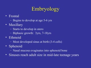
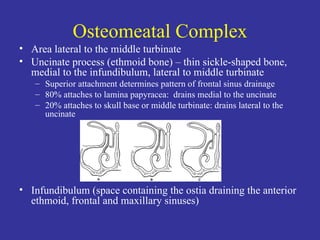
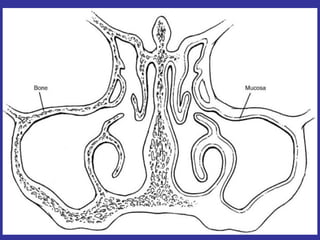
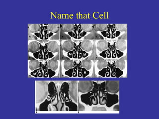
COCLIA 70: Sinus Surgery
Embryology Frontal Begins to develop at age 5-6 yrs Maxillary Starts to develop in utero Biphasic growth:  3yrs, 7-18yrs Ethmoid Most developed sinus at birth (3-4 cells) Sphenoid Nasal mucosa evaginates into sphenoid bone Sinuses reach adult size in mid-late teenage years
Osteomeatal Complex Area lateral to the middle turbinate Uncinate process (ethmoid bone) – thin sickle-shaped bone, medial to the infundibulum, lateral to middle turbinate Superior attachment determines pattern of frontal sinus drainage 80% attaches to lamina papyracea:  drains medial to the uncinate 20% attaches to skull base or middle turbinate: drains lateral to the uncinate Infundibulum (space containing the ostia draining the anterior ethmoid, frontal and maxillary sinuses)
 
Name that Cell
Name that Cell Haller cells Ethmoid cells that extend into the maxillary sinus Pneumatized medial and inferior orbital walls Onodi cells “ Sphenoethmoidal cell” A posterior ethmoidal cell that extends lateral and superior to the sphenoid sinus Can be mistaken as the sphenoid sinus Both the carotid artery and the optic nerve can be exposed within it Aggar nasi cells Most anterior of the anterior ethmoids (infundibular cells) Lies anterior-superior to the attachment of the middle turbinate to the lateral nasal wall Anterior wall of the frontal recess
FESS:  Overview Advantages Better visualization & precision No external scar Better preservation of function Disadvantages Distorted depth perception (monocular vision) One-handed technique Contraindications Osteomyelitis Inaccessible lateral frontal sinus disease
FESS:  Complications Orbital Blindness / Retrobulbar hematoma Orbital fat penetration Diplopia (medial or superior rectus muscle injury) Epiphora (lacrimal duct injury) Intracranial CSF leak Meningitis Brain abscess Hemorrhage Intranasal Synechia Anosomia
Relativity Middle meatus Lateral to the middle turbinate (medialize it) Lateral to the uncinate process (excise it) Sphenoid ostium 30-34 degrees from floor of nose 6.2-8.0 cm posterior to nasal spine Sphenoethmoidal recess at junction of superior 2/3 and inferior 1/3 of the superior turbinate Medial to middle and superior turbinates
Sphenoid Sinus Most common local causes of failure Failure to enter at the original surgery Stenosis Special considerations Intracerebral injury:  CSF leak, meningitis, abscess, injury to pituitary gland Vascular hemorrhage Retrobulbar hematoma Cranial nerve injury Cavernous sinus fistula
Nasal Antral Window Normal mucociliary flow sweeps contents from the maxillary sinus out the surgically enlarged maxillary sinus ostium A patent antral nasal window can allow secretions to reenter the maxillary sinus and are prone to becoming infected
The Middle Turbinate MTR Decreased synechia formation Higher long-term patency Improved nasal airflow Decreased nasal resistance MTP Preserve important anatomic landmark Decreased risk of alteration of nasal function,  Decreased risk of atrophic rhinitis Decreased risk of hyposmia Decreased risk of frontal recess stenosis causing sinusitis
Fate of the Middle Turbinate:  Part 1 Havas (Ann Otol Rhinol Laryngol 2000) Review of 509 pts (partial MTR) vs. 597 pts (MTP) – randomly divided, minimum one year follow-up Partial MTR: anterior inferior third of MT resected using endoscopic scissors  Important area in secretion of vasoactive sensory neuropeptides involved in hypersecretion, edema, polyposis, chronic rhinosinusitis Both groups:  most important factor of FESS success was severity of rhinosinusitis preop Partial MTR:  less synechiae, less recurrent disease requiring revision surgery – RECOMMENDED!
Fate of the Middle Turbinate:  Part 2 Giacchi (Am J Rhino 2000) Retrospective review of 50 MTR and 50 MTP sides with minimum 2 year follow-up Partial MTR:  resect anterior-inferior third to half, preserve superior and lateral attachments Not associated with increased complications (frontal recess stenosis with secondary frontal sinusitis) Consider on case-by-case basis (improved surgical access and postoperative debridement)
Whoops!  CSF Leak Refer to COCLIA page 62 (Dr. Lee 10/8/07)
External Approaches to Ethmoids Caldwell-Luc  (transantral ethmoidectomy) Trans-maxillary middle meatal antrostomy with transantral ethmoidectomy Disadvantages  Does not expose anterior ethmoids Risk of infraorbital nerve injury Open external ethmoidectomy Access through lamina payracea and lacrimal fossa Must ligate anterior ethmoid and angular arteries Disadvantages Poor exposure of anterior ethmoids Scar
Osteoplastic Flap Indications Recurrent or chronic frontal sinusitis Frontal mucoceles Procedure Make template from a Caldwell view x-ray Bicoronal incision for exposure Using template, excise periosteal and bone flap Remove diseased mucosa Obliterate cavity and plug frontal recess
Lothrop Procedure “ Chaput-Meyer” Indication Chronic frontal sinus disease Procedure Remove bilateral anterior ethmoids, middle turbinates and frontal sinus septum Creates large opening for frontal sinus drainage into the nasal cavity
Frontal Cells Anatomy May contribute to mechanical obstruction of the frontal recess Classification (Bent and Kuhn) Type I:  single cell superior to agger nasi Type II:  2 or more cells Type III:  single large that pneumatizes into the frontal sinus Type IV:  cell contained completely within the frontal sinus
 
Frontal Cells Meyer (Am J Rhino 2003) 768 CT scans met study criteria Objective:  are frontal cells associated with variants of pneumatization? Frontal cells present in 20.4% of study population Positively associated with hyperpneumatization of the frontal sinus and negatively with hypopneumatization Increased prevalence of concha bullosa In general, type III and IV individuals had increased prevalence of frontal mucosal thickening but not maxillary or ethmoid thickening
Orbital Complications Overall <0.5% serious FESS complication rate Orbital complications Pain Hemorrhage Diplopia (medial rectus injury) Blindness Minor complications using conventional instrumentation can become major complications with powered instrumentation
Post-Op Orbital Hematoma:  Part 1 Graham (Laryngoscope 2003) Case series 3 patients referred for orbital injury Not every patient with orbital hematoma requires surgical intervention  Ophthalmology consult proptosis intraocular pressure (observe if <30mmHg, poor vision likely if >40mmHg),  funduscopy (assess retinal blood flow) If retinal blood flow is normal, no immediate treatment If retinal blood flow is compromised, immediate canthotomy with upper and lower cantholysis
Post-Op Orbital Hematoma:  Part 2 Sharma (J Laryn Otol 2000) Case series 7 patients Minimize risk  Preop:  history prior sinus surgery, CT evaluation Intraop:  abort surgery if bleeding hampers visualization Recognize symptoms:  acute onset/progression orbital pain, diplopia, visual loss Assess “four P’s”  Perception of light Pupils (relative afferent pupil defect) Pallor of optic nerve Pulsatility of the central retinal artery
Treatment of Orbital Hemorrhage Globe massage Increase orbital space Cantholysis, canthotomy Medial wall decompression:  transcaruncular orbitomy, external ethmoidectomy, endoscopic Orbital floor decompression:  transconjunctival Decrease volume of orbital contents Steroids, topical beta blocker drops Mannitol, acetazolamide Paracentesis of the anterior chamber
Minimize Risk of Orbital Injury Preop:  history prior sinus surgery, CT evaluation Intraop:  abort surgery if bleeding hampers visualization Place opening of blade at 90 degrees to the medial orbital wall and dissect superiorly or inferiorly Assess vision, orbital appearance, and ocular motility during postop check
FESS Post-op Care Thaler (Arch Oto Head Neck Surg 2002) Frequent debridement Post op week 1 then weekly or greater intervals until healed (approx 4-6 weeks) Pros:  remove large crusting and clot (traps mucous leading to infection, bridge for scar formation, retained bone fragments are nidus for infection) Cons:  histologically avulses epithelium until 2 nd  postop week, pediatric FESS rarely tolerate debridement but have good outcomes Hypertonic saline irrigation Nasal splints  Endoscopic exam to assess for obstructing crust or clot
Informed Consent Recall possible complications discussed earlier Orbital Intracranial Intranasal
Balloon Sinuplasty Indications:  maxillary, sphenoid and frontal disease Safety:  less injury to mucosa, less operative bleeding, eliminates need for nasal packing Results:  not long enough follow-up

Ee3a Coclia70 Sinus Surgery

  • 1.
  • 2.
    Embryology Frontal Beginsto develop at age 5-6 yrs Maxillary Starts to develop in utero Biphasic growth: 3yrs, 7-18yrs Ethmoid Most developed sinus at birth (3-4 cells) Sphenoid Nasal mucosa evaginates into sphenoid bone Sinuses reach adult size in mid-late teenage years
  • 3.
    Osteomeatal Complex Arealateral to the middle turbinate Uncinate process (ethmoid bone) – thin sickle-shaped bone, medial to the infundibulum, lateral to middle turbinate Superior attachment determines pattern of frontal sinus drainage 80% attaches to lamina papyracea: drains medial to the uncinate 20% attaches to skull base or middle turbinate: drains lateral to the uncinate Infundibulum (space containing the ostia draining the anterior ethmoid, frontal and maxillary sinuses)
  • 4.
  • 5.
  • 6.
    Name that CellHaller cells Ethmoid cells that extend into the maxillary sinus Pneumatized medial and inferior orbital walls Onodi cells “ Sphenoethmoidal cell” A posterior ethmoidal cell that extends lateral and superior to the sphenoid sinus Can be mistaken as the sphenoid sinus Both the carotid artery and the optic nerve can be exposed within it Aggar nasi cells Most anterior of the anterior ethmoids (infundibular cells) Lies anterior-superior to the attachment of the middle turbinate to the lateral nasal wall Anterior wall of the frontal recess
  • 7.
    FESS: OverviewAdvantages Better visualization & precision No external scar Better preservation of function Disadvantages Distorted depth perception (monocular vision) One-handed technique Contraindications Osteomyelitis Inaccessible lateral frontal sinus disease
  • 8.
    FESS: ComplicationsOrbital Blindness / Retrobulbar hematoma Orbital fat penetration Diplopia (medial or superior rectus muscle injury) Epiphora (lacrimal duct injury) Intracranial CSF leak Meningitis Brain abscess Hemorrhage Intranasal Synechia Anosomia
  • 9.
    Relativity Middle meatusLateral to the middle turbinate (medialize it) Lateral to the uncinate process (excise it) Sphenoid ostium 30-34 degrees from floor of nose 6.2-8.0 cm posterior to nasal spine Sphenoethmoidal recess at junction of superior 2/3 and inferior 1/3 of the superior turbinate Medial to middle and superior turbinates
  • 10.
    Sphenoid Sinus Mostcommon local causes of failure Failure to enter at the original surgery Stenosis Special considerations Intracerebral injury: CSF leak, meningitis, abscess, injury to pituitary gland Vascular hemorrhage Retrobulbar hematoma Cranial nerve injury Cavernous sinus fistula
  • 11.
    Nasal Antral WindowNormal mucociliary flow sweeps contents from the maxillary sinus out the surgically enlarged maxillary sinus ostium A patent antral nasal window can allow secretions to reenter the maxillary sinus and are prone to becoming infected
  • 12.
    The Middle TurbinateMTR Decreased synechia formation Higher long-term patency Improved nasal airflow Decreased nasal resistance MTP Preserve important anatomic landmark Decreased risk of alteration of nasal function, Decreased risk of atrophic rhinitis Decreased risk of hyposmia Decreased risk of frontal recess stenosis causing sinusitis
  • 13.
    Fate of theMiddle Turbinate: Part 1 Havas (Ann Otol Rhinol Laryngol 2000) Review of 509 pts (partial MTR) vs. 597 pts (MTP) – randomly divided, minimum one year follow-up Partial MTR: anterior inferior third of MT resected using endoscopic scissors Important area in secretion of vasoactive sensory neuropeptides involved in hypersecretion, edema, polyposis, chronic rhinosinusitis Both groups: most important factor of FESS success was severity of rhinosinusitis preop Partial MTR: less synechiae, less recurrent disease requiring revision surgery – RECOMMENDED!
  • 14.
    Fate of theMiddle Turbinate: Part 2 Giacchi (Am J Rhino 2000) Retrospective review of 50 MTR and 50 MTP sides with minimum 2 year follow-up Partial MTR: resect anterior-inferior third to half, preserve superior and lateral attachments Not associated with increased complications (frontal recess stenosis with secondary frontal sinusitis) Consider on case-by-case basis (improved surgical access and postoperative debridement)
  • 15.
    Whoops! CSFLeak Refer to COCLIA page 62 (Dr. Lee 10/8/07)
  • 16.
    External Approaches toEthmoids Caldwell-Luc (transantral ethmoidectomy) Trans-maxillary middle meatal antrostomy with transantral ethmoidectomy Disadvantages Does not expose anterior ethmoids Risk of infraorbital nerve injury Open external ethmoidectomy Access through lamina payracea and lacrimal fossa Must ligate anterior ethmoid and angular arteries Disadvantages Poor exposure of anterior ethmoids Scar
  • 17.
    Osteoplastic Flap IndicationsRecurrent or chronic frontal sinusitis Frontal mucoceles Procedure Make template from a Caldwell view x-ray Bicoronal incision for exposure Using template, excise periosteal and bone flap Remove diseased mucosa Obliterate cavity and plug frontal recess
  • 18.
    Lothrop Procedure “Chaput-Meyer” Indication Chronic frontal sinus disease Procedure Remove bilateral anterior ethmoids, middle turbinates and frontal sinus septum Creates large opening for frontal sinus drainage into the nasal cavity
  • 19.
    Frontal Cells AnatomyMay contribute to mechanical obstruction of the frontal recess Classification (Bent and Kuhn) Type I: single cell superior to agger nasi Type II: 2 or more cells Type III: single large that pneumatizes into the frontal sinus Type IV: cell contained completely within the frontal sinus
  • 20.
  • 21.
    Frontal Cells Meyer(Am J Rhino 2003) 768 CT scans met study criteria Objective: are frontal cells associated with variants of pneumatization? Frontal cells present in 20.4% of study population Positively associated with hyperpneumatization of the frontal sinus and negatively with hypopneumatization Increased prevalence of concha bullosa In general, type III and IV individuals had increased prevalence of frontal mucosal thickening but not maxillary or ethmoid thickening
  • 22.
    Orbital Complications Overall<0.5% serious FESS complication rate Orbital complications Pain Hemorrhage Diplopia (medial rectus injury) Blindness Minor complications using conventional instrumentation can become major complications with powered instrumentation
  • 23.
    Post-Op Orbital Hematoma: Part 1 Graham (Laryngoscope 2003) Case series 3 patients referred for orbital injury Not every patient with orbital hematoma requires surgical intervention Ophthalmology consult proptosis intraocular pressure (observe if <30mmHg, poor vision likely if >40mmHg), funduscopy (assess retinal blood flow) If retinal blood flow is normal, no immediate treatment If retinal blood flow is compromised, immediate canthotomy with upper and lower cantholysis
  • 24.
    Post-Op Orbital Hematoma: Part 2 Sharma (J Laryn Otol 2000) Case series 7 patients Minimize risk Preop: history prior sinus surgery, CT evaluation Intraop: abort surgery if bleeding hampers visualization Recognize symptoms: acute onset/progression orbital pain, diplopia, visual loss Assess “four P’s” Perception of light Pupils (relative afferent pupil defect) Pallor of optic nerve Pulsatility of the central retinal artery
  • 25.
    Treatment of OrbitalHemorrhage Globe massage Increase orbital space Cantholysis, canthotomy Medial wall decompression: transcaruncular orbitomy, external ethmoidectomy, endoscopic Orbital floor decompression: transconjunctival Decrease volume of orbital contents Steroids, topical beta blocker drops Mannitol, acetazolamide Paracentesis of the anterior chamber
  • 26.
    Minimize Risk ofOrbital Injury Preop: history prior sinus surgery, CT evaluation Intraop: abort surgery if bleeding hampers visualization Place opening of blade at 90 degrees to the medial orbital wall and dissect superiorly or inferiorly Assess vision, orbital appearance, and ocular motility during postop check
  • 27.
    FESS Post-op CareThaler (Arch Oto Head Neck Surg 2002) Frequent debridement Post op week 1 then weekly or greater intervals until healed (approx 4-6 weeks) Pros: remove large crusting and clot (traps mucous leading to infection, bridge for scar formation, retained bone fragments are nidus for infection) Cons: histologically avulses epithelium until 2 nd postop week, pediatric FESS rarely tolerate debridement but have good outcomes Hypertonic saline irrigation Nasal splints Endoscopic exam to assess for obstructing crust or clot
  • 28.
    Informed Consent Recallpossible complications discussed earlier Orbital Intracranial Intranasal
  • 29.
    Balloon Sinuplasty Indications: maxillary, sphenoid and frontal disease Safety: less injury to mucosa, less operative bleeding, eliminates need for nasal packing Results: not long enough follow-up