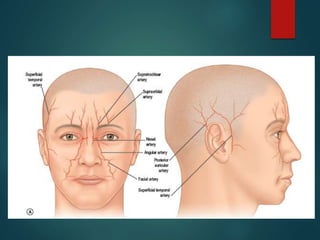
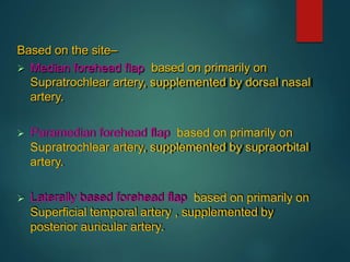
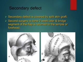
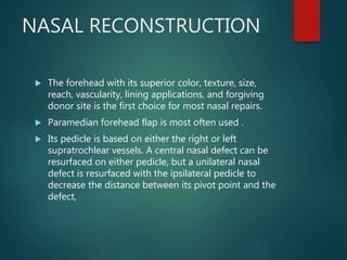
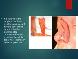
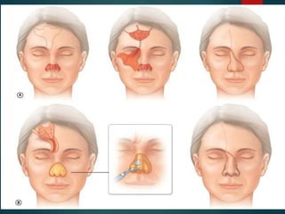
FOREHEAD FLAP
DR SAMIK SHARMA
HISTORY
 Also called as temporal flap.
 800 BC - Sushruta, described a nasal reconstruction approach based
on pedicled forehead skin flap.
 Later describe by McGregor, in 1963.
 Its axial pattern myocutaneous flap provide large area of skin &
subcutaneous tissue.
 Called as lifeboat may be raised quickly to get surgeon out of trouble…
ANATOMY
It consists of five layers:
 (S) skin,
 (C) subcutaneous tissue (superficial fascia),
 (A) occipitofrontalis (epicranius) and its aponeurosis,
 (L) subaponeurotic areolar tissue and
 (P) pericranium
 Aponeurotic layer is movable along the upper three
layers of the scalp,& can easily slide on deepest layer
is the periosteum of the skull.
 It is very easy to raise a scalp flap within the plane
between the galea and the pericranium without
compromising the blood or nerve supply of the scalp,
because all of these structures lie in the superficial
fascia.
SUPERFICIAL
TEMPORAL
ARTERY
 its smaller terminal branch of the
external carotid artery.
 It arises in the parotid gland behind the neck
of the mandible, where it is crossed by
temporal and zygomatic branches of the facial
nerve.
 Initially deep, it becomes superficial as it
passes over the posterior root of the zygomatic
process of the temporal bone, where its pulse
can be felt.
 It then runs up the scalp for 4 cm and divides
into frontal (anterior) and parietal (posterior)
branches.
 It is accompanied by corresponding veins,
& auriculotemporal nerve lies posterior to
it.
 Supratrochlear artery
 emerges from the orbit onto the face at the frontal notch. It
supplies the medial parts of the upper eyelid, forehead and
scalp.
 The supratrochlear artery anastomoses with the supraorbital
artery and with its contralateral fellow.
 Supraorbital artery
 leaves the orbit through the supraorbital notch (or foramen).
 divides into superficial and deep branches, supplying skin
and muscle of the upper eyelid, forehead and scalp.
 It anastomoses with the supratrochlear artery, frontal branch
of the superficial temporal and its contralateral fellow.
 The forehead is perfused from
 supraorbital,
 supratrochlear, the
 superficial temporal and posterior auricular arteries.
 Flaps can be based on each of these pedicles
 the median forehead flap,
 the horizontal forehead flap,
 the up-and-down flap, or the scalping flap.
 Based on the mangold
et al (1980) study on
vascular anatomy of
the forehead, he
divided forehead in
vascular territories-
Dorsal nasal artery
Supratrochlear artery
Supraorbital artery
Superficial temporal
artery.
Based on the site–
 Median forehead flap based on primarily on
Supratrochlear artery, supplemented by dorsal nasal
artery.
 Paramedian forehead flap based on primarily on
Supratrochlear artery, supplemented by supraorbital
artery.
 Laterally based forehead flap based on primarily on
Superficial temporal artery , supplemented by
posterior auricular artery.
INDICATIONS
 Used for a large number of reconstruction procedures:
 nose,
 upper eyelid,
 cheek (inside and outside),
 floor of the mouth,
 chin covering for reconstructed mandible,
 portion of tongue, and
 alveolar region.
TECHNIQUE
LATERALY BASED
FOREHEAD FLAP
 The forehead flap is
outlined.
 contour follows the eyebrows (must
not extend beyond the level of the
lateral canthus to avoid injury to the
facial nerve) to anterior border of
pinna at level of zygomatic arch and
along forehead hairline more pleasing
cosmetically.
 The incisions are beveled
to minimize the cosmetic
deformity along the
remaining edges of the
forehead and scalp
 for longer flap most often
extends to hair-line of
opposite temple.
As it is used for intraoral reconstruction a tunnel is
constructed through which flap is passed so that distal
end reaches the intra-oral defect.
Based on the route in the mouth-
 Directly through the cheek(cheek portal)
 Deep to the zygomatic arch
 Posterior part of submandibular incision of neck
dissection.
Through the
cheek
 Tunnel is made outside the cheek
avoiding facial nerve damage.
 Skin incised horizontally in front of ear
appro 1.5 below zygomatic arch(length
of incision 2/3 of the flap.
 Incision deepened to the parotid
level using scalpel then tissue
scissor thrust through the substance
of cheek in the defect.
 Ramus is dissected tunnel
is made directly through
the mouth with min
resistance of parotid.
 When ramus is intact
tunnel has to bring round in
front of bone .
 Difficult if defect is
extended both forward &
backward.
 Needs to raise longer flaps
 Drawback- salivary fistula.
 By Davis & Hoopes, 1971
 Flap is passed downward deep to the arch in to mouth
following the pathway of the temporalis muscle.
 By Millard ,1964.
 While his study on primary
bone grafting after mandibular
resection, he used forehead
flap to provide a lining to
cover the bone graft through
the submandibular incision of
neck dissection.
 Flap enter the mouth medial
to the mandible extending far
back till tongue.
 Drawback – inferior fistula
 Secondary defect is covered by split skin graft.
 Second surgery is done 3 week later & bridge
segment of the flap is returned to the temple or
forehead.
 Done after 3 weeks, flap is tunneled ,& divided it from
outside as far down the tunnel.
 Skin closure is done from outside n tunnel is kept
patent inside to drain freely.
NASAL RECONSTRUCTION
 The forehead with its superior color, texture, size,
reach, vascularity, lining applications, and forgiving
donor site is the first choice for most nasal repairs.
 Paramedian forehead flap is most often used .
 Its pedicle is based on either the right or left
supratrochlear vessels. A central nasal defect can be
resurfaced on either pedicle, but a unilateral nasal
defect is resurfaced with the ipsilateral pedicle to
decrease the distance between its pivot point and the
defect,
 Traditionally, a forehead flap is transferred in two
stages.
 The flap can be transferred as an island flap, in one
stage, but excessive bulk of the pedicle, passing under
tight glabellar skin, may jeopardize its blood supply or
distort the nasal root. Most importantly, an aesthetic
reconstruction can rarely be obtained in a single stage.
Technique of the two-stage
forehead flap
 Stage 1: flap transfer
 An exact pattern of the defect, based on the contralateral normal or
the ideal, is outlined, under the hairline, directly over the
supratrochlear artery, which lies just lateral to the frown crease. It can
be identified by Doppler.
 Pedicle width at the brow is about 1.2–1.5 cm.
 The reach of the flap is verified using a simple gauze measure,
checking the distance from the pivot point below the eyebrow to the
distal end of the flap on the forehead and most inferior aspect of the
defect.
 The flap can be lengthened by extending the design into the hairline
or, more often, the pedicle is extended inferiorly across the eyebrow
towards the medial canthus.
 Frontalis muscle and excess
subcutaneous fat are excised
within its distal 1.5–2 cm,
producing a skin flap with 2–3
mm of fat distally which will be
applied to the inferior aspect of
the nasal defect.
 The dissection then passes
under the frontalis and over
the periosteum, through the
medial brow until the flap
reaches the defect without
tension.
 It is sutured to the
recipient site, from
distal to proximal, with
a single layer of fine
suture. If the flap
blanches, stop
suturing and let the
unsutured lateral flap
edges heal secondarily
to the recipient site.
 Stage 2
 The pedicle is divided 3–4 weeks later, the proximal
aspect of the flap is re-elevated with 2–3 mm of fat,
and the underlying excess soft tissue (fat, frontalis, and
scar), which is adherent to recipient site, is excised,
contouring the superior aspect of the defect.
 The flap remains well vascularized through its distal
inset. The superior inset is completed. The proximal
pedicle is untubed and returned to the medial brow as
a small inverted “V,” discarding any excess
The three stage full-
thickness forehead flap
 Millard, in the 1970s, and Burget, in the 1990s, added
an intermediate operation between transfer and
division of the traditional two-stage forehead flap to
improve vascular safety and permit more aggressive
soft-tissue contouring.
 They thinned the flap distally at transfer but, 3 weeks
later, elevated it from its bed over the mid-nose.
 The proximal pedicle remained intact while the distal
inset was left attached over the tip, alar margins, and
columella. Underlying fat and muscle over the
dorsum and mid-vault were excised to improve mid-
vault contour.
 Menick, in the late 1990s, modified the approach by
transferring a full-thickness flap in three stages with an
intermediate operation which permitted complete flap
re-elevation and three-dimensional hard- and soft-
tissue contouring over the entire nasal surface.
Expansion of the forehead
 Pre-expansion, although not routinely helpful, can
enlarge the available surface of the forehead for
transfer to the nose.
 Forehead expansion should be considered:
 1. In an especially tight forehead with limited available
skin due to scarring or prior forehead flap harvest.
Expansion can increase the length and width of
available skin within the proposed flap.
 2. In the occasional, especially short forehead (less
than 3–4 cm in height) to increase flap length and
minimize hair transfer to the nose.
 3. To expand the donor forehead adjacent to the
proposed flap to facilitate closure of the forehead
defect
 However, there are many disadvantages to expansion –
delay in repair, increased number of operative stages,
added expense, more office visits, risk of infection and
extrusion, and recoil.
 Expanded skin, if not rigidly braced with a hard-tissue
support framework, contracts, leading to retraction
and nasal shortening. Expansion does not clinically
improve flap blood supply.
ROTATION ON FOREHEAD
FLAP
THANK YOU

Forehead flap

  • 1.
  • 2.
    HISTORY  Also calledas temporal flap.  800 BC - Sushruta, described a nasal reconstruction approach based on pedicled forehead skin flap.  Later describe by McGregor, in 1963.  Its axial pattern myocutaneous flap provide large area of skin & subcutaneous tissue.  Called as lifeboat may be raised quickly to get surgeon out of trouble…
  • 3.
  • 4.
    It consists offive layers:  (S) skin,  (C) subcutaneous tissue (superficial fascia),  (A) occipitofrontalis (epicranius) and its aponeurosis,  (L) subaponeurotic areolar tissue and  (P) pericranium
  • 6.
     Aponeurotic layeris movable along the upper three layers of the scalp,& can easily slide on deepest layer is the periosteum of the skull.  It is very easy to raise a scalp flap within the plane between the galea and the pericranium without compromising the blood or nerve supply of the scalp, because all of these structures lie in the superficial fascia.
  • 7.
    SUPERFICIAL TEMPORAL ARTERY  its smallerterminal branch of the external carotid artery.  It arises in the parotid gland behind the neck of the mandible, where it is crossed by temporal and zygomatic branches of the facial nerve.  Initially deep, it becomes superficial as it passes over the posterior root of the zygomatic process of the temporal bone, where its pulse can be felt.  It then runs up the scalp for 4 cm and divides into frontal (anterior) and parietal (posterior) branches.  It is accompanied by corresponding veins, & auriculotemporal nerve lies posterior to it.
  • 9.
     Supratrochlear artery emerges from the orbit onto the face at the frontal notch. It supplies the medial parts of the upper eyelid, forehead and scalp.  The supratrochlear artery anastomoses with the supraorbital artery and with its contralateral fellow.  Supraorbital artery  leaves the orbit through the supraorbital notch (or foramen).  divides into superficial and deep branches, supplying skin and muscle of the upper eyelid, forehead and scalp.  It anastomoses with the supratrochlear artery, frontal branch of the superficial temporal and its contralateral fellow.
  • 12.
     The foreheadis perfused from  supraorbital,  supratrochlear, the  superficial temporal and posterior auricular arteries.  Flaps can be based on each of these pedicles  the median forehead flap,  the horizontal forehead flap,  the up-and-down flap, or the scalping flap.
  • 13.
     Based onthe mangold et al (1980) study on vascular anatomy of the forehead, he divided forehead in vascular territories- Dorsal nasal artery Supratrochlear artery Supraorbital artery Superficial temporal artery.
  • 14.
    Based on thesite–  Median forehead flap based on primarily on Supratrochlear artery, supplemented by dorsal nasal artery.  Paramedian forehead flap based on primarily on Supratrochlear artery, supplemented by supraorbital artery.  Laterally based forehead flap based on primarily on Superficial temporal artery , supplemented by posterior auricular artery.
  • 16.
    INDICATIONS  Used fora large number of reconstruction procedures:  nose,  upper eyelid,  cheek (inside and outside),  floor of the mouth,  chin covering for reconstructed mandible,  portion of tongue, and  alveolar region.
  • 17.
  • 18.
    LATERALY BASED FOREHEAD FLAP The forehead flap is outlined.  contour follows the eyebrows (must not extend beyond the level of the lateral canthus to avoid injury to the facial nerve) to anterior border of pinna at level of zygomatic arch and along forehead hairline more pleasing cosmetically.
  • 19.
     The incisionsare beveled to minimize the cosmetic deformity along the remaining edges of the forehead and scalp  for longer flap most often extends to hair-line of opposite temple.
  • 20.
    As it isused for intraoral reconstruction a tunnel is constructed through which flap is passed so that distal end reaches the intra-oral defect. Based on the route in the mouth-  Directly through the cheek(cheek portal)  Deep to the zygomatic arch  Posterior part of submandibular incision of neck dissection.
  • 21.
    Through the cheek  Tunnelis made outside the cheek avoiding facial nerve damage.  Skin incised horizontally in front of ear appro 1.5 below zygomatic arch(length of incision 2/3 of the flap.  Incision deepened to the parotid level using scalpel then tissue scissor thrust through the substance of cheek in the defect.
  • 22.
     Ramus isdissected tunnel is made directly through the mouth with min resistance of parotid.  When ramus is intact tunnel has to bring round in front of bone .  Difficult if defect is extended both forward & backward.  Needs to raise longer flaps  Drawback- salivary fistula.
  • 23.
     By Davis& Hoopes, 1971  Flap is passed downward deep to the arch in to mouth following the pathway of the temporalis muscle.
  • 25.
     By Millard,1964.  While his study on primary bone grafting after mandibular resection, he used forehead flap to provide a lining to cover the bone graft through the submandibular incision of neck dissection.  Flap enter the mouth medial to the mandible extending far back till tongue.  Drawback – inferior fistula
  • 26.
     Secondary defectis covered by split skin graft.  Second surgery is done 3 week later & bridge segment of the flap is returned to the temple or forehead.
  • 27.
     Done after3 weeks, flap is tunneled ,& divided it from outside as far down the tunnel.  Skin closure is done from outside n tunnel is kept patent inside to drain freely.
  • 28.
    NASAL RECONSTRUCTION  Theforehead with its superior color, texture, size, reach, vascularity, lining applications, and forgiving donor site is the first choice for most nasal repairs.  Paramedian forehead flap is most often used .  Its pedicle is based on either the right or left supratrochlear vessels. A central nasal defect can be resurfaced on either pedicle, but a unilateral nasal defect is resurfaced with the ipsilateral pedicle to decrease the distance between its pivot point and the defect,
  • 29.
     Traditionally, aforehead flap is transferred in two stages.  The flap can be transferred as an island flap, in one stage, but excessive bulk of the pedicle, passing under tight glabellar skin, may jeopardize its blood supply or distort the nasal root. Most importantly, an aesthetic reconstruction can rarely be obtained in a single stage.
  • 30.
    Technique of thetwo-stage forehead flap  Stage 1: flap transfer  An exact pattern of the defect, based on the contralateral normal or the ideal, is outlined, under the hairline, directly over the supratrochlear artery, which lies just lateral to the frown crease. It can be identified by Doppler.  Pedicle width at the brow is about 1.2–1.5 cm.  The reach of the flap is verified using a simple gauze measure, checking the distance from the pivot point below the eyebrow to the distal end of the flap on the forehead and most inferior aspect of the defect.  The flap can be lengthened by extending the design into the hairline or, more often, the pedicle is extended inferiorly across the eyebrow towards the medial canthus.
  • 32.
     Frontalis muscleand excess subcutaneous fat are excised within its distal 1.5–2 cm, producing a skin flap with 2–3 mm of fat distally which will be applied to the inferior aspect of the nasal defect.  The dissection then passes under the frontalis and over the periosteum, through the medial brow until the flap reaches the defect without tension.
  • 33.
     It issutured to the recipient site, from distal to proximal, with a single layer of fine suture. If the flap blanches, stop suturing and let the unsutured lateral flap edges heal secondarily to the recipient site.
  • 34.
     Stage 2 The pedicle is divided 3–4 weeks later, the proximal aspect of the flap is re-elevated with 2–3 mm of fat, and the underlying excess soft tissue (fat, frontalis, and scar), which is adherent to recipient site, is excised, contouring the superior aspect of the defect.  The flap remains well vascularized through its distal inset. The superior inset is completed. The proximal pedicle is untubed and returned to the medial brow as a small inverted “V,” discarding any excess
  • 36.
    The three stagefull- thickness forehead flap  Millard, in the 1970s, and Burget, in the 1990s, added an intermediate operation between transfer and division of the traditional two-stage forehead flap to improve vascular safety and permit more aggressive soft-tissue contouring.  They thinned the flap distally at transfer but, 3 weeks later, elevated it from its bed over the mid-nose.  The proximal pedicle remained intact while the distal inset was left attached over the tip, alar margins, and columella. Underlying fat and muscle over the dorsum and mid-vault were excised to improve mid- vault contour.
  • 37.
     Menick, inthe late 1990s, modified the approach by transferring a full-thickness flap in three stages with an intermediate operation which permitted complete flap re-elevation and three-dimensional hard- and soft- tissue contouring over the entire nasal surface.
  • 40.
    Expansion of theforehead  Pre-expansion, although not routinely helpful, can enlarge the available surface of the forehead for transfer to the nose.  Forehead expansion should be considered:  1. In an especially tight forehead with limited available skin due to scarring or prior forehead flap harvest. Expansion can increase the length and width of available skin within the proposed flap.
  • 41.
     2. Inthe occasional, especially short forehead (less than 3–4 cm in height) to increase flap length and minimize hair transfer to the nose.  3. To expand the donor forehead adjacent to the proposed flap to facilitate closure of the forehead defect
  • 42.
     However, thereare many disadvantages to expansion – delay in repair, increased number of operative stages, added expense, more office visits, risk of infection and extrusion, and recoil.  Expanded skin, if not rigidly braced with a hard-tissue support framework, contracts, leading to retraction and nasal shortening. Expansion does not clinically improve flap blood supply.
  • 44.
  • 45.