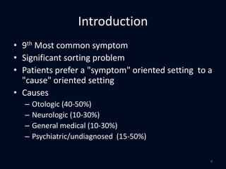
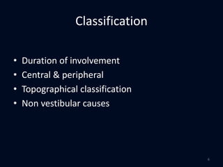
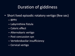
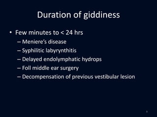
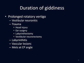
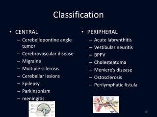
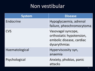
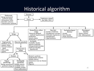
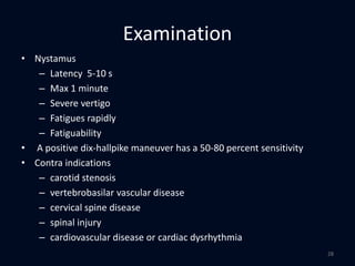
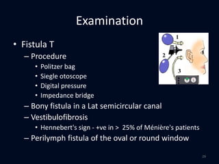
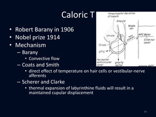
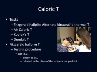
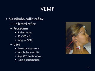
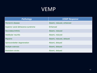
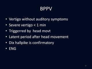
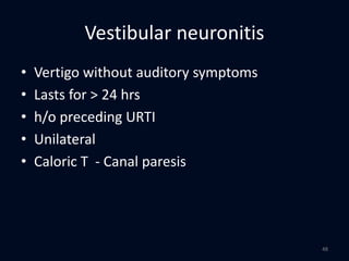
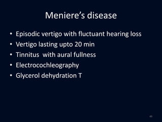
Evaluation of giddiness
1
• Introduction
• Classification of vertigo
• Evaluation
• Diagnosis
• Management
Evaluation of giddiness
2
Introduction
• Dysequilibrium, unsteadiness, vertigo, and
lightheadedness
• Vertigo is an illusory sense of motion
– Internal feeling
– Objects in the surroundings are moving or tilting
– Sense of motion
• Rotatory
• Linear
• Change in orientation relative to the vertical
3
Introduction
• 9th Most common symptom
• Significant sorting problem
• Patients prefer a "symptom" oriented setting to a
"cause" oriented setting
• Causes
– Otologic (40-50%)
– Neurologic (10-30%)
– General medical (10-30%)
– Psychiatric/undiagnosed (15-50%)
4
Classification
5
Classification
• Duration of involvement
• Central & peripheral
• Topographical classification
• Non vestibular causes
6
Duration of giddiness
• Short lived episodic rotatory vertigo (few sec)
– BPPV
– Labyrinthine fistula
– Caloric effect
– Alternobaric vertigo
– Post concussion syn
– Vertebrobasilar insufficiency
– Cervical vertigo
7
Duration of giddiness
• Few minutes to < 24 hrs
– Meniere’s disease
– Syphilitic labyrynthitis
– Delayed endolymphatic hydrops
– Foll middle ear surgery
– Decompensation of previous vestibular lesion
8
Duration of giddiness
• Prolonged rotatory vertigo
– Vestibular neuronitis
– Trauma
• Head injury
• Ear surgery
• Labyrinthectomy
• Vestibular neuronectomy
– Labyrinthitis
– Vascular lesions
– Mets at CP angle
9
Classification
• CENTRAL
– Cerebellopontine angle
tumor
– Cerebrovascular disease
– Migraine
– Multiple sclerosis
– Cerebellar lesions
– Epilepsy
– Parkinsonism
– meningitis
• PERIPHERAL
– Acute labrynthitis
– Vestibular neuritis
– BPPV
– Cholesteatoma
– Meniere’s disease
– Ostosclerosis
– Perilymphatic fistula
10
Non vestibular
System Disease
Endocrine Hypoglycaemia, adrenal
failure, pheochromocytoma
CVS Vasovagal syncope,
orthostatic hypotension,
embolic disease, cardiac
dysarythmias
Haematological Hyperviscosity syn,
anaemia
Psychological Anxiety, phobias, panic
attacks
11
Post head injury
• Post concussion
• BPPV
• Destructive labyrinth lesions
• Perilymph fistula
• Delayed endolymphatic hydrops
• Functional
12
Evaluation
13
Evaluation - history
• Define
– patient's dizziness - Vertigo, Impulsion, lightheaded, oscillopsia, ataxia, confusion.
• Timing
– (BPPV-seconds, TIA-minutes, meniere’s -hours, Vestibular Neuronitis-Days, ototoxicity-
years)
• Associations
– head motion or change in head position, hearing disturbance, headache, cognitive
symptoms, relation to stress.
• Review of systems
– especially vascular risk factors and ear surgery.
• Family History
– Similar disorder ? Migraine
• Medication History
– present and past exposures to ototoxins, antihypertensives.
• Previous studies
14
Topographical
Symptom Site of lesion
Tinnitus, hearing loss Peripheral (labyrinth / 8th CN)
Ear fullness, Tinnitus, hearing
loss
Labyrinthine
5th,6th,7th CN CP angle
EAC vesicles 7th , 8th neuritis
Diplopia, 3rd,4th,6th , facial
numbness, difficulty swallowig,
choking
Brainstem
Uni / bilateral numbness,
weakness, ataxia, long tract,
hemianopia
Cerebral hemisphere
15
Symptom Diagnosis
Aural fullness Acoustic neuroma; Ménière's disease
Ear or mastoid pain Acoustic neuroma; acute middle ear
disease (e.g., otitis media, herpes
zoster oticus)
Facial weakness Acoustic neuroma; herpes zoster oticus
Focal neurologic Cerebellopontine angle tumor;
cerebrovascular disease; multiple
sclerosis
Headache Acoustic neuroma; migraine
Associated symptoms & diagnosis
16
Symptom diagnosis
Nystagmus Peripheral or central vertigo
Photophobia Migraine
Tinnitus Acute labyrinthitis; acoustic neuroma;
Ménière's disease
Imbalance Acute vestibular neuronitis
cerebellopontine angle tumor
Hearing loss Ménière's disease; perilymphatic
fistula; acoustic neuroma;
cholesteatoma, otosclerosis; TIA or
stroke involving anterior inferior
cerebellar artery,herpes zoster oticus
Associated symptoms & diagnosis
17
Provoking Factors for Different Causes
Provoking factor Suggested diagnosis
Changes in head position Acute labyrinthitis; benign positional
paroxysmal vertigo; multiple
sclerosis; perilymphatic fistula
Spontaneous episodes Acute vestibular neuronitis;
cerebrovascular disease (stroke or
transient ischemic attack); (i.e., no
consistent provoking factors)
Ménière's disease; migraine;
multiple sclerosis
Recent upper respiratory viral
illness
Acute vestibular neuronitis
18
Provoking factor Suggested diagnosis
Stress Psychiatric or psychological
causes; migraine
Immunosuppression (e.g.,
immunosuppressive
medications, advanced age ,
stress)
Herpes zoster oticus
Changes in ear pressure,
head trauma, excessive
straining, loud noises
Perilymphatic fistula
19
Provoking Factors for Different Causes
Historical algorithm
20
Examination
21
Examination
• General Medical Examination
– Personality
– Anaemia
– Blood pressure
• Orthostatic changes in blood pressure or pulse,
Hypertensive
– Cardiac
• Arrhythmia, murmur, bruit
22
Examination
• Otologic Examination
– Middle ear pathology
– Hearing
• Neurotological examination
– Cranial nerves
– Motor power and reflexes, pathological reflexes (e.g.
Babinski)
– Sensory (proprioception)
– Cerebellar signs
23
Examination
• Cerebellar Tests
– Ataxia, atonia, and asthenia
– Intention tremor (tremor that increases on activity)
– Dyssynergia (incoordination)
– Dysmetria (overshooting or undershooting)
– Dysrhythmia (inability to repeat a rhythmic tap)
– Dysdiadochokinesis (difficulty with rapid alternating
movements)
– Dysarthria (staccato or scanning speech)
24
Examination
• Oculomotor examination
– Spontaneous nystagmus
• unilateral vestibular hypofunction +
• head is still, dampened by visual fixation
• increased or only becomes apparent when fixation is
eliminated
• Slow phase
– Alexander’s law
– Grading nysagmus
25
Examination
• Vestibular
examination
– Specific T
• Dix Hallpike T
• Fistula T
– Non Specific Test
• ENG
• Rotation T
– Otolith Function T
• Ocular counterrolling
• Parallel swing T
• Axis rotation T
– Whiplash T
• Passive neck torsion T
• Static Neck Torsion
– Vestibulospinal T
• Rhombergs T
• Untenberger T
• craniocorpography
• Posturography
• VEMP
– Others
• Caloric T
• Head shaking T
• Hyperventillation
26
Examination
• Dix hallpike T or Nylén-Bárány sign
– Procedure
• Head 45° turned
• Lowered & hyperextended -30 sec
– Rt Dix Hallpike
• Rt PSCC - Upbeat ,Torsional,
• Lt SSCC - Downbeat Torsional
– Lat SCC – modified T
• Geotropic, Ageotropic
27
Examination
• Nystamus
– Latency 5-10 s
– Max 1 minute
– Severe vertigo
– Fatigues rapidly
– Fatiguability
• A positive dix-hallpike maneuver has a 50-80 percent sensitivity
• Contra indications
– carotid stenosis
– vertebrobasilar vascular disease
– cervical spine disease
– spinal injury
– cardiovascular disease or cardiac dysrhythmia
28
Examination
• Fistula T
– Procedure
• Politzer bag
• Siegle otoscope
• Digital pressure
• Impedance bridge
– Bony fistula in a Lat semicircular canal
– Vestibulofibrosis
• Hennebert's sign - +ve in > 25% of Ménière's patients
– Perilymph fistula of the oval or round window
29
Caloric T
• Robert Barany in 1906
• Nobel prize 1914
• Mechanism
– Barany
• Convective flow
– Coats and Smith
• direct effect of temperature on hair cells or vestibular-nerve
afferents
– Scherer and Clarke
• thermal expansion of labyrinthine fluids will result in a
maintained cupular displacement
30
Caloric T
• Tests
– Fitzgerald hallpike Alternate binaural, bithermal T
– Air Caloric T
– Kobrak’s T
– Dunda’s T
• Fitzgerald hallpike T
– Testing procedure
• Lat SCC
– closest to EAC
– oriented in the plane of the temperature gradient
31
Caloric T
– Head elevated – 30 degree
– Irrigation
• 250 ml, 60 cms high, over 60 sec
• right warm, left warm, right cold, left cold
• COWS – 2-3 mins
• 10 mins - between successive irrigations
– Results
• Jonkees, Maas & philipzoon Formula
– Canal paresis
– Directional preponderance
• Significant
– UW of greater than 20%
– DP of greater than 25%
32
Caloric T
• Air caloric T
• Kobrak’s T
• Dundas Grant cold air Caloric T
– Ethylene chloride sprayed
– Cloth wrapped Coiled copper tube
– Air blown through coil
33
Examination
• Untenberger’s T
– Stepping T, 1938
– Blindfolded stretched arms
– Spot Stepping 90 steps in 1 min
– Inferance
• Displacement – 2 mts
• Angular deviation – 70- R, 50 – L
• Angular rotation – 85 – R, 60 – L
• Lateral sway – 15 cms
34
Examination
• Rhomberg’s T
– Sensory From cerebellar
– Sway > 10 cms
• Craniocorpography
– Crude Test
– Dark room
– Stepping T
– Rhomberg’s T
35
Examination
• Cervicogenic Vertigo
– Vascular theory
– Neurosensory theory
• Whiplash T
– Passive neck torsion T
• Head mobile
– Static Neck Torsion
• Body mobile
36
Lab tests
• Electronystagmography
– Defn
– Mechanism
• CRP
– Electrode placement
• 1 channel
• 2 channel
• 4 channel
– Criteria
• Eye movt to have a slow & fast phase
• Amplitude > 20 microvolts
37
ENG
• Saccade T
• Tracking T
• Optokinetic T
• Gaze T
• Positional T
• Caloric T
38
ENG
• Gaze T
– N – end point nystagmus > 40 degree
– Vertical N – CNS pathology
– Horizontal N
• BL , equal – CNS
• BL , unequal – CNS
• Unilateral - peripheral
39
VNG
• Method of oculography
• Frenzel glasses with VNG apparatus
• Video recording
– Torsional movt
– No artefacts as in ENG
40
ENG Vs VNG
• ENG
– 50 – 1000 Hz recordable
– Eyes closed / open
– Artefacts +
– Torsional Nyst -
– Calibration difficult
– Cheap
• VNG
– 60 Hz only
– Eyes open
– No Artefacts
– Torsional Nyst +
– Calibration easy
– Expensive
41
Rotatory Chair
• Principle
• Testing procedures
• Indications
– Bilateral canal paresis
– Inconclusive/equivocal
ENG reults
– Testing of special
populations (pediatric,
handicapped)
– Evaluation of vestibular
compensation
– Ototoxicity management
42
Posturography
• Nasher & Black
– Sensory organisation
– Motor coordination
• Procedure
– Sensory organisation chart
– Motor coordination T
• Sudden movement
• emg of gastrocnemius
43
VEMP
• Vestibulo-collic reflex
– Unilateral reflex
– Procedure
• 3 electrodes
• 95 -105 dB
• emg of SCM
– Uses
• Acoustic neuroma
• Vestibular neuritis
• Sup SCC dehiscence
• Tulio phenomenon
44
VEMP
Pathology VEMP Response
Meniere's disease Absent, reduced, enhanced
Superior canal dehiscence syndrome Enhanced
Neurolabyrinthitis Absent, reduced
Vestibular neuritis Absent, reduced
Migraine Absent, reduced, delayed
Spinocerebellar degeneration Absent, delayed
Multiple sclerosis Absent, delayed
Brainstem stroke Absent, delayed
45
Diagnosis
46
BPPV
• Vertigo without auditory symptoms
• Severe vertigo < 1 min
• Triggerred by head movt
• Latent period after head movement
• Dix hallpike is confirmatory
• ENG
47
Vestibular neuronitis
• Vertigo without auditory symptoms
• Lasts for > 24 hrs
• h/o preceding URTI
• Unilateral
• Caloric T - Canal paresis
48
Meniere’s disease
• Episodic vertigo with fluctuant hearing loss
• Vertigo lasting upto 20 min
• Tinnitus with aural fullness
• Electrocochleography
• Glycerol dehydration T
49
Thank you
50

13 eval of giddiness

  • 1.
  • 2.
    • Introduction • Classificationof vertigo • Evaluation • Diagnosis • Management Evaluation of giddiness 2
  • 3.
    Introduction • Dysequilibrium, unsteadiness,vertigo, and lightheadedness • Vertigo is an illusory sense of motion – Internal feeling – Objects in the surroundings are moving or tilting – Sense of motion • Rotatory • Linear • Change in orientation relative to the vertical 3
  • 4.
    Introduction • 9th Mostcommon symptom • Significant sorting problem • Patients prefer a "symptom" oriented setting to a "cause" oriented setting • Causes – Otologic (40-50%) – Neurologic (10-30%) – General medical (10-30%) – Psychiatric/undiagnosed (15-50%) 4
  • 5.
  • 6.
    Classification • Duration ofinvolvement • Central & peripheral • Topographical classification • Non vestibular causes 6
  • 7.
    Duration of giddiness •Short lived episodic rotatory vertigo (few sec) – BPPV – Labyrinthine fistula – Caloric effect – Alternobaric vertigo – Post concussion syn – Vertebrobasilar insufficiency – Cervical vertigo 7
  • 8.
    Duration of giddiness •Few minutes to < 24 hrs – Meniere’s disease – Syphilitic labyrynthitis – Delayed endolymphatic hydrops – Foll middle ear surgery – Decompensation of previous vestibular lesion 8
  • 9.
    Duration of giddiness •Prolonged rotatory vertigo – Vestibular neuronitis – Trauma • Head injury • Ear surgery • Labyrinthectomy • Vestibular neuronectomy – Labyrinthitis – Vascular lesions – Mets at CP angle 9
  • 10.
    Classification • CENTRAL – Cerebellopontineangle tumor – Cerebrovascular disease – Migraine – Multiple sclerosis – Cerebellar lesions – Epilepsy – Parkinsonism – meningitis • PERIPHERAL – Acute labrynthitis – Vestibular neuritis – BPPV – Cholesteatoma – Meniere’s disease – Ostosclerosis – Perilymphatic fistula 10
  • 11.
    Non vestibular System Disease EndocrineHypoglycaemia, adrenal failure, pheochromocytoma CVS Vasovagal syncope, orthostatic hypotension, embolic disease, cardiac dysarythmias Haematological Hyperviscosity syn, anaemia Psychological Anxiety, phobias, panic attacks 11
  • 12.
    Post head injury •Post concussion • BPPV • Destructive labyrinth lesions • Perilymph fistula • Delayed endolymphatic hydrops • Functional 12
  • 13.
  • 14.
    Evaluation - history •Define – patient's dizziness - Vertigo, Impulsion, lightheaded, oscillopsia, ataxia, confusion. • Timing – (BPPV-seconds, TIA-minutes, meniere’s -hours, Vestibular Neuronitis-Days, ototoxicity- years) • Associations – head motion or change in head position, hearing disturbance, headache, cognitive symptoms, relation to stress. • Review of systems – especially vascular risk factors and ear surgery. • Family History – Similar disorder ? Migraine • Medication History – present and past exposures to ototoxins, antihypertensives. • Previous studies 14
  • 15.
    Topographical Symptom Site oflesion Tinnitus, hearing loss Peripheral (labyrinth / 8th CN) Ear fullness, Tinnitus, hearing loss Labyrinthine 5th,6th,7th CN CP angle EAC vesicles 7th , 8th neuritis Diplopia, 3rd,4th,6th , facial numbness, difficulty swallowig, choking Brainstem Uni / bilateral numbness, weakness, ataxia, long tract, hemianopia Cerebral hemisphere 15
  • 16.
    Symptom Diagnosis Aural fullnessAcoustic neuroma; Ménière's disease Ear or mastoid pain Acoustic neuroma; acute middle ear disease (e.g., otitis media, herpes zoster oticus) Facial weakness Acoustic neuroma; herpes zoster oticus Focal neurologic Cerebellopontine angle tumor; cerebrovascular disease; multiple sclerosis Headache Acoustic neuroma; migraine Associated symptoms & diagnosis 16
  • 17.
    Symptom diagnosis Nystagmus Peripheralor central vertigo Photophobia Migraine Tinnitus Acute labyrinthitis; acoustic neuroma; Ménière's disease Imbalance Acute vestibular neuronitis cerebellopontine angle tumor Hearing loss Ménière's disease; perilymphatic fistula; acoustic neuroma; cholesteatoma, otosclerosis; TIA or stroke involving anterior inferior cerebellar artery,herpes zoster oticus Associated symptoms & diagnosis 17
  • 18.
    Provoking Factors forDifferent Causes Provoking factor Suggested diagnosis Changes in head position Acute labyrinthitis; benign positional paroxysmal vertigo; multiple sclerosis; perilymphatic fistula Spontaneous episodes Acute vestibular neuronitis; cerebrovascular disease (stroke or transient ischemic attack); (i.e., no consistent provoking factors) Ménière's disease; migraine; multiple sclerosis Recent upper respiratory viral illness Acute vestibular neuronitis 18
  • 19.
    Provoking factor Suggesteddiagnosis Stress Psychiatric or psychological causes; migraine Immunosuppression (e.g., immunosuppressive medications, advanced age , stress) Herpes zoster oticus Changes in ear pressure, head trauma, excessive straining, loud noises Perilymphatic fistula 19 Provoking Factors for Different Causes
  • 20.
  • 21.
  • 22.
    Examination • General MedicalExamination – Personality – Anaemia – Blood pressure • Orthostatic changes in blood pressure or pulse, Hypertensive – Cardiac • Arrhythmia, murmur, bruit 22
  • 23.
    Examination • Otologic Examination –Middle ear pathology – Hearing • Neurotological examination – Cranial nerves – Motor power and reflexes, pathological reflexes (e.g. Babinski) – Sensory (proprioception) – Cerebellar signs 23
  • 24.
    Examination • Cerebellar Tests –Ataxia, atonia, and asthenia – Intention tremor (tremor that increases on activity) – Dyssynergia (incoordination) – Dysmetria (overshooting or undershooting) – Dysrhythmia (inability to repeat a rhythmic tap) – Dysdiadochokinesis (difficulty with rapid alternating movements) – Dysarthria (staccato or scanning speech) 24
  • 25.
    Examination • Oculomotor examination –Spontaneous nystagmus • unilateral vestibular hypofunction + • head is still, dampened by visual fixation • increased or only becomes apparent when fixation is eliminated • Slow phase – Alexander’s law – Grading nysagmus 25
  • 26.
    Examination • Vestibular examination – SpecificT • Dix Hallpike T • Fistula T – Non Specific Test • ENG • Rotation T – Otolith Function T • Ocular counterrolling • Parallel swing T • Axis rotation T – Whiplash T • Passive neck torsion T • Static Neck Torsion – Vestibulospinal T • Rhombergs T • Untenberger T • craniocorpography • Posturography • VEMP – Others • Caloric T • Head shaking T • Hyperventillation 26
  • 27.
    Examination • Dix hallpikeT or Nylén-Bárány sign – Procedure • Head 45° turned • Lowered & hyperextended -30 sec – Rt Dix Hallpike • Rt PSCC - Upbeat ,Torsional, • Lt SSCC - Downbeat Torsional – Lat SCC – modified T • Geotropic, Ageotropic 27
  • 28.
    Examination • Nystamus – Latency5-10 s – Max 1 minute – Severe vertigo – Fatigues rapidly – Fatiguability • A positive dix-hallpike maneuver has a 50-80 percent sensitivity • Contra indications – carotid stenosis – vertebrobasilar vascular disease – cervical spine disease – spinal injury – cardiovascular disease or cardiac dysrhythmia 28
  • 29.
    Examination • Fistula T –Procedure • Politzer bag • Siegle otoscope • Digital pressure • Impedance bridge – Bony fistula in a Lat semicircular canal – Vestibulofibrosis • Hennebert's sign - +ve in > 25% of Ménière's patients – Perilymph fistula of the oval or round window 29
  • 30.
    Caloric T • RobertBarany in 1906 • Nobel prize 1914 • Mechanism – Barany • Convective flow – Coats and Smith • direct effect of temperature on hair cells or vestibular-nerve afferents – Scherer and Clarke • thermal expansion of labyrinthine fluids will result in a maintained cupular displacement 30
  • 31.
    Caloric T • Tests –Fitzgerald hallpike Alternate binaural, bithermal T – Air Caloric T – Kobrak’s T – Dunda’s T • Fitzgerald hallpike T – Testing procedure • Lat SCC – closest to EAC – oriented in the plane of the temperature gradient 31
  • 32.
    Caloric T – Headelevated – 30 degree – Irrigation • 250 ml, 60 cms high, over 60 sec • right warm, left warm, right cold, left cold • COWS – 2-3 mins • 10 mins - between successive irrigations – Results • Jonkees, Maas & philipzoon Formula – Canal paresis – Directional preponderance • Significant – UW of greater than 20% – DP of greater than 25% 32
  • 33.
    Caloric T • Aircaloric T • Kobrak’s T • Dundas Grant cold air Caloric T – Ethylene chloride sprayed – Cloth wrapped Coiled copper tube – Air blown through coil 33
  • 34.
    Examination • Untenberger’s T –Stepping T, 1938 – Blindfolded stretched arms – Spot Stepping 90 steps in 1 min – Inferance • Displacement – 2 mts • Angular deviation – 70- R, 50 – L • Angular rotation – 85 – R, 60 – L • Lateral sway – 15 cms 34
  • 35.
    Examination • Rhomberg’s T –Sensory From cerebellar – Sway > 10 cms • Craniocorpography – Crude Test – Dark room – Stepping T – Rhomberg’s T 35
  • 36.
    Examination • Cervicogenic Vertigo –Vascular theory – Neurosensory theory • Whiplash T – Passive neck torsion T • Head mobile – Static Neck Torsion • Body mobile 36
  • 37.
    Lab tests • Electronystagmography –Defn – Mechanism • CRP – Electrode placement • 1 channel • 2 channel • 4 channel – Criteria • Eye movt to have a slow & fast phase • Amplitude > 20 microvolts 37
  • 38.
    ENG • Saccade T •Tracking T • Optokinetic T • Gaze T • Positional T • Caloric T 38
  • 39.
    ENG • Gaze T –N – end point nystagmus > 40 degree – Vertical N – CNS pathology – Horizontal N • BL , equal – CNS • BL , unequal – CNS • Unilateral - peripheral 39
  • 40.
    VNG • Method ofoculography • Frenzel glasses with VNG apparatus • Video recording – Torsional movt – No artefacts as in ENG 40
  • 41.
    ENG Vs VNG •ENG – 50 – 1000 Hz recordable – Eyes closed / open – Artefacts + – Torsional Nyst - – Calibration difficult – Cheap • VNG – 60 Hz only – Eyes open – No Artefacts – Torsional Nyst + – Calibration easy – Expensive 41
  • 42.
    Rotatory Chair • Principle •Testing procedures • Indications – Bilateral canal paresis – Inconclusive/equivocal ENG reults – Testing of special populations (pediatric, handicapped) – Evaluation of vestibular compensation – Ototoxicity management 42
  • 43.
    Posturography • Nasher &Black – Sensory organisation – Motor coordination • Procedure – Sensory organisation chart – Motor coordination T • Sudden movement • emg of gastrocnemius 43
  • 44.
    VEMP • Vestibulo-collic reflex –Unilateral reflex – Procedure • 3 electrodes • 95 -105 dB • emg of SCM – Uses • Acoustic neuroma • Vestibular neuritis • Sup SCC dehiscence • Tulio phenomenon 44
  • 45.
    VEMP Pathology VEMP Response Meniere'sdisease Absent, reduced, enhanced Superior canal dehiscence syndrome Enhanced Neurolabyrinthitis Absent, reduced Vestibular neuritis Absent, reduced Migraine Absent, reduced, delayed Spinocerebellar degeneration Absent, delayed Multiple sclerosis Absent, delayed Brainstem stroke Absent, delayed 45
  • 46.
  • 47.
    BPPV • Vertigo withoutauditory symptoms • Severe vertigo < 1 min • Triggerred by head movt • Latent period after head movement • Dix hallpike is confirmatory • ENG 47
  • 48.
    Vestibular neuronitis • Vertigowithout auditory symptoms • Lasts for > 24 hrs • h/o preceding URTI • Unilateral • Caloric T - Canal paresis 48
  • 49.
    Meniere’s disease • Episodicvertigo with fluctuant hearing loss • Vertigo lasting upto 20 min • Tinnitus with aural fullness • Electrocochleography • Glycerol dehydration T 49
  • 50.