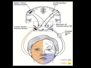
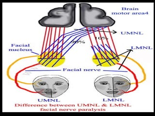
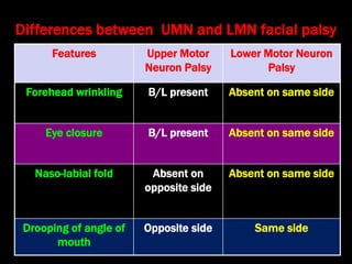
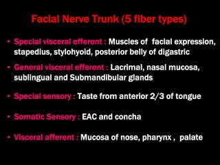
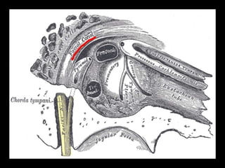
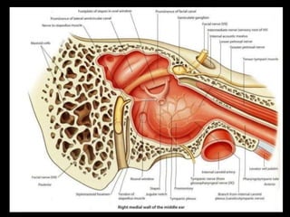
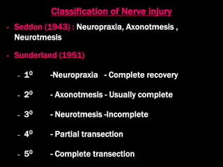
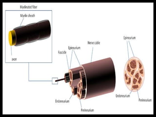
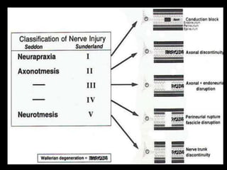
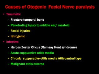
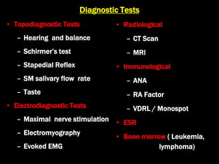
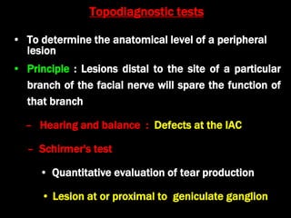
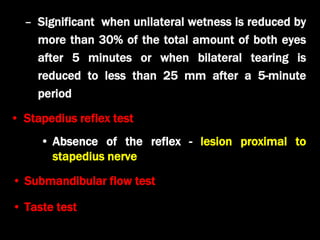
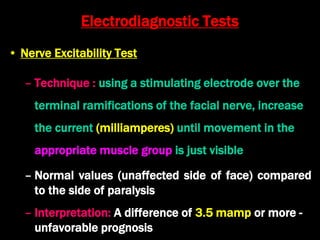
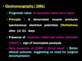
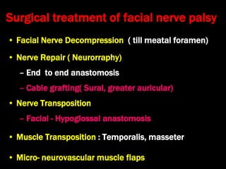
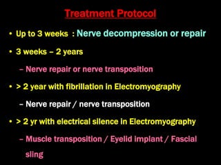
Disorders of the facial
nerve
Dr. Krishna Koirala
2018--09-04
Surgical Anatomy
• Mixed nerve having 10,000 neurons (7, 000
motor and 3,000 sensory)
• Three nuclei
–Motor nucleus : Caudal Pons
–Superior salivatory nucleus : Dorsal to motor
nucleus
–Nucleus of solitary tract : Medulla
• Superior aspect of motor nucleus has both crossed
and uncrossed input
– Upper motor neuron lesions - only the lower part of
the face on the contralateral side will be affected
due to bilateral control to the upper facial muscles
(frontalis and orbicularis oculi)
• Inferior aspect – Contralateral input
– Lower motor neuron lesions - both upper and lower
facial weakness occurs on the same side of lesion
Features Upper Motor
Neuron Palsy
Lower Motor Neuron
Palsy
Forehead wrinkling B/L present Absent on same side
Eye closure B/L present Absent on same side
Naso-labial fold Absent on
opposite side
Absent on same side
Drooping of angle of
mouth
Opposite side Same side
Differences between UMN and LMN facial palsy
Facial Nerve Trunk (5 fiber types)
• Special visceral efferent : Muscles of facial expression,
stapedius, stylohyoid, posterior belly of digastric
• General visceral efferent : Lacrimal, nasal mucosa,
sublingual and Submandibular glands
• Special sensory : Taste from anterior 2/3 of tongue
• Somatic Sensory : EAC and concha
• Visceral afferent : Mucosa of nose, pharynx , palate
Course / parts of facial nerve
F. N .Segment Location Length
(mm)
Supranuclear Cerebral cortex NA
Brain stem Motor nucleus , superior
salivatory nucleus
NA
Meatal segment Brain stem to IAC 13-15
Labyrinthine
segment
Fundus of IAC to geniculate
ganglion
3-4
Tympanic segment Geniculate ganglion to pyramidal
eminence
8-11
Mastoid segment Pyramidal eminence to
Stylomastoid foramen
10-14
Extratemporal
segment
Stylomastoid foramen to pes
anserinus
15-20
• Intracranial : Pons to porous of IAC (24 mm)
• Intratemporal
– Meatal
– Labyrinthine
•Shortest (4mm), narrowest (0.68 mm)
•From fallopian canal to geniculate ganglion (1st genu)
•Branch – greater superficial petrosal nerve
•Lacks anastomosing arterial cascades : Involved in nerve
edema in fracture temporal bone and vascular
compression ,embolic phenomena, low-flow states
• Tympanic (Horizontal) - (13 mm)
– Geniculate ganglion to Pyramidal process (2nd genu)
– Commonly dehiscent (Damaged during surgery)
• Mastoid (Vertical) - 20mm
– Pyramid to stylomastoid foramen
– Second genu lies lateral and posterior to the pyramidal
process
– Branches : Nerve to Stapedius ,Chorda tympani ,Posterior
auricular Muscular
• Extracranial /
Extratemporal
– Peripheral branches
•Temporal
•Zygomatic
•Buccal
•Marginal mandibular
•Cervical
• Processus cochleariformis : (small bony protuberance
from which tensor tympani muscle turns 900 to insert
into malleus) lies 1 mm inferior to geniculate
ganglion
• Cog: bony ridge hanging from tegmen tympani lies 1
mm above & posterior to processus cochleariformis
• Short process of incus: 2 mm below it lies the
external genu
Surgical Landmarks of facial nerve
• Lateral Semicircular Canal: 2 mm anteroinfero-
medially lies the external genu
• Oval window: 1 mm above lies the external genu
• Inferior edge of Posterior S.C.C. : 2 mm anterior &
lateral lies mastoid segment of facial nerve
• Tympano-mastoid suture in posterior canal wall: 5-8
mm medial lies mastoid segment of facial nerve
• Digastric ridge in mastoid tip: leads antero-medially to
mastoid segment of facial nerve
Classification of Nerve injury
• Seddon (1943) : Neuropraxia, Axonotmesis ,
Neurotmesis
• Sunderland (1951)
– 10 -Neuropraxia - Complete recovery
– 20 - Axonotmesis - Usually complete
– 30 - Neurotmesis -Incomplete
– 40 - Partial transection
– 50 - Complete transection
Causes of Otogenic Facial Nerve paralysis
• Traumatic
– Fracture temporal bone
– Penetrating injury to middle ear/ mastoid
– Facial injuries
– Iatrogenic
• Infective
– Herpes Zoster Oticus (Ramsay Hunt syndrome)
– Acute suppurative otitis media
– Chronic suppurative otitis media Atticoantral type
– Malignant otitis externa
• Neoplastic
– Glomus tumors / Schwannoma
– Middle ear malignancies
– Metastatic carcinoma
• Idiopathic
– Bell’s Palsy
Diagnostic Tests
• Topodiagnostic Tests
– Hearing and balance
– Schirmer’s test
– Stapedial Reflex
– SM salivary flow rate
– Taste
• Electrodiagnostic Tests
– Maximal nerve stimulation
– Electromyography
– Evoked EMG
• Radiological
– CT Scan
– MRI
• Immunological
– ANA
– RA Factor
– VDRL / Monospot
• ESR
• Bone marrow ( Leukemia,
lymphoma)
Topodiagnostic tests
• To determine the anatomical level of a peripheral
lesion
• Principle : Lesions distal to the site of a particular
branch of the facial nerve will spare the function of
that branch
– Hearing and balance : Defects at the IAC
– Schirmer's test
• Quantitative evaluation of tear production
• Lesion at or proximal to geniculate ganglion
– Significant when unilateral wetness is reduced by
more than 30% of the total amount of both eyes
after 5 minutes or when bilateral tearing is
reduced to less than 25 mm after a 5-minute
period
• Stapedius reflex test
• Absence of the reflex - lesion proximal to
stapedius nerve
• Submandibular flow test
• Taste test
Electrodiagnostic Tests
• Nerve Excitability Test
– Technique : using a stimulating electrode over the
terminal ramifications of the facial nerve, increase
the current (milliamperes) until movement in the
appropriate muscle group is just visible
– Normal values (unaffected side of face) compared
to the side of paralysis
– Interpretation: A difference of 3.5 mamp or more -
unfavorable prognosis
• Electromyography ( EMG)
– Prognostic value in traumatic facial nerve injury
– Principle : A denervated muscle produces
spontaneous electrical potentials (fibrillations)
after 14 -21 days
– Presence of voluntary motor unit action potential
(VMAP) – sign of incomplete paralysis
– Early presence of VAMP ( 10-14 days) : Better
clinical outcome suggesting no need for surgical
decompression
• Electroneurography (Evoked Electromyography)
– Interpretation: The difference in amplitude of the
potentials of the intact and involved side of the face
correlate with the percentage of degenerated motor
fibers (denervation)
– Advantage: Quantitative analysis of amount of
degeneration
– Disadvantage: Amplitudes are a 24-48 hour
delayed representation of actual events occurring
at site of lesion
Clinical applications
• Facial nerve subjected to traumatic injuries of a
magnitude requiring surgical repair undergo 90%
degeneration within six days of injury
• In cases of Bell's Palsy, a poor prognosis can be
anticipated in patients reaching 95% or more
degeneration within 14 days of onset of the palsy
Common disorders
Bell’s Palsy
• Most common cause of LMN facial palsy (80%)
• Acute, idiopathic, unilateral, peripheral LMN facial
paralysis
– ? Viral prodrome ( Herpes simplex) , ? Vascular
• No sex predilection ,no side predilection
• 5th - 6th decade-Common
• 10% family history
• Pathophysiology
– Nerve swelling within the facial canal
Clinical Features
• Unilateral LMN Facial Paralysis : Progresses to maximal deficit
over 3 to 72 hours
• Pain (50%) : Near the mastoid process
• Excess tearing (33%) ,hyperacusis, dysgeusia
• Facial weakness
– All branches of nerve : Upper & Lower , Unilateral
– Degree : Partial (30%) ; Complete (70%)
– Affected side - flat and expressionless ,twisted intact side,
palpebral fissure wide, eye does not close
• Stapedius dysfunction (33%) : Hyperacusis
• Lacrimation : Mildly affected in some patients
• Taste -- No clinically significant changes in most
patients
• Sensory loss
– Mild or None
– May be present on face or tongue on side of
paralysis
• Natural History
– Complete / Incomplete
– Recovery begins within three weeks
– Full recovery by 6 months in 84% ( 60% in HZO )
– Recurrence : 12% ( Rare IN HZO)
– Decrease in Response to electrical testing
- Peaks in 5-10 days (10-14 days In HZO)
• Herpes Zoster Oticus (Ramsay Hunt
syndrome)
• Acute LMN facial paralysis caused due to Herpes
zoster virus infection of the geniculate ganglion of
the facial nerve
• Viral prodrome
• Severe pain in and around the ear
• Vesicles in pinna, face , neck ,oral cavity (100%)
• SNHL and /or vertigo (40%)
Treatment
1)For all cases of facial paralysis
– Reassurance
– Physical Therapy : Heat, massage
– Psychosomatic Therapy
– Physiotherapy of the face
– Eye care
• Eye care
– Corneal protection
•Antibiotic eye drops e.g.. Ciprofloxacin 2 drops in the
eye TDS
•Antibiotic ointment at night
•Natural tears, isotonic saline and methylcellulose drops
•Strips of skin tape to help close the eye
•Temporary patching
•Tarsorraphy
– Comfort
2)For Bell’s Palsy
• Steroid Therapy
– Prednisone 1mg/kg/day ( 60-80 mg) to begin 24
to 48 h after onset and given for 1 wk, then
decreased gradually over the 2nd wk
•Helps to reduce residual paralysis
•Improves recovery
• Antiviral agents
– Acyclovir, famciclovir
3)For HZO (Ramsay- Hunt)
• Antiviral agents
– Acyclovir 800mg 5 times a day for 7 days
– Best results - treatment started within three days
after symptoms appear
• Steroids
• Carbamazepine : 200-600 mg TDS
• Vaccines
– Varicella vaccine
– Zostavax (helpful in preventing viral reactivation)
4) Other modalities
• Cosmetic restoration( Static Procedures)
–Fascial slings : Fascia Lata
–Tarsorraphy
–Gold weight prosthesis
–Temporalis muscle transposition
–Eyelid springs/ implants
Fascial Slings
Surgical treatment of facial nerve palsy
• Facial Nerve Decompression ( till meatal foramen)
• Nerve Repair ( Neurorraphy)
– End to end anastomosis
– Cable grafting( Sural, greater auricular)
• Nerve Transposition
– Facial - Hypoglossal anastomosis
• Muscle Transposition : Temporalis, masseter
• Micro- neurovascular muscle flaps
Treatment Protocol
• Up to 3 weeks : Nerve decompression or repair
• 3 weeks – 2 years
– Nerve repair or nerve transposition
• > 2 year with fibrillation in Electromyography
– Nerve repair / nerve transposition
• > 2 yr with electrical silence in Electromyography
– Muscle transposition / Eyelid implant / Fascial
sling

Facial nerve palsy

  • 1.
    Disorders of thefacial nerve Dr. Krishna Koirala 2018--09-04
  • 2.
    Surgical Anatomy • Mixednerve having 10,000 neurons (7, 000 motor and 3,000 sensory) • Three nuclei –Motor nucleus : Caudal Pons –Superior salivatory nucleus : Dorsal to motor nucleus –Nucleus of solitary tract : Medulla
  • 4.
    • Superior aspectof motor nucleus has both crossed and uncrossed input – Upper motor neuron lesions - only the lower part of the face on the contralateral side will be affected due to bilateral control to the upper facial muscles (frontalis and orbicularis oculi) • Inferior aspect – Contralateral input – Lower motor neuron lesions - both upper and lower facial weakness occurs on the same side of lesion
  • 7.
    Features Upper Motor NeuronPalsy Lower Motor Neuron Palsy Forehead wrinkling B/L present Absent on same side Eye closure B/L present Absent on same side Naso-labial fold Absent on opposite side Absent on same side Drooping of angle of mouth Opposite side Same side Differences between UMN and LMN facial palsy
  • 8.
    Facial Nerve Trunk(5 fiber types) • Special visceral efferent : Muscles of facial expression, stapedius, stylohyoid, posterior belly of digastric • General visceral efferent : Lacrimal, nasal mucosa, sublingual and Submandibular glands • Special sensory : Taste from anterior 2/3 of tongue • Somatic Sensory : EAC and concha • Visceral afferent : Mucosa of nose, pharynx , palate
  • 9.
    Course / partsof facial nerve
  • 11.
    F. N .SegmentLocation Length (mm) Supranuclear Cerebral cortex NA Brain stem Motor nucleus , superior salivatory nucleus NA Meatal segment Brain stem to IAC 13-15 Labyrinthine segment Fundus of IAC to geniculate ganglion 3-4 Tympanic segment Geniculate ganglion to pyramidal eminence 8-11 Mastoid segment Pyramidal eminence to Stylomastoid foramen 10-14 Extratemporal segment Stylomastoid foramen to pes anserinus 15-20
  • 12.
    • Intracranial :Pons to porous of IAC (24 mm) • Intratemporal – Meatal – Labyrinthine •Shortest (4mm), narrowest (0.68 mm) •From fallopian canal to geniculate ganglion (1st genu) •Branch – greater superficial petrosal nerve •Lacks anastomosing arterial cascades : Involved in nerve edema in fracture temporal bone and vascular compression ,embolic phenomena, low-flow states
  • 13.
    • Tympanic (Horizontal)- (13 mm) – Geniculate ganglion to Pyramidal process (2nd genu) – Commonly dehiscent (Damaged during surgery) • Mastoid (Vertical) - 20mm – Pyramid to stylomastoid foramen – Second genu lies lateral and posterior to the pyramidal process – Branches : Nerve to Stapedius ,Chorda tympani ,Posterior auricular Muscular
  • 14.
    • Extracranial / Extratemporal –Peripheral branches •Temporal •Zygomatic •Buccal •Marginal mandibular •Cervical
  • 15.
    • Processus cochleariformis: (small bony protuberance from which tensor tympani muscle turns 900 to insert into malleus) lies 1 mm inferior to geniculate ganglion • Cog: bony ridge hanging from tegmen tympani lies 1 mm above & posterior to processus cochleariformis • Short process of incus: 2 mm below it lies the external genu Surgical Landmarks of facial nerve
  • 16.
    • Lateral SemicircularCanal: 2 mm anteroinfero- medially lies the external genu • Oval window: 1 mm above lies the external genu • Inferior edge of Posterior S.C.C. : 2 mm anterior & lateral lies mastoid segment of facial nerve • Tympano-mastoid suture in posterior canal wall: 5-8 mm medial lies mastoid segment of facial nerve • Digastric ridge in mastoid tip: leads antero-medially to mastoid segment of facial nerve
  • 19.
    Classification of Nerveinjury • Seddon (1943) : Neuropraxia, Axonotmesis , Neurotmesis • Sunderland (1951) – 10 -Neuropraxia - Complete recovery – 20 - Axonotmesis - Usually complete – 30 - Neurotmesis -Incomplete – 40 - Partial transection – 50 - Complete transection
  • 22.
    Causes of OtogenicFacial Nerve paralysis • Traumatic – Fracture temporal bone – Penetrating injury to middle ear/ mastoid – Facial injuries – Iatrogenic • Infective – Herpes Zoster Oticus (Ramsay Hunt syndrome) – Acute suppurative otitis media – Chronic suppurative otitis media Atticoantral type – Malignant otitis externa
  • 23.
    • Neoplastic – Glomustumors / Schwannoma – Middle ear malignancies – Metastatic carcinoma • Idiopathic – Bell’s Palsy
  • 24.
    Diagnostic Tests • TopodiagnosticTests – Hearing and balance – Schirmer’s test – Stapedial Reflex – SM salivary flow rate – Taste • Electrodiagnostic Tests – Maximal nerve stimulation – Electromyography – Evoked EMG • Radiological – CT Scan – MRI • Immunological – ANA – RA Factor – VDRL / Monospot • ESR • Bone marrow ( Leukemia, lymphoma)
  • 25.
    Topodiagnostic tests • Todetermine the anatomical level of a peripheral lesion • Principle : Lesions distal to the site of a particular branch of the facial nerve will spare the function of that branch – Hearing and balance : Defects at the IAC – Schirmer's test • Quantitative evaluation of tear production • Lesion at or proximal to geniculate ganglion
  • 26.
    – Significant whenunilateral wetness is reduced by more than 30% of the total amount of both eyes after 5 minutes or when bilateral tearing is reduced to less than 25 mm after a 5-minute period • Stapedius reflex test • Absence of the reflex - lesion proximal to stapedius nerve • Submandibular flow test • Taste test
  • 27.
    Electrodiagnostic Tests • NerveExcitability Test – Technique : using a stimulating electrode over the terminal ramifications of the facial nerve, increase the current (milliamperes) until movement in the appropriate muscle group is just visible – Normal values (unaffected side of face) compared to the side of paralysis – Interpretation: A difference of 3.5 mamp or more - unfavorable prognosis
  • 28.
    • Electromyography (EMG) – Prognostic value in traumatic facial nerve injury – Principle : A denervated muscle produces spontaneous electrical potentials (fibrillations) after 14 -21 days – Presence of voluntary motor unit action potential (VMAP) – sign of incomplete paralysis – Early presence of VAMP ( 10-14 days) : Better clinical outcome suggesting no need for surgical decompression
  • 29.
    • Electroneurography (EvokedElectromyography) – Interpretation: The difference in amplitude of the potentials of the intact and involved side of the face correlate with the percentage of degenerated motor fibers (denervation) – Advantage: Quantitative analysis of amount of degeneration – Disadvantage: Amplitudes are a 24-48 hour delayed representation of actual events occurring at site of lesion
  • 30.
    Clinical applications • Facialnerve subjected to traumatic injuries of a magnitude requiring surgical repair undergo 90% degeneration within six days of injury • In cases of Bell's Palsy, a poor prognosis can be anticipated in patients reaching 95% or more degeneration within 14 days of onset of the palsy
  • 31.
  • 32.
    Bell’s Palsy • Mostcommon cause of LMN facial palsy (80%) • Acute, idiopathic, unilateral, peripheral LMN facial paralysis – ? Viral prodrome ( Herpes simplex) , ? Vascular • No sex predilection ,no side predilection • 5th - 6th decade-Common • 10% family history • Pathophysiology – Nerve swelling within the facial canal
  • 33.
    Clinical Features • UnilateralLMN Facial Paralysis : Progresses to maximal deficit over 3 to 72 hours • Pain (50%) : Near the mastoid process • Excess tearing (33%) ,hyperacusis, dysgeusia • Facial weakness – All branches of nerve : Upper & Lower , Unilateral – Degree : Partial (30%) ; Complete (70%) – Affected side - flat and expressionless ,twisted intact side, palpebral fissure wide, eye does not close
  • 34.
    • Stapedius dysfunction(33%) : Hyperacusis • Lacrimation : Mildly affected in some patients • Taste -- No clinically significant changes in most patients • Sensory loss – Mild or None – May be present on face or tongue on side of paralysis
  • 36.
    • Natural History –Complete / Incomplete – Recovery begins within three weeks – Full recovery by 6 months in 84% ( 60% in HZO ) – Recurrence : 12% ( Rare IN HZO) – Decrease in Response to electrical testing - Peaks in 5-10 days (10-14 days In HZO)
  • 37.
    • Herpes ZosterOticus (Ramsay Hunt syndrome) • Acute LMN facial paralysis caused due to Herpes zoster virus infection of the geniculate ganglion of the facial nerve • Viral prodrome • Severe pain in and around the ear • Vesicles in pinna, face , neck ,oral cavity (100%) • SNHL and /or vertigo (40%)
  • 39.
    Treatment 1)For all casesof facial paralysis – Reassurance – Physical Therapy : Heat, massage – Psychosomatic Therapy – Physiotherapy of the face – Eye care
  • 40.
    • Eye care –Corneal protection •Antibiotic eye drops e.g.. Ciprofloxacin 2 drops in the eye TDS •Antibiotic ointment at night •Natural tears, isotonic saline and methylcellulose drops •Strips of skin tape to help close the eye •Temporary patching •Tarsorraphy – Comfort
  • 41.
    2)For Bell’s Palsy •Steroid Therapy – Prednisone 1mg/kg/day ( 60-80 mg) to begin 24 to 48 h after onset and given for 1 wk, then decreased gradually over the 2nd wk •Helps to reduce residual paralysis •Improves recovery • Antiviral agents – Acyclovir, famciclovir
  • 42.
    3)For HZO (Ramsay-Hunt) • Antiviral agents – Acyclovir 800mg 5 times a day for 7 days – Best results - treatment started within three days after symptoms appear • Steroids • Carbamazepine : 200-600 mg TDS • Vaccines – Varicella vaccine – Zostavax (helpful in preventing viral reactivation)
  • 43.
    4) Other modalities •Cosmetic restoration( Static Procedures) –Fascial slings : Fascia Lata –Tarsorraphy –Gold weight prosthesis –Temporalis muscle transposition –Eyelid springs/ implants
  • 44.
  • 45.
    Surgical treatment offacial nerve palsy • Facial Nerve Decompression ( till meatal foramen) • Nerve Repair ( Neurorraphy) – End to end anastomosis – Cable grafting( Sural, greater auricular) • Nerve Transposition – Facial - Hypoglossal anastomosis • Muscle Transposition : Temporalis, masseter • Micro- neurovascular muscle flaps
  • 47.
    Treatment Protocol • Upto 3 weeks : Nerve decompression or repair • 3 weeks – 2 years – Nerve repair or nerve transposition • > 2 year with fibrillation in Electromyography – Nerve repair / nerve transposition • > 2 yr with electrical silence in Electromyography – Muscle transposition / Eyelid implant / Fascial sling

Editor's Notes

  • #16 Surgical landmarks of facial nerve