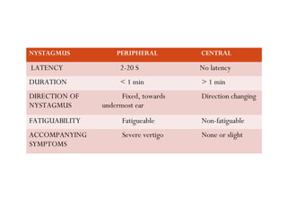
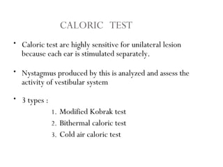
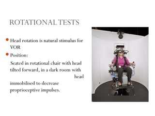
EVALUATION ANDEVALUATION AND
MANAGEMENT OF AMANAGEMENT OF A
PATIENT WITH VERTIGOPATIENT WITH VERTIGO
PRESENTER:PRESENTER:
DR ROHINI R. NAIRDR ROHINI R. NAIR
DEFINITION: VERTIGO
Episodic illusion of movement, either rotatory or linear, of
oneself or environment.
Pathological involvement from labyrinth upto vestibular
cortex.
Dizziness is the 9th
most common cause of presentation to
clinician.
Shunting between general physician, neurologist, ENT
specialist and psychiatrists.
To be differentiated from other causes of dizziness.
TYPES OF DIZZINESS
TYPES DEFINITION CAUSES
Light headedness Feeling of fainting about to
occur
Hypotension,
hypoglycaemia, anxiety.
Presyncope/ syncope Impending loss of
consciousnss
Cerebral perfusion of brain
falls below a critical level.
Vertigo Illusion of movement of
oneself or surroundings.
Disturbance of peripheral
vestibular system or their
central projections.
Disequilibrium Sensation of being off
balance.
Non-vestibular
Dec. somatosensation or
weakness of lower
extremity.
Oscillopsia Stationary object in the
visual environment appears
moving
Vestibular hypofunction.
Others Vague, ill-defined c/o
dizziness
Psychological.
CAUSES OF VERTIGO
PERIPHERALPERIPHERAL: Lesion of vestibular end organs and their first
order neuron(i.e.vestibular nerve)
Responsible for 85% of all cases of vertigo.
CENTRALCENTRAL : Lesion in central neural pathways(vestibular
nuclei, brainstem, cerebellum, vestibular cortex).
CAUSES
PERIPHERAL VERTIGO CENTRAL VERTIGO
• Meniere’s disease
• Benign paroxysmal positional
vertigo
• Vestibular neuronitis
• Labyrinthitis
• Vestibulotoxic drugs
• Head trauma
• Perilymph fistula
• Syphilis
• Acoustic neuroma
• Vertebrobasilar insufficiency
• Posterior inferior cerebellar
artery syndrome.
• Basilar migraine
• Cerebellar disease
• Multiple sclerosis
• Tumours of brainstem and 4th
ventricle
• Epilepsy
• Cervical vertigo
NYSTAGMUS PERIPHERAL CENTRAL
LATENCY 2-20 S No latency
DURATION < 1 min > 1 min
DIRECTION OF
NYSTAGMUS
Fixed, towards
undermost ear
Direction changing
FATIGUABILITY Fatigueable Non-fatiguable
ACCOMPANYING
SYMPTOMS
Severe vertigo None or slight
HISTORY:
1) Nature of sensation.
2) Timing of initial spell.
3) Duration and frequency of symptoms:
Short term spellsShort term spells: BPPV, Perilymph fistula, LSSC fistula.
Medium length spellsMedium length spells: Meniere’s disease, hypoglycemia,
arrythmias.
Longer spellsLonger spells: Vestibular migraine.
BEDSIDE EVALUATION FOR
VESTIBULAR SYSTEM
4) Precipitating factors:
Rolling over bed/Tilting backwards: BPPVBPPV
Sound or pressure stimuli: Superior canal dehiscence syndromeSuperior canal dehiscence syndrome.
Exposure to light or sound/Certain food: Vestibular migraineVestibular migraine.
5) Associated symptoms:
• Aural fullness and tinnitus: Meniere’s DiseaseMeniere’s Disease.
• Headache or visual symptoms: Vestibular migraineVestibular migraine.
• Other cranial nerve involvement: CP angle tumourCP angle tumour.
• Ataxia: Cerebellar DysfunctionCerebellar Dysfunction.
• Sweating, dyspnoea, palpitations: Panic attacks.Panic attacks.
• Otosclerosis : Hearing lossHearing loss.
6) Medical conditions : CVA, head trauma.
7) Drug history:
• Anticonvulsant
-Barbiturates
-Phenytoin
-Carbamazepine
• Alcohol
• Salicylates
• Cinchona alkaloids-quinine
• Aminoglycosides
8) Family History: Otosclerosis/ Meniere’s disease/
Migraine
EXAMINATION
Complete head and neck examination
Otoscopy & audiology
Cranial nerve assessment
Visual acuity
Vestibular function test.
Cerebellar function test.
VESTIBULAR FUNCTION TEST
Examination of eye movements
Examination of postural balance
Otolith function test
1) NYSTAGMUS
• Spontaneous nystagmus
• Gaze evoked nystagmus
• Pursuit Movements
• Saccadic Movements
1) DOLL’S EYE MANOEUVRE
2) FISTULA TEST
3) HEAD THRUST TEST
4) POSITIONAL MANOEUVRE
• Dix Hallpike
1) OCULOGRAPHY
• Electro-nystagmography
• Video-Oculography
1) CALORIC TEST
• Modified Kobrak Test
• Fitzgerald Hallpike Test
(Bithermal Caloric Test)
1) ROTATIONAL TEST
• EXAMINATION OF GAIT,
POSTURE AND BALANCE
• ROMBERG TEST
• UNTERBERGER’S STEPPING
TEST
• WOFEC TEST
• POSTUROGRAPHY
• LINEAR ACCELERATION TEST
• SUBJECTIVE VISUAL VERTICAL
• CLICK – EVOKED VESTIBULAR
MYOGENIC POTENTIAL
NYSTAGMUS
To-and-fro beating of the eyes with slow and fast
component.
Caused by disorder in the physiology of vestibulo-ocular
reflex.
VOR is a three-neuron arc; vestibular afferent neuron,
interneuron, oculomotor neuron.
Fastest reflex in the body, latency 7ms, <5% error rate.
Alexander’s law.
Ewald’s law.
EWALD’S LAW
E.J. Richard Ewald(1892), German physiologist.
Experiments on SCC of pigeons.
1ST
LAW:The axis of nystagmus parallels the anatomic axis of
semi-circular canal that generated it.
2ND
LAW:Ampullopetal endolymphatic flow produces a
stronger response than ampullofugal flow, in the horizontal
canal.
ALEXANDER’S LAW:
GustavAlexander(1912),Austrian otolaryngologist.
FIRST LAW:After an acute vestibular impairment,
spontaneous nystagmus has the first phase directed towards
healthy ear.
SECOND LAW: Nystagmus is maximum when gaze is
directed towards healthy ear, attentuated at central gaze and
may be absent at gaze towards impaired ear.
THIRD LAW: Spontaneous nystagmus with central gaze is
augmented, when vision is denied.
SPONTANEOUS NYSTAGMUS
Nystagmus in the primary, straight ahead position of the eyes.
Usually seen during acute vertigo attack.
Waveform, direction of beat, site of lesion.
Severity of nystagmus
-1st
degree: weak nystagmus, present only when pt.look in
direction of fast component.
- 2nd
degree: moderate nystagmus, present when pt. looks
straight ahead.
- 3rd
degree: strong nystagmus, present when pt. looks in
direction of slow component.
GAZE EVOKED NYSTAGMUS
Patient is asked to look 30 degree from the primary position, in
any direction.
SMOOTH PURSUIT MOVEMENTS
-Tracking movement of the eye, when the target moves slowly
(<10-15 deg/sec).
-Velocity limited.
-Normal pursuit rules out central vestibular disorder.
SACCADIC EYE MOVEMENTS
-Fast movements of eyes(200-500 deg/sec)which allows to shift
gaze from one object of interest to another.
DOLL’S EYETEST
Patient in sitting position
↓
Asked to fixate on nose of examiner
↓
Examiner oscillates patient’s head
from side to side(0.5-1Hz).
↓
1)Absence ofVOR - Eye movement
will not be smooth and interrupted
by catchup saccades towards fixation
target.
2) Decrease in dynamic visual
activity.
3) On opthalmoscopy, small
amplitude nystagmus can be seen.
FISTULA TEST
Stimulus – intermittent pressure on tragus or with use of Siegle’s
speculum.
Response :
Normal- No vertigo and nystagmus.
Positive- vertigo and nystagmus.
Mechanism – in presence of fistula or vestibulofibrous bands,
applied pressure causes deviation of cupula.
Positive in HSC fistula, Meniere’s disease etc
To makeVOR deficit more
apparent.
Sudden discrete brisk and
unpredicatable head turn(10-
15deg)
↓
Catchup Saccades seen
Useful for identifying acute U/L
peripheral vestibular deficits.
HEAD THRUST TEST
POSITIONAL MANOEUVRE
Dix-Hall pike manoeuvre:
Used for identification of posterior canal BPPV
Features of Nystagmus:
1.Latency of 2-10sec
2.Increase in amplitude in 10s and decline in 30s.
3.Fatuigability present.
4.Direction of Nystagmus-Vertical torsional nystagmus.
HCVariant of BPPV- Exclusively horizontal, lasts longer.
ACVariant of BPPV- Rare, vertical torsional nystagmus.
LABORATORYTESTS OF EYE MOVEMENTS
ELECTRONYSTAGMOGRAPHY
PRINCIPLE:
• Retina is negatively charged as compared
to cornea, creating an electric field k/a
corneoretinal potential
• Movement of eye Movement of→
electric field Change in electric→
potential detected by surface electrodes.
INDICATIONS
Assessment of eye movements in dark (avoid visual
suppression).
Nystagmus waveform (to d/b congenital and acquired).
Quantification to monitor progression and recovery
from vestibular disorders.
OTHERTECHNIQUES
VIDEO NYSTAGMOGRAPHY
-Small video camera mount on goggles used.
-2D Systems record horizontal and vertical nystagmus.
-3D Records torsional nystagmus.
INFRARED OCULOGRAPHY.
SCLERAL SEARCH COILTECHNIQUE.
CALORICTEST
Principle: Changes in temperature in external auditory canal
influence the activity of vestibular labyrinth.
Position of Patient: Supine with head raised 30 degree above
horizontal-which places HSC in vertical position.
Mechanism:Thermal changes induce convection currents in
horizontal canal when placed vertically and thus, cupular deflections
and change the discharge rate of vestibular nerves.
CALORIC TEST
• Caloric test are highly sensitive for unilateral lesion
because each ear is stimulated separately.
• Nystagmus produced by this is analyzed and assess the
activity of vestibular system
• 3 types :
1. Modified Kobrak test
2. Bithermal caloric test
3. Cold air caloric test
• Fitzgerald – Hallpike test.
• Pt. lies supine with head tilted 30 degree forward so that
HSCC is vertical.
• Ear is irrigated for 40 sec, with water at 30 degree and 44
degree and nystagmus are noted till its end point.
• If no nystagmus appeared from any ear, test is repeated with
water at 20 degree water for 4 minute before labeling the
labyrinth dead.
BITHERMAL CALORIC TEST
MODIFIED KOBRAKTEST
• Pt. seated with head tilted 60 degree backwards to place H S C C
in vertical position.
• Ear irrigated by ice cool water for 60 sec with 5/ 10/20/40 ml.
• Response seen with 5ml of water towards opposite ear - Normal
• Response seen with 5 to 40 ml water – Hypoactive labyrinth.
• No response seen with 40 ml – Dead labyrinth.
COLDAIR CALORICTEST
• Test done in perforated TM because irrigation is
contraindicated.
• Done with DUNDAS GRANT TUBE , coiled copper
tube wrapped in cloth, air is cooled by mixing with
ethyl chloride and then blown to ear.
• “Varioair” is used these days which has precise control
over amount and temperature of air.
INTERPRETATION
Calculating the velocity of each of the slow phase component of
nystagmus.
UW = (RW+RC)-(LW+LC) × 100
(RW+RC)+(LW+LC)
is a sign of decrease responsiveness of horizontal canal.
DP= (RW+LC)-(LW+RC) × 100
(RW+RC)+(LW+LC)
i.e duration of nystagmus to R/L, irrespective of its side of
elicitation.
UW > 20%,DP > 25% considered significant
ABNORMALITIES :
 Bilateral absence of Caloric NystagmusBilateral absence of Caloric Nystagmus: Ototoxicity or
post meningitis
 Unilateral canal paresisUnilateral canal paresis: U/LVestibular Neuritis, U/L
vestibular schwannoma
 Direction PreponderanceDirection Preponderance: non-specific.
 AbnormalVOR suppressionAbnormalVOR suppression: central disorder of eye
movement.
 Perverted nystagmusPerverted nystagmus: posterior fossa disease
ROTATIONALTESTS
Head rotation is natural stimulus for
VOR
Position:
Seated in rotational chair with head
tilted forward, in a dark room with
head
immobilised to decrease
proprioceptive impulses.
TECHNIQUES
VELOCITY STEP
UP/IMPULSIVE ROTATIONAL
TEST
SINUSOIDAL ROTATION
Stimulus- Sudden increase in chair velocity
( acceleration)
↓
Full chair velocity maintained for 60-90 sec
until the nystagmus stops.
↓
Chair suddenly stopped.(de-acceleration)
↓
Nystagmus in opposite direction.
↓
The whole procedure repeated in opposite
direction
Right rotation induces right beating
nystagmus.
Sudden stop leads to left beating Nystagmus
Velocity of chair is sinusoidly
modulated and peak velocity of
stimulus is kept constant
↓
Acceleration increases progressively
• The computer compares head velocity, slow phase eye velocity
and calculates phase, gain, symmetry for each of the test
frequency.
• Gain: Slow eye velocity /head velocity. Reduction in gain seen
in B/L vestibular disease.
• Phase angle: Temporal relationship between eye and head
velocity.
Increase phase - Peripheral vestibular system
Decreased phase - Cerebellar lesion.
• Symmetry: Ratio of rightward to leftward slow phase eye
velocity.
EXAMINATION OF POSTURAL
BALANCE
UNTERBERGER’S STEPPING TEST:
• Visual and proprioceptive inputs are cut off
• Pt deviate/rotate to side of weaker hypoactive vestibule.
• Parameters evaluated in test:
 Displacement
 Angular deviation
 Angular rotation
 Breadth of lateral sway
ROMBERG TEST :
 Pt. blind folded and stands erect with feet close together for 1
minute.
 Max. breadth of sway is 10 cm .
 >10cm - abnormal - indicates a central lesion.
WOFEC TEST :
 Pt is asked to walk on floor on a imaginary straight line with
tandem walk and eyes closed.
 In central lesion and acute peripheral vestibular lesion, patient
falls repeatedly.
POSTUROGRAPHY
Quantitative measurements of processes that maintain
upright stance under static and dynamic conditions.
The most commonly used posturography paradigm is
sensory organization test.
Equilibrium score measures patient’s sway when standing
upright in 6 conditions.
RESULT:
 Eyes closed, stable surface(2):Eyes closed, stable surface(2): Loss of proprioceptive
functions.
 Eyes open, sway referenced surface(4)Eyes open, sway referenced surface(4):: Loss of visual
contribution to balance.
 Eyes closed, sway referenced surface(5)Eyes closed, sway referenced surface(5):: Loss of
Vestibular function.
USES:
Planning the course ofVestibular rehabilitation and
modulating the response in patients with vestibular
hypofunction.
Documentation of postural responses in suspected
malingering, exaggeration of disability, conversion disorder.
EVALUATION OF OTOLITH FUNCTION
Vestibular Evoked Myogenic Potential Responses
Short latency responses
measured from tonically
contracting SCM that relax
in response to I/L presentation
of loud click.
VEMP responses probably
originate in sacculesaccule.
Each ear can be tested separately.
Clinical applications of this test are still
developing, main diagnostic value in
SSC dehiscence syndrome.
OTHERS:
Subjective visual vertical.
Linear acceleration test.
Perceptual and subjective vertical testing.
MANAGEMENT
Pharmacological treatment.
Vestibular rehabilitation.
Surgical treatment.
Adjunctive treatment.
PHARMACOLOGICAL TREATMENT
Suppress symptoms during acute vestibular attack i.e
vestibular sedatives.
Specific treatment of condition that causes vestibular
symptoms i.e. Meniere,migraine.
General medical t/t of other coexisting or underlying
condition.
Experimental drugs- accelerate compensation.
VESTIBULAR SEDATIVES
ANTI-CHOLINERGICS
 Targets efferent feedback from brainstem to vestibular labyrinth and
muscarinic receptor antagonist, increases motion tolerance.
 Hyoscine: most effective drug for t/t of motion sickness.
 Scopolamine.
 S/E: Dry mouth, dilated pupils, sedation.
ANTI-HISTAMINICS
 Competitive antagonists at H1 receptors, increases firing of MVN
through H2 receptors.
 PHENOTHIAZINES: Promethazine.
 PIPERAZINES: Cyclizine, dimenhydrinate.
VESTIBULAR SEDATIVES
DOPAMINE ANTAGONISTS
 D2 receptor antagonist, acts through its anti-emetic action.
 Prochlorperazine( STEMETIL)
 S/E: Sedation, extrapyramidal side effects.
CALCIUM CHANNEL BLOCKERS
 Mechnism of action- unclear
 Inhibits influx of Ca2+ intracellularly
 Flunarizine, nimodipine.
VESTIBULAR SEDATIVES
OTHERS
 Diazepam: modulates GABA receptors, acting centrally to suppress
vestibular responses.
 Ondansetron : anti-emetic, 5-HT3 antagonist. Limited use in vertigo.
SPECIFIC DRUGS
BETAHISTINE(VERTIN):
MOA: Reduces endolymphatic pressure through improved
microvascular circulation in stria vascularis of cochlea.
Inhibits activity of vestibular nuclei.
Reduces vertigo, possibly tinnitus, no effect on hearing loss.
Prescribed initially 16 mg tds, maintenance dose 24-48 mg.
S/E: GI disturbance, headaches, rashes, pruritis.
Caution in asthma, PUD, pregnancy, breast-feeding.
SPECIFIC DRUGS
DIURETICS:
Hydrochlorothiazide, acetazolamide and co-triamterzide.
Reduced the accumulation of endolymph.
Used in Meniere’s disease.
INTRATYMPANIC INJECTIONS OF
AMINOGLYCOSIDES.
Chemical labyrinthectomy
Partially ablative procedure for Meniere’s disease.
STEROIDS
EXPERIMENTAL DRUGS
Aims to enhance vestibular compensation and target specific
central vestibular neurotransmitters.
Gingko biloba, melanotropic peptides, opiates.
Homeopathic remedies.
VESTIBULAR REHABILITATION
Based on the concept of capacity of vestibular system for
adaptation, habituation and plasticity for recalibration
of vestibular reflexes by substitution of sensory input,
motor responses and strategies,in order to achieve
symptomatic recovery following a vestibular lesion.
Includes:
Repositioning exercises.
Adaptation exercises.
Habituation exercises.
General exercises to increase muscle force, balance and gait.
GENERAL PRINCIPLES OF VRT
Decrease centrally sedating or vestibular suppressant drugs
Exercise must provoke vertigo
Initiate as early as possible
Exercise should simulate real life situations
Maintenance exercises to recurrence of symptoms
KEY COMPONENTS OF VRT
Explain the rationale behind the exercises and patient
motivation.
Choice of exercise.
Graded approach, to facilitate learning.
Diligence and perservance.
PHYSICAL EXERCISE REGIMENS
Targets adaptation, sensory substitution, habituation and
balance retraining.
Includes:
 Systematic preset exercise programmes.
 Cawthorne-Cooksey exercises.
 Vestibular habituation exercises: Norre’s approach.
 Customized exercise programmes.
 University of Michigan’s approach.
 The John Hopkins/University of Miami programme.
PRESET EXERCISES
CAWTHORNE-COOKSEY VETIBULAR HABITUATION
TRAINING(NORRE)
Based on empirical observations.
Eye-head coordination tasks,
balance tasks.
Tailored to symptoms
Based on stimulus-specific
‘error-signal’ driven adaptation.
Positional manoeuvres.
Tailored to symptoms.
CUSTOMISED EXERCISE
PROGRAMMES
UNIVERSITY OF MICHIGAN’S
APPROACH
JOHN HOPKINS/UNIVERSITY OF
MIAMI PROGRAMME
Aims to optimise compensation, to
reduce risk of falls and to educate the
patient.
Habituation tasks, balance and gait
retraining.
Selected on basis of posturography, gait
evaluation and motion sensitivity
quotient.
Aims to improve balance and vision
during locomotion, to reduce social
isolation and sensation of disequilibrium.
Adaptation, substitution, habituation,
cawthorne-Cooksey exercises and
functional activities.
Selected on basis of medical history and
diagnosis.
REPOSITIONING MANOEUVRES
AND EXERCISES
Based on the canalithiasis theory of free-floating debris in the
SCC, by John Epley(1991).
To move the displaced canaliths from the canal, into the
vestibule, causing signs and symptoms to resolve.
Involve a series of specifically patterned head and trunk
movements performed by a trained professional, while
carefully observing the eye movements.
SPECIFIC TREATMENT FOR p-BPPV
1)EPLEY’S REPOSITIONING MANOEUVRE
P-BPPV
2) SEMONT’S LIBERATORY MANOEUVRE (92%)
P-BPPV
3)BRANDT-DAROFF POSITIONAL EXERCISE
Based on Schuknecht’s hypothesis of cupulolithiasis
Self treatment, in the form of exercises, patient should
perform for 15 minutes- 3 times daily.
Authors reported resolution of symptoms in 98% of patients
within 3-14 days.
Recent controlled trial, only 23% after 1 week.
BRANDT DAROFF EXERCISE
SPECIFIC TREATMENT OF h-BPPV
FORCED PROLONGED POSITION
Vannuchi et al, recommended, patients with h-BPPV to lie
down on healthy side for 12 hours
Allows otolith debris to gravitate to vestibule, by maintaining
affected h-SCC uppermost.
74.3% recovery rate within 3days.
h-BPPV  I/L p-BPPV; treated with Semont’s.
270 ˚ BARBECUE MANOEUVRE
Adaptation of Epley’s manoeuvre.
H-BPPV
360˚YAW ROTATION:
360 deg yaw rotation performed in 90 deg steps at 30 sec
intervals.
Devised by Baloh.
75% recovery in 1 week.
LIBERATORY MANOEUVRES:
 Meilleure et al.Meilleure et al. used a manoeuvre, with 100% success rate.
 Pt. in supine head lifted by 30 deg  turned to affected
side, maintained for 5 mins head turned to 180 deg,
maintained for 5 mins.
Modified Semont’sModified Semont’s by Casani et al.
SPECIFIC TREATMENT OF a-BPPV
MODIFIED EPLEY’S MANOEUVRE:
Head turned 45 deg away from the affected ear, brought to
30 deg below the horizontal, for 30 secs.
Head elevated while for 1 minute, maintaining supine
position.
Seated position assumed with chin bent forwards at 30 deg.
96.7% efficacy reported.
SURGICAL INTERVENTION
Surgical options contemplated with long standing peripheral
vestibular disorders, that fail medical treatment.
BPPV:
Singular neurectomy:Transcanal approach, RW niche
exposed Scala tympani visualised Drilling at post margin
of membrane, 1-2 mm deep, where singular nerve is identified
and severed.
Posterior SCC occlusion: Mastoidectomy, bony posterior SCC
identified Occluded with bone chips and fascia, at farthest
point from ampulla, inferior to region bisected by HSCC.
SURGICAL INTERVENTION
MENIERE’S DISEASE:
Decompression of endolymphatic sac:
 FICK’S OPERATION
 CODY’S TACK PROCEDURE
 OTIC-PERIOTIC SHUNT
Endolymphatic shunt operation.
Vestibular neurectomy via middle fossa or retrosigmoid
approach.
Labyrinthectomy: Membranous labyrinth is completely
destroyed via transmastoid or transcanal approach.
Local overpressure therapy(Meniett device)
SURGICAL INTERVENTION
SUPERIOR CANAL DEHISCENCE SYNDROME:
Absence of bone over superior SCC; third mobile window.
Surgical plugging of affected canal, with fascia or bone chips’
Approach: middle cranial fossamiddle cranial fossa or transmastoid.transmastoid.
ADJUNCTIVE TREATMENT
Aims to address issues relating to vision and proprioception.
Includes environmental modifications and safety measures.
Psychological and psychiatric interventions like cognitive
behaviour therapy, relaxing and breathing exercises.
THANKYOU!

Vertigo

  • 1.
    EVALUATION ANDEVALUATION AND MANAGEMENTOF AMANAGEMENT OF A PATIENT WITH VERTIGOPATIENT WITH VERTIGO PRESENTER:PRESENTER: DR ROHINI R. NAIRDR ROHINI R. NAIR
  • 2.
    DEFINITION: VERTIGO Episodic illusionof movement, either rotatory or linear, of oneself or environment. Pathological involvement from labyrinth upto vestibular cortex. Dizziness is the 9th most common cause of presentation to clinician. Shunting between general physician, neurologist, ENT specialist and psychiatrists. To be differentiated from other causes of dizziness.
  • 3.
    TYPES OF DIZZINESS TYPESDEFINITION CAUSES Light headedness Feeling of fainting about to occur Hypotension, hypoglycaemia, anxiety. Presyncope/ syncope Impending loss of consciousnss Cerebral perfusion of brain falls below a critical level. Vertigo Illusion of movement of oneself or surroundings. Disturbance of peripheral vestibular system or their central projections. Disequilibrium Sensation of being off balance. Non-vestibular Dec. somatosensation or weakness of lower extremity. Oscillopsia Stationary object in the visual environment appears moving Vestibular hypofunction. Others Vague, ill-defined c/o dizziness Psychological.
  • 4.
    CAUSES OF VERTIGO PERIPHERALPERIPHERAL:Lesion of vestibular end organs and their first order neuron(i.e.vestibular nerve) Responsible for 85% of all cases of vertigo. CENTRALCENTRAL : Lesion in central neural pathways(vestibular nuclei, brainstem, cerebellum, vestibular cortex).
  • 5.
    CAUSES PERIPHERAL VERTIGO CENTRALVERTIGO • Meniere’s disease • Benign paroxysmal positional vertigo • Vestibular neuronitis • Labyrinthitis • Vestibulotoxic drugs • Head trauma • Perilymph fistula • Syphilis • Acoustic neuroma • Vertebrobasilar insufficiency • Posterior inferior cerebellar artery syndrome. • Basilar migraine • Cerebellar disease • Multiple sclerosis • Tumours of brainstem and 4th ventricle • Epilepsy • Cervical vertigo
  • 6.
    NYSTAGMUS PERIPHERAL CENTRAL LATENCY2-20 S No latency DURATION < 1 min > 1 min DIRECTION OF NYSTAGMUS Fixed, towards undermost ear Direction changing FATIGUABILITY Fatigueable Non-fatiguable ACCOMPANYING SYMPTOMS Severe vertigo None or slight
  • 7.
    HISTORY: 1) Nature ofsensation. 2) Timing of initial spell. 3) Duration and frequency of symptoms: Short term spellsShort term spells: BPPV, Perilymph fistula, LSSC fistula. Medium length spellsMedium length spells: Meniere’s disease, hypoglycemia, arrythmias. Longer spellsLonger spells: Vestibular migraine. BEDSIDE EVALUATION FOR VESTIBULAR SYSTEM
  • 8.
    4) Precipitating factors: Rollingover bed/Tilting backwards: BPPVBPPV Sound or pressure stimuli: Superior canal dehiscence syndromeSuperior canal dehiscence syndrome. Exposure to light or sound/Certain food: Vestibular migraineVestibular migraine. 5) Associated symptoms: • Aural fullness and tinnitus: Meniere’s DiseaseMeniere’s Disease. • Headache or visual symptoms: Vestibular migraineVestibular migraine. • Other cranial nerve involvement: CP angle tumourCP angle tumour. • Ataxia: Cerebellar DysfunctionCerebellar Dysfunction. • Sweating, dyspnoea, palpitations: Panic attacks.Panic attacks. • Otosclerosis : Hearing lossHearing loss.
  • 9.
    6) Medical conditions: CVA, head trauma. 7) Drug history: • Anticonvulsant -Barbiturates -Phenytoin -Carbamazepine • Alcohol • Salicylates • Cinchona alkaloids-quinine • Aminoglycosides 8) Family History: Otosclerosis/ Meniere’s disease/ Migraine
  • 10.
    EXAMINATION Complete head andneck examination Otoscopy & audiology Cranial nerve assessment Visual acuity Vestibular function test. Cerebellar function test.
  • 11.
    VESTIBULAR FUNCTION TEST Examinationof eye movements Examination of postural balance Otolith function test
  • 12.
    1) NYSTAGMUS • Spontaneousnystagmus • Gaze evoked nystagmus • Pursuit Movements • Saccadic Movements 1) DOLL’S EYE MANOEUVRE 2) FISTULA TEST 3) HEAD THRUST TEST 4) POSITIONAL MANOEUVRE • Dix Hallpike 1) OCULOGRAPHY • Electro-nystagmography • Video-Oculography 1) CALORIC TEST • Modified Kobrak Test • Fitzgerald Hallpike Test (Bithermal Caloric Test) 1) ROTATIONAL TEST
  • 13.
    • EXAMINATION OFGAIT, POSTURE AND BALANCE • ROMBERG TEST • UNTERBERGER’S STEPPING TEST • WOFEC TEST • POSTUROGRAPHY
  • 14.
    • LINEAR ACCELERATIONTEST • SUBJECTIVE VISUAL VERTICAL • CLICK – EVOKED VESTIBULAR MYOGENIC POTENTIAL
  • 15.
    NYSTAGMUS To-and-fro beating ofthe eyes with slow and fast component. Caused by disorder in the physiology of vestibulo-ocular reflex. VOR is a three-neuron arc; vestibular afferent neuron, interneuron, oculomotor neuron. Fastest reflex in the body, latency 7ms, <5% error rate. Alexander’s law. Ewald’s law.
  • 16.
    EWALD’S LAW E.J. RichardEwald(1892), German physiologist. Experiments on SCC of pigeons. 1ST LAW:The axis of nystagmus parallels the anatomic axis of semi-circular canal that generated it. 2ND LAW:Ampullopetal endolymphatic flow produces a stronger response than ampullofugal flow, in the horizontal canal.
  • 17.
    ALEXANDER’S LAW: GustavAlexander(1912),Austrian otolaryngologist. FIRSTLAW:After an acute vestibular impairment, spontaneous nystagmus has the first phase directed towards healthy ear. SECOND LAW: Nystagmus is maximum when gaze is directed towards healthy ear, attentuated at central gaze and may be absent at gaze towards impaired ear. THIRD LAW: Spontaneous nystagmus with central gaze is augmented, when vision is denied.
  • 18.
    SPONTANEOUS NYSTAGMUS Nystagmus inthe primary, straight ahead position of the eyes. Usually seen during acute vertigo attack. Waveform, direction of beat, site of lesion. Severity of nystagmus -1st degree: weak nystagmus, present only when pt.look in direction of fast component. - 2nd degree: moderate nystagmus, present when pt. looks straight ahead. - 3rd degree: strong nystagmus, present when pt. looks in direction of slow component.
  • 19.
    GAZE EVOKED NYSTAGMUS Patientis asked to look 30 degree from the primary position, in any direction. SMOOTH PURSUIT MOVEMENTS -Tracking movement of the eye, when the target moves slowly (<10-15 deg/sec). -Velocity limited. -Normal pursuit rules out central vestibular disorder. SACCADIC EYE MOVEMENTS -Fast movements of eyes(200-500 deg/sec)which allows to shift gaze from one object of interest to another.
  • 20.
    DOLL’S EYETEST Patient insitting position ↓ Asked to fixate on nose of examiner ↓ Examiner oscillates patient’s head from side to side(0.5-1Hz). ↓ 1)Absence ofVOR - Eye movement will not be smooth and interrupted by catchup saccades towards fixation target. 2) Decrease in dynamic visual activity. 3) On opthalmoscopy, small amplitude nystagmus can be seen.
  • 21.
    FISTULA TEST Stimulus –intermittent pressure on tragus or with use of Siegle’s speculum. Response : Normal- No vertigo and nystagmus. Positive- vertigo and nystagmus. Mechanism – in presence of fistula or vestibulofibrous bands, applied pressure causes deviation of cupula. Positive in HSC fistula, Meniere’s disease etc
  • 22.
    To makeVOR deficitmore apparent. Sudden discrete brisk and unpredicatable head turn(10- 15deg) ↓ Catchup Saccades seen Useful for identifying acute U/L peripheral vestibular deficits. HEAD THRUST TEST
  • 23.
    POSITIONAL MANOEUVRE Dix-Hall pikemanoeuvre: Used for identification of posterior canal BPPV Features of Nystagmus: 1.Latency of 2-10sec 2.Increase in amplitude in 10s and decline in 30s. 3.Fatuigability present. 4.Direction of Nystagmus-Vertical torsional nystagmus. HCVariant of BPPV- Exclusively horizontal, lasts longer. ACVariant of BPPV- Rare, vertical torsional nystagmus.
  • 25.
    LABORATORYTESTS OF EYEMOVEMENTS ELECTRONYSTAGMOGRAPHY PRINCIPLE: • Retina is negatively charged as compared to cornea, creating an electric field k/a corneoretinal potential • Movement of eye Movement of→ electric field Change in electric→ potential detected by surface electrodes.
  • 26.
    INDICATIONS Assessment of eyemovements in dark (avoid visual suppression). Nystagmus waveform (to d/b congenital and acquired). Quantification to monitor progression and recovery from vestibular disorders.
  • 27.
    OTHERTECHNIQUES VIDEO NYSTAGMOGRAPHY -Small videocamera mount on goggles used. -2D Systems record horizontal and vertical nystagmus. -3D Records torsional nystagmus. INFRARED OCULOGRAPHY. SCLERAL SEARCH COILTECHNIQUE.
  • 28.
    CALORICTEST Principle: Changes intemperature in external auditory canal influence the activity of vestibular labyrinth. Position of Patient: Supine with head raised 30 degree above horizontal-which places HSC in vertical position. Mechanism:Thermal changes induce convection currents in horizontal canal when placed vertically and thus, cupular deflections and change the discharge rate of vestibular nerves.
  • 29.
    CALORIC TEST • Calorictest are highly sensitive for unilateral lesion because each ear is stimulated separately. • Nystagmus produced by this is analyzed and assess the activity of vestibular system • 3 types : 1. Modified Kobrak test 2. Bithermal caloric test 3. Cold air caloric test
  • 30.
    • Fitzgerald –Hallpike test. • Pt. lies supine with head tilted 30 degree forward so that HSCC is vertical. • Ear is irrigated for 40 sec, with water at 30 degree and 44 degree and nystagmus are noted till its end point. • If no nystagmus appeared from any ear, test is repeated with water at 20 degree water for 4 minute before labeling the labyrinth dead. BITHERMAL CALORIC TEST
  • 31.
    MODIFIED KOBRAKTEST • Pt.seated with head tilted 60 degree backwards to place H S C C in vertical position. • Ear irrigated by ice cool water for 60 sec with 5/ 10/20/40 ml. • Response seen with 5ml of water towards opposite ear - Normal • Response seen with 5 to 40 ml water – Hypoactive labyrinth. • No response seen with 40 ml – Dead labyrinth.
  • 32.
    COLDAIR CALORICTEST • Testdone in perforated TM because irrigation is contraindicated. • Done with DUNDAS GRANT TUBE , coiled copper tube wrapped in cloth, air is cooled by mixing with ethyl chloride and then blown to ear. • “Varioair” is used these days which has precise control over amount and temperature of air.
  • 33.
    INTERPRETATION Calculating the velocityof each of the slow phase component of nystagmus. UW = (RW+RC)-(LW+LC) × 100 (RW+RC)+(LW+LC) is a sign of decrease responsiveness of horizontal canal. DP= (RW+LC)-(LW+RC) × 100 (RW+RC)+(LW+LC) i.e duration of nystagmus to R/L, irrespective of its side of elicitation. UW > 20%,DP > 25% considered significant
  • 34.
    ABNORMALITIES :  Bilateralabsence of Caloric NystagmusBilateral absence of Caloric Nystagmus: Ototoxicity or post meningitis  Unilateral canal paresisUnilateral canal paresis: U/LVestibular Neuritis, U/L vestibular schwannoma  Direction PreponderanceDirection Preponderance: non-specific.  AbnormalVOR suppressionAbnormalVOR suppression: central disorder of eye movement.  Perverted nystagmusPerverted nystagmus: posterior fossa disease
  • 35.
    ROTATIONALTESTS Head rotation isnatural stimulus for VOR Position: Seated in rotational chair with head tilted forward, in a dark room with head immobilised to decrease proprioceptive impulses.
  • 36.
    TECHNIQUES VELOCITY STEP UP/IMPULSIVE ROTATIONAL TEST SINUSOIDALROTATION Stimulus- Sudden increase in chair velocity ( acceleration) ↓ Full chair velocity maintained for 60-90 sec until the nystagmus stops. ↓ Chair suddenly stopped.(de-acceleration) ↓ Nystagmus in opposite direction. ↓ The whole procedure repeated in opposite direction Right rotation induces right beating nystagmus. Sudden stop leads to left beating Nystagmus Velocity of chair is sinusoidly modulated and peak velocity of stimulus is kept constant ↓ Acceleration increases progressively
  • 37.
    • The computercompares head velocity, slow phase eye velocity and calculates phase, gain, symmetry for each of the test frequency. • Gain: Slow eye velocity /head velocity. Reduction in gain seen in B/L vestibular disease. • Phase angle: Temporal relationship between eye and head velocity. Increase phase - Peripheral vestibular system Decreased phase - Cerebellar lesion. • Symmetry: Ratio of rightward to leftward slow phase eye velocity.
  • 38.
    EXAMINATION OF POSTURAL BALANCE UNTERBERGER’SSTEPPING TEST: • Visual and proprioceptive inputs are cut off • Pt deviate/rotate to side of weaker hypoactive vestibule. • Parameters evaluated in test:  Displacement  Angular deviation  Angular rotation  Breadth of lateral sway
  • 39.
    ROMBERG TEST : Pt. blind folded and stands erect with feet close together for 1 minute.  Max. breadth of sway is 10 cm .  >10cm - abnormal - indicates a central lesion. WOFEC TEST :  Pt is asked to walk on floor on a imaginary straight line with tandem walk and eyes closed.  In central lesion and acute peripheral vestibular lesion, patient falls repeatedly.
  • 40.
    POSTUROGRAPHY Quantitative measurements ofprocesses that maintain upright stance under static and dynamic conditions. The most commonly used posturography paradigm is sensory organization test. Equilibrium score measures patient’s sway when standing upright in 6 conditions.
  • 42.
    RESULT:  Eyes closed,stable surface(2):Eyes closed, stable surface(2): Loss of proprioceptive functions.  Eyes open, sway referenced surface(4)Eyes open, sway referenced surface(4):: Loss of visual contribution to balance.  Eyes closed, sway referenced surface(5)Eyes closed, sway referenced surface(5):: Loss of Vestibular function.
  • 43.
    USES: Planning the courseofVestibular rehabilitation and modulating the response in patients with vestibular hypofunction. Documentation of postural responses in suspected malingering, exaggeration of disability, conversion disorder.
  • 44.
    EVALUATION OF OTOLITHFUNCTION Vestibular Evoked Myogenic Potential Responses Short latency responses measured from tonically contracting SCM that relax in response to I/L presentation of loud click. VEMP responses probably originate in sacculesaccule. Each ear can be tested separately. Clinical applications of this test are still developing, main diagnostic value in SSC dehiscence syndrome.
  • 45.
    OTHERS: Subjective visual vertical. Linearacceleration test. Perceptual and subjective vertical testing.
  • 46.
  • 47.
    PHARMACOLOGICAL TREATMENT Suppress symptomsduring acute vestibular attack i.e vestibular sedatives. Specific treatment of condition that causes vestibular symptoms i.e. Meniere,migraine. General medical t/t of other coexisting or underlying condition. Experimental drugs- accelerate compensation.
  • 48.
    VESTIBULAR SEDATIVES ANTI-CHOLINERGICS  Targetsefferent feedback from brainstem to vestibular labyrinth and muscarinic receptor antagonist, increases motion tolerance.  Hyoscine: most effective drug for t/t of motion sickness.  Scopolamine.  S/E: Dry mouth, dilated pupils, sedation. ANTI-HISTAMINICS  Competitive antagonists at H1 receptors, increases firing of MVN through H2 receptors.  PHENOTHIAZINES: Promethazine.  PIPERAZINES: Cyclizine, dimenhydrinate.
  • 49.
    VESTIBULAR SEDATIVES DOPAMINE ANTAGONISTS D2 receptor antagonist, acts through its anti-emetic action.  Prochlorperazine( STEMETIL)  S/E: Sedation, extrapyramidal side effects. CALCIUM CHANNEL BLOCKERS  Mechnism of action- unclear  Inhibits influx of Ca2+ intracellularly  Flunarizine, nimodipine.
  • 50.
    VESTIBULAR SEDATIVES OTHERS  Diazepam:modulates GABA receptors, acting centrally to suppress vestibular responses.  Ondansetron : anti-emetic, 5-HT3 antagonist. Limited use in vertigo.
  • 51.
    SPECIFIC DRUGS BETAHISTINE(VERTIN): MOA: Reducesendolymphatic pressure through improved microvascular circulation in stria vascularis of cochlea. Inhibits activity of vestibular nuclei. Reduces vertigo, possibly tinnitus, no effect on hearing loss. Prescribed initially 16 mg tds, maintenance dose 24-48 mg. S/E: GI disturbance, headaches, rashes, pruritis. Caution in asthma, PUD, pregnancy, breast-feeding.
  • 52.
    SPECIFIC DRUGS DIURETICS: Hydrochlorothiazide, acetazolamideand co-triamterzide. Reduced the accumulation of endolymph. Used in Meniere’s disease. INTRATYMPANIC INJECTIONS OF AMINOGLYCOSIDES. Chemical labyrinthectomy Partially ablative procedure for Meniere’s disease. STEROIDS
  • 53.
    EXPERIMENTAL DRUGS Aims toenhance vestibular compensation and target specific central vestibular neurotransmitters. Gingko biloba, melanotropic peptides, opiates. Homeopathic remedies.
  • 54.
    VESTIBULAR REHABILITATION Based onthe concept of capacity of vestibular system for adaptation, habituation and plasticity for recalibration of vestibular reflexes by substitution of sensory input, motor responses and strategies,in order to achieve symptomatic recovery following a vestibular lesion. Includes: Repositioning exercises. Adaptation exercises. Habituation exercises. General exercises to increase muscle force, balance and gait.
  • 55.
    GENERAL PRINCIPLES OFVRT Decrease centrally sedating or vestibular suppressant drugs Exercise must provoke vertigo Initiate as early as possible Exercise should simulate real life situations Maintenance exercises to recurrence of symptoms
  • 56.
    KEY COMPONENTS OFVRT Explain the rationale behind the exercises and patient motivation. Choice of exercise. Graded approach, to facilitate learning. Diligence and perservance.
  • 57.
    PHYSICAL EXERCISE REGIMENS Targetsadaptation, sensory substitution, habituation and balance retraining. Includes:  Systematic preset exercise programmes.  Cawthorne-Cooksey exercises.  Vestibular habituation exercises: Norre’s approach.  Customized exercise programmes.  University of Michigan’s approach.  The John Hopkins/University of Miami programme.
  • 58.
    PRESET EXERCISES CAWTHORNE-COOKSEY VETIBULARHABITUATION TRAINING(NORRE) Based on empirical observations. Eye-head coordination tasks, balance tasks. Tailored to symptoms Based on stimulus-specific ‘error-signal’ driven adaptation. Positional manoeuvres. Tailored to symptoms.
  • 59.
    CUSTOMISED EXERCISE PROGRAMMES UNIVERSITY OFMICHIGAN’S APPROACH JOHN HOPKINS/UNIVERSITY OF MIAMI PROGRAMME Aims to optimise compensation, to reduce risk of falls and to educate the patient. Habituation tasks, balance and gait retraining. Selected on basis of posturography, gait evaluation and motion sensitivity quotient. Aims to improve balance and vision during locomotion, to reduce social isolation and sensation of disequilibrium. Adaptation, substitution, habituation, cawthorne-Cooksey exercises and functional activities. Selected on basis of medical history and diagnosis.
  • 60.
    REPOSITIONING MANOEUVRES AND EXERCISES Basedon the canalithiasis theory of free-floating debris in the SCC, by John Epley(1991). To move the displaced canaliths from the canal, into the vestibule, causing signs and symptoms to resolve. Involve a series of specifically patterned head and trunk movements performed by a trained professional, while carefully observing the eye movements.
  • 61.
    SPECIFIC TREATMENT FORp-BPPV 1)EPLEY’S REPOSITIONING MANOEUVRE
  • 67.
  • 68.
    P-BPPV 3)BRANDT-DAROFF POSITIONAL EXERCISE Basedon Schuknecht’s hypothesis of cupulolithiasis Self treatment, in the form of exercises, patient should perform for 15 minutes- 3 times daily. Authors reported resolution of symptoms in 98% of patients within 3-14 days. Recent controlled trial, only 23% after 1 week.
  • 69.
  • 70.
    SPECIFIC TREATMENT OFh-BPPV FORCED PROLONGED POSITION Vannuchi et al, recommended, patients with h-BPPV to lie down on healthy side for 12 hours Allows otolith debris to gravitate to vestibule, by maintaining affected h-SCC uppermost. 74.3% recovery rate within 3days. h-BPPV  I/L p-BPPV; treated with Semont’s. 270 ˚ BARBECUE MANOEUVRE Adaptation of Epley’s manoeuvre.
  • 71.
    H-BPPV 360˚YAW ROTATION: 360 degyaw rotation performed in 90 deg steps at 30 sec intervals. Devised by Baloh. 75% recovery in 1 week. LIBERATORY MANOEUVRES:  Meilleure et al.Meilleure et al. used a manoeuvre, with 100% success rate.  Pt. in supine head lifted by 30 deg  turned to affected side, maintained for 5 mins head turned to 180 deg, maintained for 5 mins. Modified Semont’sModified Semont’s by Casani et al.
  • 72.
    SPECIFIC TREATMENT OFa-BPPV MODIFIED EPLEY’S MANOEUVRE: Head turned 45 deg away from the affected ear, brought to 30 deg below the horizontal, for 30 secs. Head elevated while for 1 minute, maintaining supine position. Seated position assumed with chin bent forwards at 30 deg. 96.7% efficacy reported.
  • 73.
    SURGICAL INTERVENTION Surgical optionscontemplated with long standing peripheral vestibular disorders, that fail medical treatment. BPPV: Singular neurectomy:Transcanal approach, RW niche exposed Scala tympani visualised Drilling at post margin of membrane, 1-2 mm deep, where singular nerve is identified and severed. Posterior SCC occlusion: Mastoidectomy, bony posterior SCC identified Occluded with bone chips and fascia, at farthest point from ampulla, inferior to region bisected by HSCC.
  • 74.
    SURGICAL INTERVENTION MENIERE’S DISEASE: Decompressionof endolymphatic sac:  FICK’S OPERATION  CODY’S TACK PROCEDURE  OTIC-PERIOTIC SHUNT Endolymphatic shunt operation. Vestibular neurectomy via middle fossa or retrosigmoid approach. Labyrinthectomy: Membranous labyrinth is completely destroyed via transmastoid or transcanal approach. Local overpressure therapy(Meniett device)
  • 75.
    SURGICAL INTERVENTION SUPERIOR CANALDEHISCENCE SYNDROME: Absence of bone over superior SCC; third mobile window. Surgical plugging of affected canal, with fascia or bone chips’ Approach: middle cranial fossamiddle cranial fossa or transmastoid.transmastoid.
  • 76.
    ADJUNCTIVE TREATMENT Aims toaddress issues relating to vision and proprioception. Includes environmental modifications and safety measures. Psychological and psychiatric interventions like cognitive behaviour therapy, relaxing and breathing exercises.
  • 77.