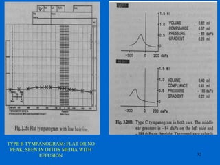
OTITIS MEDIA WITH
EFFUSION
1
• Definition
• Introduction
• Epidemiology
• Aetio pathogenesis
• Clinical presentation
• Diagnosis
• Treatment
• Sequelae
2
Otitis media with effusion (OME)
Defined as Chronic accumulation of mucus
within middle ear and sometimes mastoid air
cell system.
Time for fluid to be present – 12 weeks
3
Synonyms
•Otitis media catarrhal
•Sub acute catarrh of middle ear
•Mucous aural catarrh
•Exudative catarrh
•Adhesive catarrh
•Chronic middle ear exudate catarrh
•Secretory otitis media
•Chronic non suppurative otitis
media
4
Introduction
•Serous otitis media
•Otitis media with effusion
•The hypersecretory ear
•Tympanic hydrops
•Glue ear
•Otitis media ex vacuo
•Exudative otitis media
•Indolent otitis media
•Sero mucinous otitis media
•Mucoid otitis media
• $3.5 billion in expenditures in USA
• Most common reason for visit to pediatrician
• In the developed world AOM is the most common cause
for prescribing antibiotics in children and accounts for over
90% of all antimicrobial consumption during the first 2
years of life (Dagan, 1995).
• Tympanostomy tube placement is 2nd most common
surgical procedure in children
• Development of multidrug-resistant bacteria
• Most common cause of acquired HL in pediatric age
Introduction : the burden of otitis media
5
• Egyptian mummies have perforations of TM and mastoid
destruction
• 450 BC: Hippocrates described the disease
• 1704: Valsalva maneuver and Toynbee – ET dysfunction
with negative ME pressure
• 1869: Politzer described OM catarrahalis & its treatment
as insufflation of air & paracentesis of middle ear
• Jean Riolan: Inadvertant puncture of TM-
tympanocentesis. Fell into disrepute for decades.
• 20th century: Schwartz reintroduces tympanocentesis
• 20th century: Armstrong – use of tympanostomy tubes
• Post World war II- Nasopharyngeal radium irradiation.
Given up for potential for malignancies
Introduction : History of otitis media
6
Epidemiology : risk factors
I. Host-Related factors (intrinsic or genetic)
• Age
• Gender : males more prone to persistent OME
• Race : American natives and Eskimos > whites >
blacks
• Cleft palate/craniofacial abnormality/Down Syndrome
• genetic
• Allergy (no support to prove)
7
2. Environmental or aetiological factors
(extrinsic)
• season and upper respiratory infection
• day care / home care
• siblings
• passive smoking (negligible risk.)
• breast feeding / bottle feeding
• socio-economic status
8
Aetio pathogenesis
No single cause for OME in children
• Viral URI
• Eustachian tube dysfunction
• Middle ear gas composition
• Nasopharyngeal disproportion
9
Developmental Differences between Infants
and Adults in Anatomy of the Eustachian Tube*
• Adults
– ant 2/3-
cartilaginous
– post 1/3- bony
– 45 degree angle
– nasopharyngeal
orifice 8-9 mm
• Children
– longer bony portion
– 10 degree angle
– nasopharyngeal
orifice 4-5 mm in
infants
10
Eustachian tube
• Usually closed
• Opens during swallowing, yawning, and
sneezing for 2 sec
• Opening involves cartilaginous portion
• Tensor veli palatini responsible for active
tubal opening
• No constrictor function
11
Role of the Eustachian Tube
1. Pressure regulation of
middle ear
2. Clearance ‘Drainage’ of
middle ear secretions
mucociliary
muscular
3. Protection from sound and
secretion
anatomic
immunologic and mucociliairy
12
Eustachian Tube Dysfunction
• Palatal abnormalities
• Cleft palate
• Sub mucous cleft palate
• Altered mucociliary system
• Infection
• Allergy
• Immunological factors
• Surfactant deficiency
• Cilial abnormalities
• Hormonal factors 13
Aetio pathogenesis
Aetio pathogenesis
• Middle ear gas composition
– Acts as gas pocket
– Spectrometry shows middle ear gas partial
pressures
• PN2 – 82.4 percent
• PO2 – 7.6 percent
• PCO2 – 10.0 percent
– Closely resemble mixed venous blood and not
atmospheric air
– Theories of middle ear gas deficiency
14
Theories of middle ear gas deficiencies
• Hydrops ex vacuo theory
• Ventilation deficiency theory
• Sniff theory
• Excess diffusion theory
15
Anatomic obstruction
Intrinsic
Inflammation
Extrinsic
adenoid
Tumor
ObstructionWeak TVP
Open
Functional obstruction
Ventilation deficiency theory
16
Theories of middle ear gas deficiencies
• Hydrops ex vacuo theory
• Ventilation deficiency theory
• Sniff theory
• Excess diffusion theory
17
Aetio pathogenesis
• Nasopharyngeal disproportion
• Cranio facial abnormalities
• Adenoids and nasopharynx
– Mechanical blockage of ET
– Focus of infection
18
Related Clinical Conditions
• Barotrauma
• Diving / flying / HBO therapy - OM even in normal ET
function
• NP tumors
• Radiation therapy
• Inadequate antibiotic therapy
• Gross DNS
• Neoplasms
• Syndromic diseases
19
Aetio pathogenesis
Pathology
• Normal flat cuboidal middle ear and
mastoid epithelium – thickened
pseudostratified mucus secreting epithelium
• Goblet cells , mucus secreting glands
• Ciliary lining less efficient
• Defective mucociliary clearance
• Submucosa – oedematous – with dilated
blood vessels and increase in no. of
macrophages and lymphocytes
20
Resolution of acute inflammation and bacterial
infection -- a failure of the middle ear clearance
mechanism allows OME to persist.
Factors implicated in the failure of the clearance
mechanism:
•ciliary dysfunction
•mucosal edema
•hyperviscosity of the effusion
•possibly, an unfavorable pressure gradient.
23
Composition of fluid
• Fluid in middle ear – mixture of epithelial cells,
goblet cells, mucous glands alongwith
inflammatory exudate/ transudate – intercellular
spaces from the inflamed submucosa
• Bacteria isolated in 1/3rd of cases
• Biochemical: increase protein concentration.
• OME: sclerotic mastoids
– Children – smaller mastoids
– Pneumatization less with recurrent episodes
24
Pathogenesis of OM
Infection ET dysfunction
Host response
Liberation of inflammatory mediators
Increase of vascular permeability
Increase of glandular secretion
Inflammation Mucosal proliferation
26
• Quiescent phase – Asymptomatic
• Covert or overt hearing loss
• Impaired speech and language development
• Behavioral changes
• Indirect symptoms of hearing loss
• Otalgia – due to secondary infection
• Features of associated URI, Nasal pathologies
• Symptoms of palatal abnormality or syndromic
conditions with cranio facial abnormalities
27
Clinical features
• History
• Otoscopy
• Tympanometry
• Pure tone audiometry
• Myringotomy and aspiration of fluid (Gold
standard)
28
Diagnosis
Medical history
Hearing loss
Otalgia : ear pulling, irritability
Otorrhoea
Fever
Preceding upper respiratory tract infection
Purulent conjunctivitis (Haemophilus influenzae)
Vertigo : not common as a complaint, unilateral disease,
clumsiness
Nystagmus : labyrinthitis
Tinnitus
Swelling about the ear : DD mastoiditis, external otitis, adenitis
Facial paralysis 29
Examination
• Tympanic Membrane – otoscopy
• Nasopharynx / nose –especially in adults
• Adequate examination of the head and neck
30
• Audiological evaluation-
– TFT
– Tympanometry
– PTA-Conductive Loss of 10-40dB
• Radiology -Mastoids
-Adenoids
31
Examination
TYPE B TYMPANOGRAM: FLAT OR NO
PEAK, SEEN IN OTITIS MEDIA WITH
EFFUSION 32
• Audiological evaluation-
– TFT
– Tympanometry
– PTA-Conductive Loss of 10-40dB
• Myringotomy and aspiration of fluid (Gold
standard)
• Radiology -Mastoids
-Adenoids
33
Examination
I. Counselling and hearing tactics
• Parents of children of OME often misinformed-both
overpessimistic and overoptimistic.
• Counsel about benign nature and high spontaneous
resolution rates as well as natural history of disease.
• Concern about hearing – Impairement associated with
OME variable and mild or moderate at most.
• Minimise disability by Hearing tactics
TREATMENT
34
II. Medical therapy
• Antibiotic therapy - modest impact on short-term and
impact on long-term resolution is smaller.
• Steroid therapy and antihistamine-decongestant therapy
- no proven effect
• Ventilate the ET- No URTI
• Valsalva
• Chewing gums
• Politzerization
• Auto inflation using otovent balloons-older children
• Mucolytic agents – no long term effect.
• Management of associated nasal condition / allergy
• Homeopathy – not shown to be of benefit
TREATMENT
35
III. Surgical therapy
• (a) Myringotomy and aspiration
• (b) Ventilation tubes
• (c) Adenoidectomy
• (d) Rarely explorative tympanotomy with/without mastoidectomy
• No improvement > 90 days of treatment
• Persistent OME with hearing loss in children
• Chronic effusion
• Reassessment at pre admission clinic for
persistence
36
(b). Myringotomy with ventilation tubes
• Diagnostic
• OME –therapeutic
• Types of ventilation tubes
• Procedure –
• Outcomes
37
Tympanostomy Tubes
• Silicone rubber
• Teflon
• Plastic
• Precautions-flange
– Not too near the annulus
38
Tube Choice
• Standard grommets (Armstrong, Shepard, collar button)
– Persistent perforation rates 3-5%
– Persist 6-18 months
• Goode T-tubes
– Persistent perforation rates 10 of 64 in best study
– Persist 2-4 yrs
• Bioabsorbable/bacteriostatic grommets
– New choice in future
– Made of bacteriostatic polylactic acid
– Similar material to resorbable miniplates
– Mix can be customized to persist a given amount of time
39
Myringotomy with ventilation tubes
• Diagnostic
• OME –therapeutic
• Types of ventilation tubes
• Procedure –
• Outcomes
40
• Complications -
(c). Adenoidectomy
• Rationale
• Recommendation
• Effect on hearing improvement
41
IV. Hearing Aids
100
90
80
70
60
50
40
30
20
10
0
0 1 2 3 4 5 6 7 8 9 10
Proportion(%)withfluidremaining
Survival
functions
for time to
fluid Clearance
as Confirmed
by otoscopy
• No surgery (n=77)
• Ventilation tube only
(n=77)
• Adenoidectomy only
• Adenoidectomy
and tube (n=136)
Years
Maw et al, 1994
42
Treatment Recurrent Otitis media
• Chemoprophylaxis
– Sulfisoxazole, amoxicillin, ampicillin
– less efficacy for intermittent propylaxis
• Myringotomy and tube insertion
– Decreased frequency and severity of AOM
– otorrhoea and other complications
– may require prophylaxis if severe
• Adenoidectomy
– 28% and 35% fewer episodes of AOM at first and
second years
43
Sequelae Of OME
• Scarring
• Atelectasis
• Attic retraction
• Tympanosclerosis
• Otorrhoea
• Perforation
• COM / cholesteatoma
• SNHL
• Behavioural/ developmental 44
Follow-up
• Child should demonstrate symptomatic benefit
within 72 hours of antibiotic
• Failure to show improvement - re-evaluation.
• A follow-up examination - one month after the
diagnosis and should include:
- Inspection of the tympanic membrane
- Assessment of hearing
• The purpose of the follow-up - identify persistent
otitis media or persistent middle ear effusion
• Children with persistent otitis media or persistent
middle ear effusion - seen on a monthly basis
until their exam is normal 45
Future solution
• Immunisation with Haemophilus influenzae
type B vaccine
• Heptavalent pneumococcal conjugate
vaccine PCV7
• Attempt to produce vaccine for nontypable
H. influenzae and M. catarrhalis
• Surfactants
46
Bibliography
47
• Scott Brown Otorhinolaryngology, Head
and Neck surgery, 6th and 7th edition
• Ballenger’s Otorhinolaryngology, Head and
Neck surgery, 17th edition
• Ludman and Wright diseases of the ear, 6th
edition
Thank You
48

05 ome

  • 1.
  • 2.
    • Definition • Introduction •Epidemiology • Aetio pathogenesis • Clinical presentation • Diagnosis • Treatment • Sequelae 2
  • 3.
    Otitis media witheffusion (OME) Defined as Chronic accumulation of mucus within middle ear and sometimes mastoid air cell system. Time for fluid to be present – 12 weeks 3
  • 4.
    Synonyms •Otitis media catarrhal •Subacute catarrh of middle ear •Mucous aural catarrh •Exudative catarrh •Adhesive catarrh •Chronic middle ear exudate catarrh •Secretory otitis media •Chronic non suppurative otitis media 4 Introduction •Serous otitis media •Otitis media with effusion •The hypersecretory ear •Tympanic hydrops •Glue ear •Otitis media ex vacuo •Exudative otitis media •Indolent otitis media •Sero mucinous otitis media •Mucoid otitis media
  • 5.
    • $3.5 billionin expenditures in USA • Most common reason for visit to pediatrician • In the developed world AOM is the most common cause for prescribing antibiotics in children and accounts for over 90% of all antimicrobial consumption during the first 2 years of life (Dagan, 1995). • Tympanostomy tube placement is 2nd most common surgical procedure in children • Development of multidrug-resistant bacteria • Most common cause of acquired HL in pediatric age Introduction : the burden of otitis media 5
  • 6.
    • Egyptian mummieshave perforations of TM and mastoid destruction • 450 BC: Hippocrates described the disease • 1704: Valsalva maneuver and Toynbee – ET dysfunction with negative ME pressure • 1869: Politzer described OM catarrahalis & its treatment as insufflation of air & paracentesis of middle ear • Jean Riolan: Inadvertant puncture of TM- tympanocentesis. Fell into disrepute for decades. • 20th century: Schwartz reintroduces tympanocentesis • 20th century: Armstrong – use of tympanostomy tubes • Post World war II- Nasopharyngeal radium irradiation. Given up for potential for malignancies Introduction : History of otitis media 6
  • 7.
    Epidemiology : riskfactors I. Host-Related factors (intrinsic or genetic) • Age • Gender : males more prone to persistent OME • Race : American natives and Eskimos > whites > blacks • Cleft palate/craniofacial abnormality/Down Syndrome • genetic • Allergy (no support to prove) 7
  • 8.
    2. Environmental oraetiological factors (extrinsic) • season and upper respiratory infection • day care / home care • siblings • passive smoking (negligible risk.) • breast feeding / bottle feeding • socio-economic status 8
  • 9.
    Aetio pathogenesis No singlecause for OME in children • Viral URI • Eustachian tube dysfunction • Middle ear gas composition • Nasopharyngeal disproportion 9
  • 10.
    Developmental Differences betweenInfants and Adults in Anatomy of the Eustachian Tube* • Adults – ant 2/3- cartilaginous – post 1/3- bony – 45 degree angle – nasopharyngeal orifice 8-9 mm • Children – longer bony portion – 10 degree angle – nasopharyngeal orifice 4-5 mm in infants 10
  • 11.
    Eustachian tube • Usuallyclosed • Opens during swallowing, yawning, and sneezing for 2 sec • Opening involves cartilaginous portion • Tensor veli palatini responsible for active tubal opening • No constrictor function 11
  • 12.
    Role of theEustachian Tube 1. Pressure regulation of middle ear 2. Clearance ‘Drainage’ of middle ear secretions mucociliary muscular 3. Protection from sound and secretion anatomic immunologic and mucociliairy 12
  • 13.
    Eustachian Tube Dysfunction •Palatal abnormalities • Cleft palate • Sub mucous cleft palate • Altered mucociliary system • Infection • Allergy • Immunological factors • Surfactant deficiency • Cilial abnormalities • Hormonal factors 13 Aetio pathogenesis
  • 14.
    Aetio pathogenesis • Middleear gas composition – Acts as gas pocket – Spectrometry shows middle ear gas partial pressures • PN2 – 82.4 percent • PO2 – 7.6 percent • PCO2 – 10.0 percent – Closely resemble mixed venous blood and not atmospheric air – Theories of middle ear gas deficiency 14
  • 15.
    Theories of middleear gas deficiencies • Hydrops ex vacuo theory • Ventilation deficiency theory • Sniff theory • Excess diffusion theory 15
  • 16.
  • 17.
    Theories of middleear gas deficiencies • Hydrops ex vacuo theory • Ventilation deficiency theory • Sniff theory • Excess diffusion theory 17
  • 18.
    Aetio pathogenesis • Nasopharyngealdisproportion • Cranio facial abnormalities • Adenoids and nasopharynx – Mechanical blockage of ET – Focus of infection 18
  • 19.
    Related Clinical Conditions •Barotrauma • Diving / flying / HBO therapy - OM even in normal ET function • NP tumors • Radiation therapy • Inadequate antibiotic therapy • Gross DNS • Neoplasms • Syndromic diseases 19 Aetio pathogenesis
  • 20.
    Pathology • Normal flatcuboidal middle ear and mastoid epithelium – thickened pseudostratified mucus secreting epithelium • Goblet cells , mucus secreting glands • Ciliary lining less efficient • Defective mucociliary clearance • Submucosa – oedematous – with dilated blood vessels and increase in no. of macrophages and lymphocytes 20
  • 21.
    Resolution of acuteinflammation and bacterial infection -- a failure of the middle ear clearance mechanism allows OME to persist. Factors implicated in the failure of the clearance mechanism: •ciliary dysfunction •mucosal edema •hyperviscosity of the effusion •possibly, an unfavorable pressure gradient. 23
  • 22.
    Composition of fluid •Fluid in middle ear – mixture of epithelial cells, goblet cells, mucous glands alongwith inflammatory exudate/ transudate – intercellular spaces from the inflamed submucosa • Bacteria isolated in 1/3rd of cases • Biochemical: increase protein concentration. • OME: sclerotic mastoids – Children – smaller mastoids – Pneumatization less with recurrent episodes 24
  • 23.
    Pathogenesis of OM InfectionET dysfunction Host response Liberation of inflammatory mediators Increase of vascular permeability Increase of glandular secretion Inflammation Mucosal proliferation 26
  • 24.
    • Quiescent phase– Asymptomatic • Covert or overt hearing loss • Impaired speech and language development • Behavioral changes • Indirect symptoms of hearing loss • Otalgia – due to secondary infection • Features of associated URI, Nasal pathologies • Symptoms of palatal abnormality or syndromic conditions with cranio facial abnormalities 27 Clinical features
  • 25.
    • History • Otoscopy •Tympanometry • Pure tone audiometry • Myringotomy and aspiration of fluid (Gold standard) 28 Diagnosis
  • 26.
    Medical history Hearing loss Otalgia: ear pulling, irritability Otorrhoea Fever Preceding upper respiratory tract infection Purulent conjunctivitis (Haemophilus influenzae) Vertigo : not common as a complaint, unilateral disease, clumsiness Nystagmus : labyrinthitis Tinnitus Swelling about the ear : DD mastoiditis, external otitis, adenitis Facial paralysis 29
  • 27.
    Examination • Tympanic Membrane– otoscopy • Nasopharynx / nose –especially in adults • Adequate examination of the head and neck 30
  • 28.
    • Audiological evaluation- –TFT – Tympanometry – PTA-Conductive Loss of 10-40dB • Radiology -Mastoids -Adenoids 31 Examination
  • 29.
    TYPE B TYMPANOGRAM:FLAT OR NO PEAK, SEEN IN OTITIS MEDIA WITH EFFUSION 32
  • 30.
    • Audiological evaluation- –TFT – Tympanometry – PTA-Conductive Loss of 10-40dB • Myringotomy and aspiration of fluid (Gold standard) • Radiology -Mastoids -Adenoids 33 Examination
  • 31.
    I. Counselling andhearing tactics • Parents of children of OME often misinformed-both overpessimistic and overoptimistic. • Counsel about benign nature and high spontaneous resolution rates as well as natural history of disease. • Concern about hearing – Impairement associated with OME variable and mild or moderate at most. • Minimise disability by Hearing tactics TREATMENT 34
  • 32.
    II. Medical therapy •Antibiotic therapy - modest impact on short-term and impact on long-term resolution is smaller. • Steroid therapy and antihistamine-decongestant therapy - no proven effect • Ventilate the ET- No URTI • Valsalva • Chewing gums • Politzerization • Auto inflation using otovent balloons-older children • Mucolytic agents – no long term effect. • Management of associated nasal condition / allergy • Homeopathy – not shown to be of benefit TREATMENT 35
  • 33.
    III. Surgical therapy •(a) Myringotomy and aspiration • (b) Ventilation tubes • (c) Adenoidectomy • (d) Rarely explorative tympanotomy with/without mastoidectomy • No improvement > 90 days of treatment • Persistent OME with hearing loss in children • Chronic effusion • Reassessment at pre admission clinic for persistence 36
  • 34.
    (b). Myringotomy withventilation tubes • Diagnostic • OME –therapeutic • Types of ventilation tubes • Procedure – • Outcomes 37
  • 35.
    Tympanostomy Tubes • Siliconerubber • Teflon • Plastic • Precautions-flange – Not too near the annulus 38
  • 36.
    Tube Choice • Standardgrommets (Armstrong, Shepard, collar button) – Persistent perforation rates 3-5% – Persist 6-18 months • Goode T-tubes – Persistent perforation rates 10 of 64 in best study – Persist 2-4 yrs • Bioabsorbable/bacteriostatic grommets – New choice in future – Made of bacteriostatic polylactic acid – Similar material to resorbable miniplates – Mix can be customized to persist a given amount of time 39
  • 37.
    Myringotomy with ventilationtubes • Diagnostic • OME –therapeutic • Types of ventilation tubes • Procedure – • Outcomes 40 • Complications -
  • 38.
    (c). Adenoidectomy • Rationale •Recommendation • Effect on hearing improvement 41 IV. Hearing Aids
  • 39.
    100 90 80 70 60 50 40 30 20 10 0 0 1 23 4 5 6 7 8 9 10 Proportion(%)withfluidremaining Survival functions for time to fluid Clearance as Confirmed by otoscopy • No surgery (n=77) • Ventilation tube only (n=77) • Adenoidectomy only • Adenoidectomy and tube (n=136) Years Maw et al, 1994 42
  • 40.
    Treatment Recurrent Otitismedia • Chemoprophylaxis – Sulfisoxazole, amoxicillin, ampicillin – less efficacy for intermittent propylaxis • Myringotomy and tube insertion – Decreased frequency and severity of AOM – otorrhoea and other complications – may require prophylaxis if severe • Adenoidectomy – 28% and 35% fewer episodes of AOM at first and second years 43
  • 41.
    Sequelae Of OME •Scarring • Atelectasis • Attic retraction • Tympanosclerosis • Otorrhoea • Perforation • COM / cholesteatoma • SNHL • Behavioural/ developmental 44
  • 42.
    Follow-up • Child shoulddemonstrate symptomatic benefit within 72 hours of antibiotic • Failure to show improvement - re-evaluation. • A follow-up examination - one month after the diagnosis and should include: - Inspection of the tympanic membrane - Assessment of hearing • The purpose of the follow-up - identify persistent otitis media or persistent middle ear effusion • Children with persistent otitis media or persistent middle ear effusion - seen on a monthly basis until their exam is normal 45
  • 43.
    Future solution • Immunisationwith Haemophilus influenzae type B vaccine • Heptavalent pneumococcal conjugate vaccine PCV7 • Attempt to produce vaccine for nontypable H. influenzae and M. catarrhalis • Surfactants 46
  • 44.
    Bibliography 47 • Scott BrownOtorhinolaryngology, Head and Neck surgery, 6th and 7th edition • Ballenger’s Otorhinolaryngology, Head and Neck surgery, 17th edition • Ludman and Wright diseases of the ear, 6th edition
  • 45.