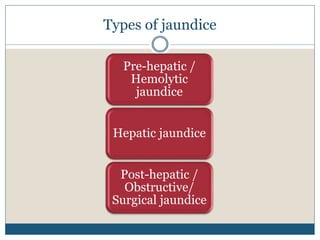
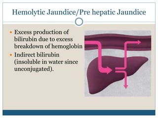
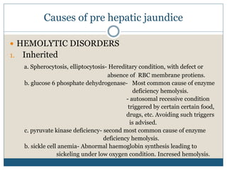
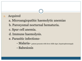
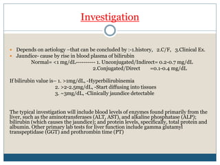
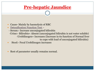
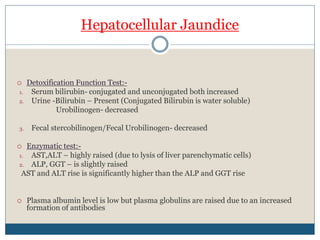
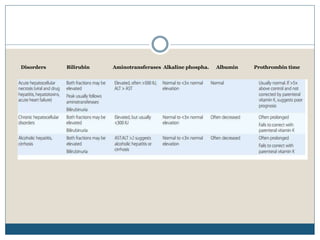
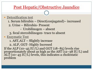
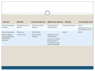
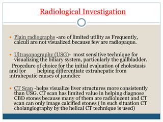
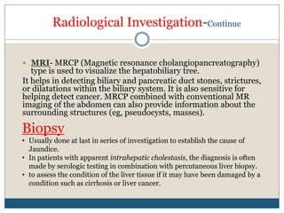
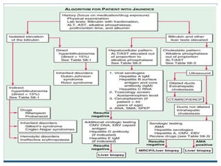
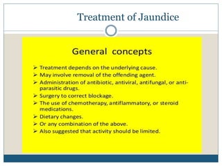
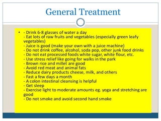
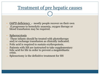
I T I S T H E Y E L L O W I S H D I S C O L O R A T I O N O F T H E
T I S S U E S D U E T O D E P O S I T I O N O F B I L I R U B I N
W H I C H O C C U R S I N P R E S E N C E O F
H Y P E R B I L I R U B I N E M I A
JAUNDICE
Clinically jaundice is evident when serum bilirubin
crosses 3 mg/DL
Jaundice is latent I.e., clinically non evident (only
detected by serum analysis) when serum bilirubin
is in between 1 - 3 mg/DL.
Unconjugated hyperbilirubinemia – when direct
bilirubin level is less than 15% of total serum
bilirubin.
Conjugated hyperbilirubinemia – when direct
bilirubin level is greater than 15%
Sites to be examined
Upper bulbar conjuctiva (contains elastin which
has hisj affinity for bilirubin)
Base of tongue
Mucous membrane of palate (specially soft palate)
Palms and soles
General skin surface
Differential diagnosis
Carotenoderma (discoloration is limited to palms,
sole, forehead, nasolabial fold with sparing of
sclera)
intake of drug quinacrine (only skin, urine and eyes
are yellow)
excessive exposure to phenols.
Hemolytic Jaundice/Pre hepatic Jaundice
Excess production of
bilirubin due to excess
breakdown of hemoglobin
Indirect bilirubin
(insoluble in water since
unconjugated).
Causes of pre hepatic jaundice
HEMOLYTIC DISORDERS
1. Inherited
a. Spherocytosis, elliptocytosis- Hereditary condition, with defect or
absence of RBC membrane protiens.
b. glucose 6 phosphate dehydrogenase- Most common cause of enzyme
deficiency hemolysis.
- autosomal recessive condition
triggered by certain certain food,
drugs, etc. Avoiding such triggers
is advised.
c. pyruvate kinase deficiency- second most common cause of enzyme
deficiency hemolysis.
b. sickle cell anemia- Abnormal haemoglobin synthesis leading to
sickeling under low oxygen condition. Incresed hemolysis.
2. Acquired
a. Microangiopathic haemolytic anemias
b. Paroxysmal nocturnal hematuria.
c. Spur cell anemia.
d. Immune haemolysis.
e. Parasitic infections-
- Malaria- patient presents with fever chills rigor, hepatosplenomegaly
- Babesiosis
INEFFECTIVE ERYTHROPOIESIS
1. Cobalamin, Folate deficiency- Megaloblastic anemia
2. severe iron deficiencies- Microcytic hypochromic anemia
3. Thalassemia
• INCREASED BILIRUBIN PRODUCTION
1. Massive blood transfusion
2. Resorption of hematoma.
• DRUGS
1. Rifampicin, ribavirin, Probenecid.
Hepatic Jaundice
Liver’s ability to conjugate or
excrete bilirubin is affected
Increased level of conjugated
and unconjugated bilirubin
present
Causes of hepatic jaundice
VIRAL HEPATITIS
features HAV HBV HCV HDV HEV
transmission Feco-
oral
Parentral
,sexual,
perinatal
parentral Parentral,
sexual,
perinatal
Feco oral
Carrier none .1-30% 1.5-3.2% variable none
chronicity none occasional common common none
cancer none + + +- none
prognosis excellent Worsen with
age
moderate Acute-good
Chronic-poor
good
• Other viruses- Epstein barr, CMV, HSV
• ALCOHOLIC HEPATITIS- Balloon degeneration, fibrosis of
parenchymal tissue.
- 160g/day for 10-20yrs(women are more
susceptible)
• DRUG TOXICITY
1. Acetaminophen- Predictable, Dose dependent
2. Isoniazid- Unpredictable, Idiosyncratic.
• ENVIRONMENTAL TOXINS
- Vinyl chloride, Jamaica bush tea-pyrrolizidine
alkaloids , kava kava, Wild mushrooms.
WILSON’S DISEASE- hepato lenticular degeneration
due to copper accumulation in liver tissue.
AUTOIMMUNE HEPATITIS
INHERITED CONDITIONS-
Indirect hyperbilirubinemia
- Gilbert syndrome- Enzyme(UDP glucoronyl transferase) deficiency- 10-33%
activity only.
- Crigler-najjar syndrome type 1- Enzyme – absent
type2- enzyme activity- 0-10%
Direct hyperbilirubinemia
- Dubin Johnson syndrome- mutation in MRP2 gene. Defect in canalicular
transport of organic anions
- Rotor syndrome- defect in bilirubin storage.
Obstructive Jaundice
Bilirubin formation rate is
normal
Conjugation is normal =
direct bilirubin
Any intrahepatic or
extrahepatic condition leading
Obstruction to the flow of bile.
Causes of post hepatic / obstructive jaundice
INTRAHEPATIC CAUSES
1. Viral hepatitis-
a. Fibrosing cholestatic hepatitis- hep.B and C
b. hep A, Epstein barr, cytomegalovirus infection.
2. Alcoholic hepatitis
3. Drug Toxicity-
a. Pure cholestasis- anabolic and contraceptive steroids
b. Choleststic hepatitis- chlorpromazine, erythromycin estolate
c. Chronic cholestasis- prochlorperazine and chlorpromazine
Investigation
Depends on aetiology –that can be concluded by :-1.history, 2.C/F, 3.Clinical Ex.
Jaundice- cause by rise in blood plasma of bilirubin
Normal= <1 mg/dL---------- 1. Unconjugated/Indirect= 0.2-0.7 mg/dL
2.Conjugated/Direct =0.1-0.4 mg/dL
If bilirubin value is– 1. >1mg/dL, -Hyperbilirubinemia
2. >2-2.5mg/dL, -Start diffusing into tissues
3. ~3mg/dL, -Clinically jaundice detectable
The typical investigation will include blood levels of enzymes found primarily from the
liver, such as the aminotransferases (ALT, AST), and alkaline phosphatase (ALP);
bilirubin (which causes the jaundice); and protein levels, specifically, total protein and
albumin. Other primary lab tests for liver function include gamma glutamyl
transpeptidase (GGT) and prothrombin time (PT)
Pre-hepatic Jaundice
Cause- Mainly by haemolysis of RBC
Detoxification Function Test –
- Serum:- Increase unconjugated bilirubin
-Urine:- Bilirubin= Absent (unconjugated bilirubin is not water soluble)
-Urobilinogen= Increases (Increase in 6x function of Normal liver
to cope with load of unconjugated bilirubin)
Stool:- Fecal Urobilinogen increases
Rest of parameter usually remains normal.
Hepatocellular Jaundice
Detoxification Function Test:-
1. Serum bilirubin- conjugated and unconjugated both increased
2. Urine -Bilirubin – Present (Conjugated Bilirubin is water soluble)
Urobilinogen- decreased
3. Fecal stercobilinogen/Fecal Urobilinogen- decreased
Enzymatic test:-
1. AST,ALT – highly raised (due to lysis of liver parenchymatic cells)
2. ALP, GGT – is slightly raised
AST and ALT rise is significantly higher than the ALP and GGT rise
Plasma albumin level is low but plasma globulins are raised due to an increased
formation of antibodies
Post Hepatic/Obstructive Jaundice
Detoxification test
1. Serum bilirubin – Direct(conjugated)– increased
2. Urine – Bilirubin- Present
- Urobilinogen – absent
3. fecal stercobilinogen- trace to absent
Enzymatic Test
1. AST,ALT – Slightly increase
2. ALP, GGT- Highly Incresed
If the ALP (10–45 IU/L) and GGT (18–85) levels rise
proportionately about as high as the AST (12–38 IU/L) and
ALT (10–45 IU/L) levels, this indicates a cholestatic
problem
Radiological Investigation
Plain radiographs -are of limited utility as Frequently,
calculi are not visualized because few are radiopaque.
Ultrasonography (USG)- most sensitive technique for
visualizing the biliary system, particularly the gallbladder.
Procedure of choice for the initial evaluation of cholestasis
and for helping differentiate extrahepatic from
intrahepatic causes of jaundice
CT Scan -helps visualize liver structures more consistently
than USG. CT scan has limited value in helping diagnose
CBD stones because many of them are radiolucent and CT
scan can only image calcified stones ( in such situation CT
cholangiography by the helical CT technique is used)
Radiological Investigation-Continue
MRI- MRCP (Magnetic resonance cholangiopancreatography)
type is used to visualize the hepatobiliary tree.
It helps in detecting biliary and pancreatic duct stones, strictures,
or dilatations within the biliary system. It is also sensitive for
helping detect cancer. MRCP combined with conventional MR
imaging of the abdomen can also provide information about the
surrounding structures (eg, pseudocysts, masses).
Biopsy
• Usually done at last in series of investigation to establish the cause of
Jaundice.
• In patients with apparent intrahepatic cholestasis, the diagnosis is often
made by serologic testing in combination with percutaneous liver biopsy.
• to assess the condition of the liver tissue if it may have been damaged by a
condition such as cirrhosis or liver cancer.
Treatment of pre hepatic causes
• G6PD deficiency - mostly people recover on their own
o if progresses to hemolytic anaemia, oxygen therapy or
blood transfusion may be required.
• Spherocytosis
o These infants should be treated with phototherapy
and/or exchange transfusion as clinically indicated.
o Folic acid is required to sustain erythropoiesis.
o Patients with HS are instructed to take supplementary
folic acid for life in order to prevent a megaloblastic
crisis.
o Splenectomy is the definitive treatment for HS
• Sickle cell anaemia
o Treatments may include medications to reduce pain and
prevent complications, blood transfusions and supplemental
oxygen, as well as a bone marrow transplant.
o Antibiotics -Children with sickle cell anemia may begin taking
the antibiotic penicillin when they're about 2 months of age and
continue taking it until they're at least 5 years old.
• Immune related hemolysis – corticosteroids, folic acid is
main line of treatment
• Parasitic Infections like malaria are treated with
antimalarial drugs like chloroquine, artesunate,
lumefantrine,amodiaquine
• Ineffective erythropoiesis- iron and folic acid
supplementation, vit B12 tablets given and repeated blood
transfusions
Treatment of hepatic causes
• Viral hepatitis
Hepatitis A is mostly self limiting no treatment is
required, but in some cases 0.02 ml/kg administration
of anti-HAV Ig can be given.
Hepatitis B treated with combination of HBIG and Hep
b vaccines.
Recombivax and Engerix-B are 2 vaccines for
hepatitis B
Hepatitis C is treated with interferons.
• Other Viral infections like EBV, CMV, HSV are treated
with Antiviral medications like acyclovir , ganciclovir
and foscarnet.
Discriminant function - determines the prognosis of the person suffering from
alcoholic liver disease. Given by Maddrey.
It is calculated by a simple formula:
(4.6 x (PT test - control))+ S.Bilirubin in mg/dl
A value more than 32 implies poor outcome.
MELD – model for end stage liver disease
scoring system for assessing the severity of chronic liver disease
MELD uses the patient's values for serum bilirubin, serum creatinine, and the
international normalized ratio for prothrombin time (INR) to predict survival.
• Wilsons disease- pharmacologic
treatment with chelating agents such as
D-penicillamine and Other agents
include sodium dimercaptosuccinate,
dimercaptosuccinic acid
SUPPORTIVE MANAGEMENT
Preoperative biliary decompression (ERCP or PTC)
Intravenous admistration of 5% dextrose saline followed by 10%mannitol
or loop diuretics to prevent hepatorenal syndrome/ renal failure(12 to 24
hours prior to surgery)
catheterization to monitor output
Broad spectrum antibiotic prophylaxis with 3rd generation cephalosporins
Parenteral vitamin K +/- fresh frozen plasma
Need careful fluid balance to correct dehydration
Correction of hypokalemia and other electrolyte imbalance.
Cholestyramine and antihistamine for symptomatic relief of pruritis
44
Definitive treatment depends upon the
cause-
• Choledocholithiasis- cholecystectomy is done
• Carcinoma of head of pancreas- whipple resection
done
• Ca gall bladder- whipple resection done but if
unoperable radiological stenting is done.
• Choledochal cyst- excision of the cyst with
reconstruction of extrahepatic biliary tree.
• Stricture- endoscopic stenting. Standard care is
surgery by roux-en-y choledocojejunostomy.