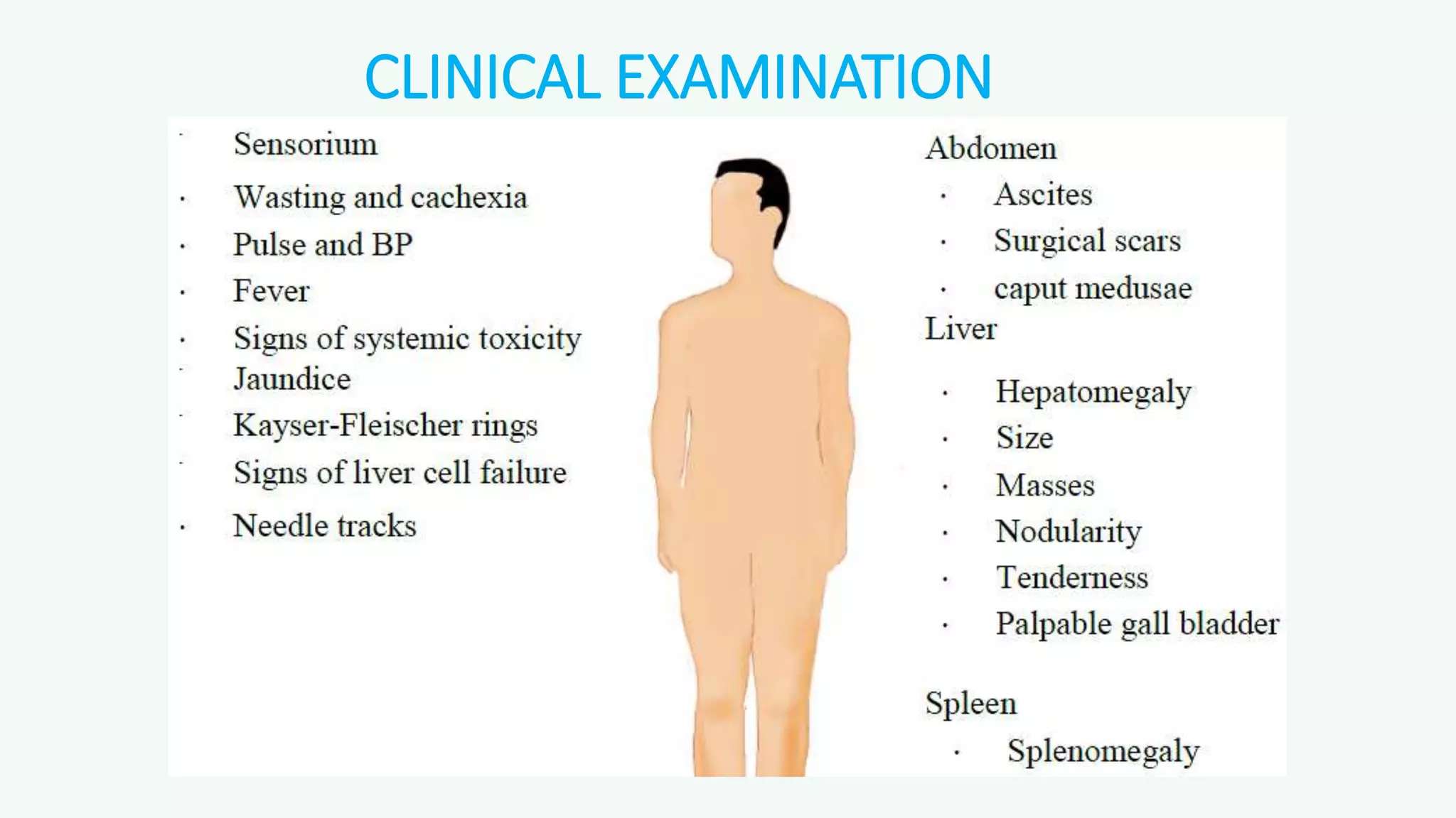
APPROACH TO
JAUNDICE
Dr.Navas Shareef .P .P
DNB Family med resident
Overview of discussion
I. Definitions
II. Jaundice etio pathogenesis
III. Evaluation of a patient with jaundice
a. History
b. Clinical examination
c. Investigations
d. Algorithms
JAUNDICE
• Yellow discoloration of skin, sclera, and mucous membranes
due to increased level of serum bilirubin.
• NB: In carotinemia, sclera is never yellow
SERUM BILIRUBIN LEVELS
• Normal: 0 to 1mg/dL
• Unconjugated/free/indirect (bilirubin-albumin complex): 0.2 to 1 mg/dL
• Conjugated/direct:0.1 to 0.2 mg/dL
• Latent jaundice: Above 1 mg/Dl
• Clinical Jaundice: Above 2 mg/dL
• Used to determine liver’s ability to clear endogenous/exogenous substances
from the circulation
• Derived mainly from hemoglobin (95%)
• Continuous production (300 mg daily)
Bilirubin production
and metabolism
UDP Glucoronosyl
transferase 1g Hb=34mg Br
90%
10%
Direct Bilirubin
• conjugated
• water soluble
• polar
• seen in urine
• elevated with biliary
obstruction and
hepatocellular disease.
Indirect Bilirubin
• unconjugated
• lipid soluble
• non-polar
• not in urine
• Elevated with hemolysis,
hepatic disease
Major Metabolic Functions of the Liver
SYNTHETIC FUNCTIONS
• Proteins: plasma proteins albumin, globulins, coagulation factors…etc
• Lipids: cholesterol, triglycerides, fatty acids, phospholipids & lipoproteins (VLDL)
• Carbohydrates: glycogen synthesis & gluconeogenesis
DETOXIFICATION & EXCRETION FUNCTIONS
• Detoxication of ammonia to urea (urea cycle)
• Billirubin conjugation & excretion into bile
• Excretion of cholesterol into bile
• Drug detoxication & excretion into bile
STORAGE FUNCTIONS
• Storage of fat soluble vitamins (vitamins A, D, E & K)Storage of vitamin B12
Production of bile salts
• Help digestion of lipids
BILE JUICE COMPOSITION
• Water (85%)
• Bile salts (10%)(Cholic, chenodeoxycholic, deoxycholic and lithocholic
acid)
• Mucus
• Bile Pigments (3%)e.g bilirubin glucuronide
• Fats (1%), such as Phospholipids (lecithin) , cholesterol.
• Inorganic salts. 0.7%
Classification of Jaundice
According to etiology:
Hemolytic
Increased rate of RBC destruction
Increased Hb breakdown to bilirubin in RES cells
This exceeds the capacity of conjugation in liver.
Hepatocellular Inability of hepatocytes to conjugate and/or excrete bilirubin.
Obstructive
Failure of excretion of conjugated bilirubin into the intestine,
causing its regurgitation in circulation.
Urinary and fecal urobilinogen are decreased,
Classification of Jaundice
According to the main type of bilirubin increased in plasma:
UNCONJUGATED
HYPERBILIRUBINEMIA
CONJUGATED
HYPERBILIRUBINEMIA
Indirect or unconjugated bilirubin is >
85% of total
Direct or conjugated bilirubin is
>15% of total
Causes include Causes include
Hemolysis Viral and alcoholic Hepatitis
Resorption of a large Cirrhosis
hematoma, Cholestasis
Ineffective erythropoiesis Drug (anabolic steroids, oral
(Thalassemia, Megaloblastic contraceptives)
anemia) Toxins
Physiologic jaundice of Congenital
newborn.
Congenital
Dubin-Johnson Syndrome
Rotor syndrome.
Gilbert’sSyndrome
Crigler-Najjar syndrome.
Classification of JaundiceAccording to site of disease:
Class of Jaundice Type of Bilirubin Etiology
Pre-hepatic or Hemolytic Unconjugated
 Hemolysis/Abnormal RBCs
 Antibodies
 drugs and toxins
 Ineffective erythropoiesis
(thalassemia, B12 Deficiency).
 Hemoglobinopathies
Hepatic or Hepatocellular
Unconjugated &
conjugated
Viral hepatitis
Toxic hepatitis
Intrahepatic cholestasis
Gilbert’s Syndrome
Crigler-Najjar Syndrome
Post-hepatic or Obstructive Conjugated
Extrahepatic cholestasis
Gallstones
Tumors of the bile duct
Carcinoma of pancreas
Carcinoma of ampulla of Vater
CHOLESTATIC PATTERN
INTRAHEPATIC
• Drugs
• Hepatitis A, B, C
• Alcoholic hepatitis
• TPN
• Primary Sclerosing Cholangitis
• Primary Biliary Cirrhosis
EXTRAHEPATIC
• Gall stones
• Primary Sclerosing Cholangitis
• Malignancy
• Chronic pancreatitis
• HIV cholangiopathy
Congenital Hyperbilirubinemias
Gilbert’s Syndrome
Most common inherited cause of unconjugated hyperbilirubinemia.
It is a familial, benign disease with autosomal dominant inheritance.
Affects 5% of general population.
Mild deficiency or decreased hepatic levels of Bilirubin glucoronosyltransferase
jaundice is mild and fluctuating, goes unnoticed for years; often related to
stress/fasting/infection.
Mildly elevated serum bilirubin is the sole abnormality.
Not associated with any morbidity.
Congenital Hyperbilirubinemias
Crigler Najjar Syndromes
Autosomal recessive Unconjugated hyperbilirubinemia.
Type I -
• Serum bilirubin > 20 mg/dl
• Complete absence of Bilirubin UDP-glucoronosyltransferase1-A1
expression detected in liver tissue.
• Fatal due to kernicterus – causes rapid death in neonate.
Type II (Arias Syndrome) –
• Serum bilirubin < 20mg/dl.
• Reduced levels of detectable Bilirubin UDP-glucoronosyltransferase1-
A1 expression in liver tissue owing to single base pair mutations.
• Bile is pigmented instead of pale or dark as normal.
• No risk of kernicterus.
Congenital Hyperbilirubinemias
Dubin Johnson Syndrome
• Benign autosomal recessive disorder.
• Conjugated hyperbilirubinemia
• Decreased hepatocellular bilirubin excretion due to a defect in the
canalicular cationic transport protein.
Gross : black pigmentation of the liver due to accumulation of
polymerized epinephrine metabolites
Lab - Bromosulphthalein excretion test is abnormal.
• No clinical consequences
Congenital Hyperbilirubinemias
Rotor Syndrome
Autosomal recessive disorder.
Conjugated hyperbilirubinemia.
Decreased intraheptic binding and intake to unsaturated hepatocytes.
Similar to Dubin-Johnson but without liver pigmentation.
Bromosulphthalein test is normal.
No clinical consequences.
Primary biliary cirrhosis
probably autoimmune characterised by inflammation & granulomatous destruction of
intrahepatic bile ducts.
Males: Females = 1:10, age – 30-65 yrs.
Presentation : middle-aged women with obstructive jaundice: xanthomas, xanthelasmas,
increased serum cholesterol, fatigue, cirrhosis.
Laboratory -Diagnosis
 Increased conjugated bilirubin
 Increased alkaline phosphatase
 Increased 5’ nucleotidase.
 Antimitochondrial Antibody(AMA)
Primary sclerosing cholangitis
CLD of unknown etiology characterized by segmental inflammation & fibrosing
destruction of intra-& extrahepatic bile ducts.
Males: Females = 2:1, age – 20-40yrs; most associated with ulcerative colitis.
Presentation – similar to Primary Biliary Cirrhosis.
Microscopy –
• Concentric fibrosis around bile ducts.
• Segmental stenosis of the bile ducts.
Beaded appearance of bile ducts on cholangiogram.
ANCA is a Marker for Primary Sclerosing Cholangitis.
HISTORY
• Age at onset
• Was it acute or chronic?
• Previous episodes of jaundice
• Painless jaundice
• Pain associated with jaundice
• Prodrome of nausea, vomiting, low grade fever few days prior to jaundice
• Pale stools (clay-coloured)
• Itching with deepening of jaundice
• High fever, right upper quadrant pain, jaundice (Charcot‘s triad)
HISTORY
• Symptoms of anemia like fatigue, pallor, congestive failure and jaundice
• Fever, jaundice, hemorrhagic conjunctiva, skin, renal failure
• Fever and jaundice in malaria endemic zones
• Jaundice, heavy alcohol intake +/- signs of liver cell failure
• Jaundice, vomiting, hepatotoxic drug intake.
• History of blood transfusion
CLINICAL EXAMINATION
CLINICAL EXAMINATION-SIGNS OF LIVER CELL FAILURE
• Spider naevi
• Gynecomastia
• Testicular atrophy
• Loss of pubic and axillary hair
• Palmar erythema
• Parotid enlargement
• Dupuytrens contracture
• Leukonychia
• Jaundice
• Ascites
• Bleeding tendencies-
petechiae,purpura
• Pallor(B12 and nutritional)
• Asterixis
• clubbing
CLINICAL EXAMINATION-SIGNS OF PORTAL HTN
• Splenomegaly
• Caput medusa
• Esophageal varices-hematemesis
• Ascites
• Pedal edema
Red flags
The following findings are of particular concern:
• Marked abdominal pain and tenderness
• Altered mental status
• GI bleeding (occult or gross)
• Painless jaundice in an older patient (Carcinoma of the head of pancreas)
INVESTIGATIONS
• LFT
• URINE FOR BILE PIGMENTS,BILE SALTS,UROBILINOGEN
• PT,APTT-INR
• Hemoglobin, MCV, Retic count, DCT
• Platelet count.
• Peripheral smear
• LDH
• USG ABDOMEN
• CT ABDOMEN, ERCP, GASTROSCOPY,FIBROSCAN IF INDICATED
• FOR CONFIRMATION OF AETIOLOGY-LIVER BIOPSY, VIRAL MARKERS, ANA,AMA,ANCA
Liver Function Test
• Total protein, Albumin, Globulin
• Bilirubin:
• Total Bilirubin
• Direct Bilirubin (conjugated bilirubin)
• Serum aminotransferases
• Aspartate aminotransferase (AST)
• Alanine aminotransferase (ALT)
• Alkaline Phosphatase
• GGT
• Prothrombin time
Albumin
• Accounts for 65% of protein in serum
• Half-life ~20 days
• Useful indicator of synthetic function of liver in c/c dis
• Hypoalbuminemia
Decreased intake: malnutrition.
Decreased absorption: malabsorption syndromes.
Decreased synthesis: liver disease, sepsis, chronic infections.
Increased catabolism: thyrotoxicosis, fever, malignancy, infections.
Increased loss: nephrotic syndrome, severe burns, protein-losing enteropathies, ascites,
sepsis
Increased blood volume: pregnancy, congestive cardiac failure.
Aminotransferases
Aspartate aminotransferase (AST)/SGOT
• in cytosol and mitochondria
• liver > heart > skeletal muscle > kidneys > brain >
pancreas > lungs > WBCs > RBCs
• half-life 17hrs
Alanine aminotransferase (ALT)/SGPT
• in cytosol
• predominantly liver
• more sensitive and specific than AST
• Half-life 47hrs
MOST MARKED ELEVATIONS OF ALT AND AST (>15 times normal)
• acute viral hepatitis, Acute Budd-Chiari
• toxin-induced hepatocellular damage (e.g. carbon tetrachloride)
• centrilobular necrosis due to ischemia (congestive cardiac failure).
MODERATE ELEVATIONS (5-15 times)
• acute and chronic V.hepatitis, autoimmune hepatitis, alcoholic hepatitis
• acute biliary tract obstruction can briefly be in the 1000–2000 IU/L range
• drug-induced hepatitis.
MILD ELEVATIONS (1-3 times)
• cirrhosis
• nonalcoholic steatosis
• cholestasis(Exception: acute phase of biliary obstruction)
Aminotransferases
• Acute hepatocellular disorders
ALT is higher than or equal to the AST.
• Ethanol, fulminant hepatitis and pregnancy
 AST > ALT
• Alcoholic liver disease
AST:ALT ratio >2:1 .
The AST in alcoholic liver disease is rarely >300 IU/L, and
the ALT is often normal.
A low level of ALT in the serum is due to an alcohol
induced deficiency of pyridoxal phosphate
“Hit and Run” pattern of liver enzymes
(AST 17h, ALT 47h):
ischemic, toxic, CBD stone acute obstruction
AST
ALT
ALKALINE PHOSPHATASE
• ALP mainly comes from surface of bile duct epithelial cells.
Cholestasis enhances synthesis and release of ALP
• Since half life is 1week ; ALP rise late in bile obstruction and
decrease slowly after resolution
• Found in:
Liver
Bone
Intestine
3rd trimester placenta
Kidney
ALKALINE PHOSPHATASE
• increases seen with cell injury or obstruction
slight to moderate (1-2x) – usually hepatocellular
(infiltrative dis of liver-TB, sarcoidosis, amyloidosis,
malignancy)
large increases (3-10x) – obstruction or cholestasis
(Ca head of pancreas, CBD gall stone, biliary atresia,
Primary sclerosing cholangitis)
GAMMA-GLUTAMYL TRANSPEPTIDASE (GGT)
• GGT – To confirm hepatic source of ALP
• elevated ALP of Liver origin: Elevated GGT
elevated ALP of Bone origin: Normal GGT
• Normal levels=0-45 IU/L
• Last enzyme to return to normal following a/c hepatitis
• Non specific as Causes of elevations include
liver disease » pancreatic disease
alcohol » renal disease etc
drugs – GGT is “inducible”
• phenobarbital anticoagulants
• dilantin oral contraceptives
• acetaminophen tricyclic antidepressants
PROTHROMBIN TIME (PT)
• Liver is in charge of the synthesis of many clotting factors :
Factor I (fibrinogen) , II (prothrombin) ,V ,VII , IX , X , XII and XIII
• PT measures the rate of conversion of prothrombin to thrombin
(requiring factors II, V, VII, and X) and thus reflects a vital synthetic
function of the liver
• Elevated PT may be reflection of decreased synthetic activity of liver.
Coagulopathy in Hepatobiliary disease
• Coagulopathy in patients with liver disease results from impairments in the
clotting and fibrinolytic systems, as well as from thrombocytopenia.
• The high fibrinogen concentrations in patients with chronic hepatitis,
cholestasis and HCC do not result in increased clot formation, as these are
dysfunctional fibrinogen.
• But there is increased thrombotic risk in cirrhotic patients which is likely
attributable to increased factor VIII and von Willebrand factor.
Prolonged prothrombin time
due to hepatocellular
disease?
due to chronic cholestasis
with fat malabsorption?
administration of vitamin K administration of vitamin K
PT remains high PT comes to normal
Give FFP/CRYOPRECIPITATE
PT comes to normal
• ANA(antinuclear antibody)
 SLE, autoimmune hepatitis etc…
• ASMA (anti smooth muscle antibody)
autoimmune hepatitis….
• AMA (anti mitochondrial antibody)
primary biliary cirrhosis….
URINE TEST
HEMOLYTIC
JAUNDICE
HEPATOCELLULAR
JAUNDICE
OBSTRUCTIVE
JAUNDICE
Bile pigments
(Fouchet’s test)
Absent Present ++ Present ++++
Urobilinogen
(Ehrlich’s aldehyde test)
Increased++++ Increased/normal Absent
Bile salt
(Hay’s test)
Absent Nil / + ++++
Clinical Jaundice
serum bilirubin-
conjugated vs unconjugated
Conjugated
hyperbilirubinemia
Predominant
elevation of AST,
ALT
-Hemolytic anemia
-Ineffective
erythropoiesis
ALT,AST,ALP
-Gilbert syndrome
-Crigler najar
-Dubin Johnson
-Rotor syndrome
Cholestatic or
infiltrative
A/C Hepatocellular
Injury
-Predominant
elevation of ALP
-GGT elevated.
Normal
AbsentPresent
Unconjugated
hyperbilirubinemia
Evidence of hemolysis
Hb, Peripheral smear,
Retic count
A/C Hepatocellular
Injury
A/C VIRAL
HEPATITIS
ALCOHOLIC HEPATITIS-
HISTORY & AST/ALT>2
TOXIC or ISCHEMIC
>3000 IU/L
A/C HEP C-
Anti HCV,HCV RNA
DRUG INDUCED-
HISTORY & NEGATIVE
VIRAL MARKERS
A/C HEP B-
IgM anti HBc/HBsAg
A/C HEP A-
IgM anti HAV
THANK YOU

jaundice approach

  • 1.
    APPROACH TO JAUNDICE Dr.Navas Shareef.P .P DNB Family med resident
  • 2.
    Overview of discussion I.Definitions II. Jaundice etio pathogenesis III. Evaluation of a patient with jaundice a. History b. Clinical examination c. Investigations d. Algorithms
  • 3.
    JAUNDICE • Yellow discolorationof skin, sclera, and mucous membranes due to increased level of serum bilirubin. • NB: In carotinemia, sclera is never yellow
  • 4.
    SERUM BILIRUBIN LEVELS •Normal: 0 to 1mg/dL • Unconjugated/free/indirect (bilirubin-albumin complex): 0.2 to 1 mg/dL • Conjugated/direct:0.1 to 0.2 mg/dL • Latent jaundice: Above 1 mg/Dl • Clinical Jaundice: Above 2 mg/dL • Used to determine liver’s ability to clear endogenous/exogenous substances from the circulation • Derived mainly from hemoglobin (95%) • Continuous production (300 mg daily)
  • 5.
    Bilirubin production and metabolism UDPGlucoronosyl transferase 1g Hb=34mg Br 90% 10%
  • 6.
    Direct Bilirubin • conjugated •water soluble • polar • seen in urine • elevated with biliary obstruction and hepatocellular disease. Indirect Bilirubin • unconjugated • lipid soluble • non-polar • not in urine • Elevated with hemolysis, hepatic disease
  • 7.
    Major Metabolic Functionsof the Liver SYNTHETIC FUNCTIONS • Proteins: plasma proteins albumin, globulins, coagulation factors…etc • Lipids: cholesterol, triglycerides, fatty acids, phospholipids & lipoproteins (VLDL) • Carbohydrates: glycogen synthesis & gluconeogenesis DETOXIFICATION & EXCRETION FUNCTIONS • Detoxication of ammonia to urea (urea cycle) • Billirubin conjugation & excretion into bile • Excretion of cholesterol into bile • Drug detoxication & excretion into bile STORAGE FUNCTIONS • Storage of fat soluble vitamins (vitamins A, D, E & K)Storage of vitamin B12 Production of bile salts • Help digestion of lipids
  • 8.
    BILE JUICE COMPOSITION •Water (85%) • Bile salts (10%)(Cholic, chenodeoxycholic, deoxycholic and lithocholic acid) • Mucus • Bile Pigments (3%)e.g bilirubin glucuronide • Fats (1%), such as Phospholipids (lecithin) , cholesterol. • Inorganic salts. 0.7%
  • 9.
    Classification of Jaundice Accordingto etiology: Hemolytic Increased rate of RBC destruction Increased Hb breakdown to bilirubin in RES cells This exceeds the capacity of conjugation in liver. Hepatocellular Inability of hepatocytes to conjugate and/or excrete bilirubin. Obstructive Failure of excretion of conjugated bilirubin into the intestine, causing its regurgitation in circulation. Urinary and fecal urobilinogen are decreased,
  • 10.
    Classification of Jaundice Accordingto the main type of bilirubin increased in plasma: UNCONJUGATED HYPERBILIRUBINEMIA CONJUGATED HYPERBILIRUBINEMIA Indirect or unconjugated bilirubin is > 85% of total Direct or conjugated bilirubin is >15% of total Causes include Causes include Hemolysis Viral and alcoholic Hepatitis Resorption of a large Cirrhosis hematoma, Cholestasis Ineffective erythropoiesis Drug (anabolic steroids, oral (Thalassemia, Megaloblastic contraceptives) anemia) Toxins Physiologic jaundice of Congenital newborn. Congenital Dubin-Johnson Syndrome Rotor syndrome. Gilbert’sSyndrome Crigler-Najjar syndrome.
  • 11.
    Classification of JaundiceAccordingto site of disease: Class of Jaundice Type of Bilirubin Etiology Pre-hepatic or Hemolytic Unconjugated  Hemolysis/Abnormal RBCs  Antibodies  drugs and toxins  Ineffective erythropoiesis (thalassemia, B12 Deficiency).  Hemoglobinopathies Hepatic or Hepatocellular Unconjugated & conjugated Viral hepatitis Toxic hepatitis Intrahepatic cholestasis Gilbert’s Syndrome Crigler-Najjar Syndrome Post-hepatic or Obstructive Conjugated Extrahepatic cholestasis Gallstones Tumors of the bile duct Carcinoma of pancreas Carcinoma of ampulla of Vater
  • 12.
    CHOLESTATIC PATTERN INTRAHEPATIC • Drugs •Hepatitis A, B, C • Alcoholic hepatitis • TPN • Primary Sclerosing Cholangitis • Primary Biliary Cirrhosis EXTRAHEPATIC • Gall stones • Primary Sclerosing Cholangitis • Malignancy • Chronic pancreatitis • HIV cholangiopathy
  • 13.
    Congenital Hyperbilirubinemias Gilbert’s Syndrome Mostcommon inherited cause of unconjugated hyperbilirubinemia. It is a familial, benign disease with autosomal dominant inheritance. Affects 5% of general population. Mild deficiency or decreased hepatic levels of Bilirubin glucoronosyltransferase jaundice is mild and fluctuating, goes unnoticed for years; often related to stress/fasting/infection. Mildly elevated serum bilirubin is the sole abnormality. Not associated with any morbidity.
  • 14.
    Congenital Hyperbilirubinemias Crigler NajjarSyndromes Autosomal recessive Unconjugated hyperbilirubinemia. Type I - • Serum bilirubin > 20 mg/dl • Complete absence of Bilirubin UDP-glucoronosyltransferase1-A1 expression detected in liver tissue. • Fatal due to kernicterus – causes rapid death in neonate. Type II (Arias Syndrome) – • Serum bilirubin < 20mg/dl. • Reduced levels of detectable Bilirubin UDP-glucoronosyltransferase1- A1 expression in liver tissue owing to single base pair mutations. • Bile is pigmented instead of pale or dark as normal. • No risk of kernicterus.
  • 15.
    Congenital Hyperbilirubinemias Dubin JohnsonSyndrome • Benign autosomal recessive disorder. • Conjugated hyperbilirubinemia • Decreased hepatocellular bilirubin excretion due to a defect in the canalicular cationic transport protein. Gross : black pigmentation of the liver due to accumulation of polymerized epinephrine metabolites Lab - Bromosulphthalein excretion test is abnormal. • No clinical consequences
  • 16.
    Congenital Hyperbilirubinemias Rotor Syndrome Autosomalrecessive disorder. Conjugated hyperbilirubinemia. Decreased intraheptic binding and intake to unsaturated hepatocytes. Similar to Dubin-Johnson but without liver pigmentation. Bromosulphthalein test is normal. No clinical consequences.
  • 17.
    Primary biliary cirrhosis probablyautoimmune characterised by inflammation & granulomatous destruction of intrahepatic bile ducts. Males: Females = 1:10, age – 30-65 yrs. Presentation : middle-aged women with obstructive jaundice: xanthomas, xanthelasmas, increased serum cholesterol, fatigue, cirrhosis. Laboratory -Diagnosis  Increased conjugated bilirubin  Increased alkaline phosphatase  Increased 5’ nucleotidase.  Antimitochondrial Antibody(AMA)
  • 18.
    Primary sclerosing cholangitis CLDof unknown etiology characterized by segmental inflammation & fibrosing destruction of intra-& extrahepatic bile ducts. Males: Females = 2:1, age – 20-40yrs; most associated with ulcerative colitis. Presentation – similar to Primary Biliary Cirrhosis. Microscopy – • Concentric fibrosis around bile ducts. • Segmental stenosis of the bile ducts. Beaded appearance of bile ducts on cholangiogram. ANCA is a Marker for Primary Sclerosing Cholangitis.
  • 19.
    HISTORY • Age atonset • Was it acute or chronic? • Previous episodes of jaundice • Painless jaundice • Pain associated with jaundice • Prodrome of nausea, vomiting, low grade fever few days prior to jaundice • Pale stools (clay-coloured) • Itching with deepening of jaundice • High fever, right upper quadrant pain, jaundice (Charcot‘s triad)
  • 20.
    HISTORY • Symptoms ofanemia like fatigue, pallor, congestive failure and jaundice • Fever, jaundice, hemorrhagic conjunctiva, skin, renal failure • Fever and jaundice in malaria endemic zones • Jaundice, heavy alcohol intake +/- signs of liver cell failure • Jaundice, vomiting, hepatotoxic drug intake. • History of blood transfusion
  • 21.
  • 22.
    CLINICAL EXAMINATION-SIGNS OFLIVER CELL FAILURE • Spider naevi • Gynecomastia • Testicular atrophy • Loss of pubic and axillary hair • Palmar erythema • Parotid enlargement • Dupuytrens contracture • Leukonychia • Jaundice • Ascites • Bleeding tendencies- petechiae,purpura • Pallor(B12 and nutritional) • Asterixis • clubbing
  • 23.
    CLINICAL EXAMINATION-SIGNS OFPORTAL HTN • Splenomegaly • Caput medusa • Esophageal varices-hematemesis • Ascites • Pedal edema
  • 24.
    Red flags The followingfindings are of particular concern: • Marked abdominal pain and tenderness • Altered mental status • GI bleeding (occult or gross) • Painless jaundice in an older patient (Carcinoma of the head of pancreas)
  • 25.
    INVESTIGATIONS • LFT • URINEFOR BILE PIGMENTS,BILE SALTS,UROBILINOGEN • PT,APTT-INR • Hemoglobin, MCV, Retic count, DCT • Platelet count. • Peripheral smear • LDH • USG ABDOMEN • CT ABDOMEN, ERCP, GASTROSCOPY,FIBROSCAN IF INDICATED • FOR CONFIRMATION OF AETIOLOGY-LIVER BIOPSY, VIRAL MARKERS, ANA,AMA,ANCA
  • 26.
    Liver Function Test •Total protein, Albumin, Globulin • Bilirubin: • Total Bilirubin • Direct Bilirubin (conjugated bilirubin) • Serum aminotransferases • Aspartate aminotransferase (AST) • Alanine aminotransferase (ALT) • Alkaline Phosphatase • GGT • Prothrombin time
  • 27.
    Albumin • Accounts for65% of protein in serum • Half-life ~20 days • Useful indicator of synthetic function of liver in c/c dis • Hypoalbuminemia Decreased intake: malnutrition. Decreased absorption: malabsorption syndromes. Decreased synthesis: liver disease, sepsis, chronic infections. Increased catabolism: thyrotoxicosis, fever, malignancy, infections. Increased loss: nephrotic syndrome, severe burns, protein-losing enteropathies, ascites, sepsis Increased blood volume: pregnancy, congestive cardiac failure.
  • 28.
    Aminotransferases Aspartate aminotransferase (AST)/SGOT •in cytosol and mitochondria • liver > heart > skeletal muscle > kidneys > brain > pancreas > lungs > WBCs > RBCs • half-life 17hrs Alanine aminotransferase (ALT)/SGPT • in cytosol • predominantly liver • more sensitive and specific than AST • Half-life 47hrs
  • 29.
    MOST MARKED ELEVATIONSOF ALT AND AST (>15 times normal) • acute viral hepatitis, Acute Budd-Chiari • toxin-induced hepatocellular damage (e.g. carbon tetrachloride) • centrilobular necrosis due to ischemia (congestive cardiac failure). MODERATE ELEVATIONS (5-15 times) • acute and chronic V.hepatitis, autoimmune hepatitis, alcoholic hepatitis • acute biliary tract obstruction can briefly be in the 1000–2000 IU/L range • drug-induced hepatitis. MILD ELEVATIONS (1-3 times) • cirrhosis • nonalcoholic steatosis • cholestasis(Exception: acute phase of biliary obstruction)
  • 30.
    Aminotransferases • Acute hepatocellulardisorders ALT is higher than or equal to the AST. • Ethanol, fulminant hepatitis and pregnancy  AST > ALT • Alcoholic liver disease AST:ALT ratio >2:1 . The AST in alcoholic liver disease is rarely >300 IU/L, and the ALT is often normal. A low level of ALT in the serum is due to an alcohol induced deficiency of pyridoxal phosphate
  • 31.
    “Hit and Run”pattern of liver enzymes (AST 17h, ALT 47h): ischemic, toxic, CBD stone acute obstruction AST ALT
  • 33.
    ALKALINE PHOSPHATASE • ALPmainly comes from surface of bile duct epithelial cells. Cholestasis enhances synthesis and release of ALP • Since half life is 1week ; ALP rise late in bile obstruction and decrease slowly after resolution • Found in: Liver Bone Intestine 3rd trimester placenta Kidney
  • 34.
    ALKALINE PHOSPHATASE • increasesseen with cell injury or obstruction slight to moderate (1-2x) – usually hepatocellular (infiltrative dis of liver-TB, sarcoidosis, amyloidosis, malignancy) large increases (3-10x) – obstruction or cholestasis (Ca head of pancreas, CBD gall stone, biliary atresia, Primary sclerosing cholangitis)
  • 35.
    GAMMA-GLUTAMYL TRANSPEPTIDASE (GGT) •GGT – To confirm hepatic source of ALP • elevated ALP of Liver origin: Elevated GGT elevated ALP of Bone origin: Normal GGT • Normal levels=0-45 IU/L • Last enzyme to return to normal following a/c hepatitis • Non specific as Causes of elevations include liver disease » pancreatic disease alcohol » renal disease etc drugs – GGT is “inducible” • phenobarbital anticoagulants • dilantin oral contraceptives • acetaminophen tricyclic antidepressants
  • 36.
    PROTHROMBIN TIME (PT) •Liver is in charge of the synthesis of many clotting factors : Factor I (fibrinogen) , II (prothrombin) ,V ,VII , IX , X , XII and XIII • PT measures the rate of conversion of prothrombin to thrombin (requiring factors II, V, VII, and X) and thus reflects a vital synthetic function of the liver • Elevated PT may be reflection of decreased synthetic activity of liver.
  • 37.
    Coagulopathy in Hepatobiliarydisease • Coagulopathy in patients with liver disease results from impairments in the clotting and fibrinolytic systems, as well as from thrombocytopenia. • The high fibrinogen concentrations in patients with chronic hepatitis, cholestasis and HCC do not result in increased clot formation, as these are dysfunctional fibrinogen. • But there is increased thrombotic risk in cirrhotic patients which is likely attributable to increased factor VIII and von Willebrand factor.
  • 38.
    Prolonged prothrombin time dueto hepatocellular disease? due to chronic cholestasis with fat malabsorption? administration of vitamin K administration of vitamin K PT remains high PT comes to normal Give FFP/CRYOPRECIPITATE PT comes to normal
  • 39.
    • ANA(antinuclear antibody) SLE, autoimmune hepatitis etc… • ASMA (anti smooth muscle antibody) autoimmune hepatitis…. • AMA (anti mitochondrial antibody) primary biliary cirrhosis….
  • 40.
    URINE TEST HEMOLYTIC JAUNDICE HEPATOCELLULAR JAUNDICE OBSTRUCTIVE JAUNDICE Bile pigments (Fouchet’stest) Absent Present ++ Present ++++ Urobilinogen (Ehrlich’s aldehyde test) Increased++++ Increased/normal Absent Bile salt (Hay’s test) Absent Nil / + ++++
  • 41.
    Clinical Jaundice serum bilirubin- conjugatedvs unconjugated Conjugated hyperbilirubinemia Predominant elevation of AST, ALT -Hemolytic anemia -Ineffective erythropoiesis ALT,AST,ALP -Gilbert syndrome -Crigler najar -Dubin Johnson -Rotor syndrome Cholestatic or infiltrative A/C Hepatocellular Injury -Predominant elevation of ALP -GGT elevated. Normal AbsentPresent Unconjugated hyperbilirubinemia Evidence of hemolysis Hb, Peripheral smear, Retic count
  • 42.
    A/C Hepatocellular Injury A/C VIRAL HEPATITIS ALCOHOLICHEPATITIS- HISTORY & AST/ALT>2 TOXIC or ISCHEMIC >3000 IU/L A/C HEP C- Anti HCV,HCV RNA DRUG INDUCED- HISTORY & NEGATIVE VIRAL MARKERS A/C HEP B- IgM anti HBc/HBsAg A/C HEP A- IgM anti HAV
  • 44.