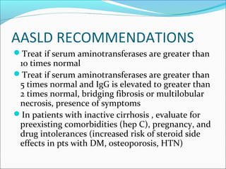
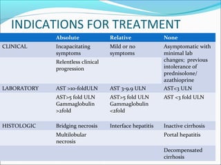
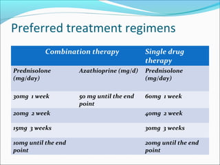
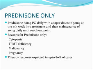
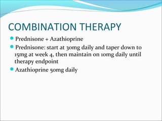
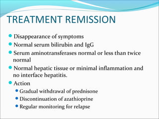
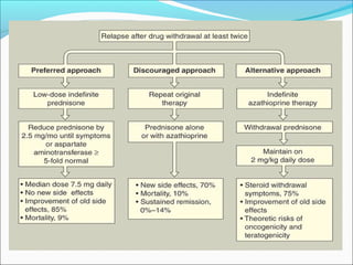
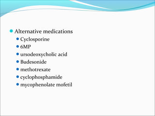
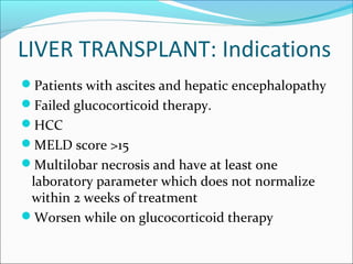
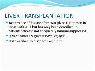
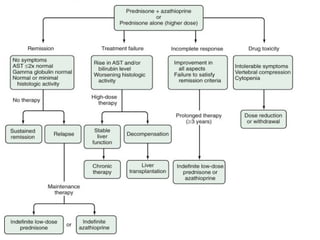
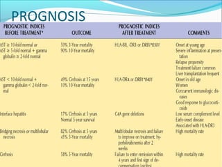
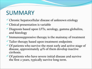
CHAIRED BY : Dr. Raminderpal Singh Sibia
PRESENTED BY : Dr. Rintu Sharma
Immunity
Case scenarios
HISTORY AND CLINICAL FINDINGS:
Case 1: A 46-year old female patient presented with a recently
occurred icterus of unknown origin as well as dark urine and
decolored stool. No diseases were found in the patient's medical
history. Clinical examination showed no other findings except
from the icterus.
Case 2: A 48-year old female patient was admitted to hospital
with epigastric pain and icterus. Similar symptoms reoccurred
regularly since several years.
She reported chronic pain in her finger joints and appearance of
haematomas without adequate trauma.
CLINICAL INVESTIGATIONS:
Case 1: Highly elevated liver enzymes and bilirubin.
Ultrasound examination was unremarkable.
Negative serology for hepatitis A, B and C
Marked immunoglobulin G (IgG) elevation and
hypergammaglobulinaemia.
ANA and smooth muscle antibodies (SMA)+++
Case 2:
reduced liver function with low albumin and prothrombin time
moderate elevation of liver enzymes and a high bilirubin.
Ultrasound examination revealed hepatic parenchymal changes,
splenomegaly, and ascites.
Oesophagogastroduodenoscopy showed oesophageal varices I°.
Serology for hepatitis A, B, and C was negative.
a marked IgG elevation and hypergammaglobulinaemia
Autoimmune antibodies (ANA and SMA)++
AUTOIMMUNE HEPATITIS
Continuing/unresolving hepatocellular
inflammation and necrosis of unknown etiology
Can progress to cirrhosis
Exclusion of other chronic liver diseases
Characteristics include:
presence of autoimmune antibody
evidence of hepatitis (interface being
characteristic)
elevation of serum globulins
OTHER NAMES
Active chronic hepatitis or chronic active hepatitis
Chronic aggressive hepatitis
Lupoid hepatitis
Plasma cell hepatitis
Autoimmune chronic active hepatitis
BACKGROUND
First described in 1950’s
Accounts for 5.6% of liver transplants in the US
Affects women more than men (3.6:1)
If untreated approximately 40% die within 6 months
40% develop cirrhosis
54% develop esophageal varices
20% die of hemorrhage
EPIDEMIOLOGY
Incidence: 1.9 cases per 100,000 persons per yr
Frequency of AIH among patients with chronic
liver disease in North America is between 11%-
22%
Accounts for 5.6% of liver transplants in the US
Prevalence greatest among northern European
white persons
 Japenese have a lower frequency
Scand J Gastroenterol1998;33:99.
PATHOGENESIS
Unknown mechanism but several proposed
mechanisms
Genetically predisposed individual with exposure to an
environmental agent triggers the autoimmune
pathogenic process
Genetic predisposing factors:
HLA-DR3: early onset, severe form
HLA-DR4: caucasian, late onset, better response to steroids,
higher incidence of extrahepatic manifestations
 Triggering factors
Infections (HAV, HBV, HCV, HSV, EBV, measles)?
Medications (ABX, statins, NSAIDs etc.)?
Toxins?
Molecular mimicry?
Recognition of antigen-MHC II complex by
uncommitted CD4 cells
 Cytokine release from TH1 and TH2 CD4 cells
IL-12 and IL-2: proliferation of CD8 cells
IL-4 and IL-10: proliferation of B cells
Evidence supporting autoimmune
pathogenesis
Histopathological lesions composed of cytotoxic Tcells
and plama cells
Circulating autoantibodies
Hyperglobulinemia
Other autoimmune disorders: thyroiditis, RA ,
autoimmune hemolytic anemia, ulcerative colitis,
membranoproliferative glomerulonephritis, diabetes
mellitus, celiac disease, sjogren’s syndrome
Histocompatibility haplotypes assosciations
Response to steroids and immunosuppression.
CLASSIFICATION
TYPE 1
TYPE 2
TYPE 3
OVERLAP SYNDROMES
TYPE 1
Classically in young females
ANA or Anti-Smooth Muscle antibody
positive
Titer usually > 1:100
10% will have an antibody to Soluble
Liver antigens (SLA)
Other Antibodies: anti-DNA, pANCA,
Anti-mitochondrial, Anti-Actin (AAA),
cytoskeletal antibody, nuclear envelope
proteins lamin A and C, plasma
membrane sulfatides
Anti-actin antibodies have greater
specificity
TYPE 1
Bimodal Age distribution (ages 10-20 and 45-70)
Female:male (3.6:1)
HLA DR3 or DR4 assosciation
Associated with extrahepatic
manifestations(38%):
Autoimmune thyroiditis, Graves disease, Chronic UC
Less commonly with RA, pernicious anemia, systemic
sclerosis, ITP, SLE,coombs positive hemolytic anemia,
leucocytoclastic vasculitis, erythema nodosum
40% present with acute onset of symptoms
similar to toxic hepatitis or acute viral hepatitis
TYPE 2
Seen in children (2-14 years)in
Meditteranean population
HLA DR1 or DQB1 assosciation
Presence of anti-Liver/Kidney
Microsome Antibodies (anti-
LKM1 )directed against
cytochrome p450 2D6 {same as
LKM seen in patients with
chronic hepatitis C}
Anti-Liver Cytosol antibody
(ALC-1)
Acute or fulminant presentation
possible
TYPE 3
Antibodies to soluble liver
antigen / liver pancreas antigen
Lack ANA and anti- LKM 1
antibodies
More in women, part of
spectrum of type 1 AIH
% Concurrent autoimmune
disease: 58
Elevated gamma-globulin: ++
Steroid responsive: +++
% progression to cirrhosis: 75
OVERLAP SYNDROMES
Primary Biliary Cirrhosis
Primary Sclerosing Cholangitis
5% of patients with chronic hepatitis C will have an
ANA titer of >1:100
A homogeneous pattern of staining is more common
in ANA positive autoimmune hepatitis compared to
that of ANA positive chronic hepatitis C
Autoimmune hepatitis and
Chronic hepatitis C
8% of white North American adults have Concurrent
infection with HCV
52% of chronic hepatitis C patients have
autoantibodies
Interferon therapy can enhance immune
manifestations of AIH and concurrent HCV infection
Immunosuppressive treatment can increase serum
viral levels in patients with chr hepatitis C
Treatment should be appropriate for predominant
disease , based on nature of concurrent immune
disease
Autoimmune hepatitis and
Chronic hepatitis C
CLINICAL PRESENTATION
Similar as chronic hepatitis
May be confused with acute hepatitis
Can hav acute severe or fulminant presntation;
history of recurrent bouts
Asymptomatic in 34%-45% cases
Symptoms:
Fatigue, Malaise- 86%
Jaundice- 77%
abdominal pain- 48%
Pruritis – 36%
Anorexia- 30%
Amenorrhea
Arthralgias
nausea, vomiting
myalgias
 Fever
Arthritis
maculopapular eruptions
erythema nodosum, colitis
anemia
Features of concurrent
immune disease
Signs
Hepatomegaly- 78%
Jaundice- 69%
Splenomegaly -32%
Concurrent immune disease- < 38%
Ascites- 20 %
Encephalopathy – 14%
Stigmata of chronic liver disease
Lab findings
Similar to chronic viral hepatitis
May not corelate with clinical or histological
severity
Elevated AST 100%
Hypergammaglobulinemia 92%
Inc immunoglobulin G level 91%
Hyperbilirubinemia 83%
Alk Phos >2 times 33%
Immunoserological markers:
 SMA,ANA, anti-LKM1- 100 %
pANCA- 92 % ( type 1 only)
anti-asialoglycoprotein- 82 %
AAA- 74 %
anti-liver cytosol – 32% ( type 2 only )
anti-soluble liver antigen- 11-17 %
Clinical criteria
Presence of characteristic clinical features
Liver histology
Exclusion of other diseases
Scoring criteria
Assess the strength of the diagnosis
Pretreatment and post-treatment
Helpful with variant or atypical forms of AIH
Diagnostic criteria
Diagnostic criteria
Simplified scoring system
Greater specificity vs original scoring system ( 90% vs
73%)
Greater predictability ( 92% vs 82% )
Useful for excluding AIH in patients with other
conditions and concurrent immune features
Less sensitivity (95% vs 100 %)
DIAGNOSTIC
ALGORITHM
FOR AUTOIMMUNE
HEPATITIS
HISTOLOGY
Chronic hepatitis with marked piecemeal necrosis
and lobular involvement
Numerous plasma cells
Interface hepatitis: hallmark finding
Necroinflammatory activity
Evidence of hepatocellular regeneration (“rosette
formation” , regenerative “pseudolobules”)
Bile duct injuries and granulomas are uncommon
DIFFERENTIAL DIAGNOSIS
Primary biliary cirrhosis
Post-necrotic cryptogenic cirrhosis
Primary sclerosing cholangitis
Acute viral hepatitis
Mild chronic viral hepatitis
Wilsons disease
Haemochromatosis
Alcoholic hepatitis
Non alcoholic fatty liver disease
TREATMENT
Should be based on:
Severity of symptoms
Degree of elevation in transaminases and IgG
Histologic findings
Potential side effects of treatment
AASLD RECOMMENDATIONS
Treat if serum aminotransferases are greater than
10 times normal
Treat if serum aminotransferases are greater than
5 times normal and IgG is elevated to greater than
2 times normal, bridging fibrosis or multilobular
necrosis, presence of symptoms
In patients with inactive cirrhosis , evaluate for
preexisting comorbidities (hep C), pregnancy, and
drug intolerances (increased risk of steroid side
effects in pts with DM, osteoporosis, HTN)
INDICATIONS FOR TREATMENT
Absolute Relative None
CLINICAL Incapacitating
symptoms
Mild or no
symptoms
Asymptomatic with
minimal lab
changes; previous
intolerance of
prednisolone/
azathioprine
Relentless clinical
progression
LABORATORY AST >10-foldULN AST 3-9.9 ULN AST<3 ULN
AST>5 fold ULN
Gammaglobulin
>2fold
AST>5 fold ULN
Gammaglobulin
<2fold
AST <3 fold ULN
HISTOLOGIC Bridging necrosis Interface hepatitis Inactive cirrhosis
Multilobular
necrosis
Portal hepatitis
Decompensated
cirrhosis
Preferred treatment regimens
Combination therapy Single drug
therapy
Prednisolone
(mg/day)
Azathioprine (mg/d) Prednisolone
(mg/day)
30mg 1 week 50 mg until the end
point
60mg 1 week
20mg 2 week 40mg 2 week
15mg 3 weeks 30mg 3 weeks
10mg until the end
point
20mg until the end
point
PREDNISONE ONLY
Prednisone 60mg PO daily with a taper down to 30mg at
the 4th week into treatment and then maintenance of
20mg daily until reach endpoint
Reasons for Prednisone only:
Cytopenia
TPMT deficiency
Malignancy
Pregnancy
Therapy response expected in upto 80% of cases
COMBINATION THERAPY
Prednisone + Azathioprine
Prednisone: start at 30mg daily and taper down to
15mg at week 4, then maintain on 10mg daily until
therapy endpoint
Azathioprine 50mg daily
Side effects : Prednisone
Side effects : Azathioprine
TREATMENT REMISSION
Disappearance of symptoms
Normal serum bilirubin and IgG
Serum aminotransferases normal or less than twice
normal
Normal hepatic tissue or minimal inflammation and
no interface hepatitis.
Action
Gradual withdrawal of prednisone
Discontinuation of azathioprine
Regular monitoring for relapse
TREATMENT FAILURE
Worsening clinical, laboratory and histologic findings
despite compliance with therapy
Onset of ascites or encephalopathy
Increase in aminotransferases by >67%
Action
Pred 60 mg/d or pred 30 mg/d with aza 150 mg/d x 1
month
Reduction of the dose each month of improvement
until maintenance levels
TREATMENT FAILURE
Treatment failures are frequent in patients with
established cirrhosis, HLA-DR3 or in patients who
present with disease at a younger age and with a
longer duration of symptoms
INCOMPLETE RESPONSE
Some or no improvement in clinical, laboratory or
histologic features that does not satisfy remission
criteria
Failure to achieve remission after 3 years
Action
Reduction of dose to lowest levels possible to prevent
worsening
Indefinite treatment
RELAPSE
An exacerbation after drug withdrawal in patients who
enter remission
Reappearance of histological disease
AST >3 folds ULN
Cirrhosis develops commonly
Reinstitute original treatment: azathioprine continued
indefinitely
Liver transplantation
Alternative medications
Cyclosporine
6MP
ursodeoxycholic acid
Budesonide
methotrexate
cyclophosphamide
mycophenolate mofetil
LIVER TRANSPLANT: Indications
Patients with ascites and hepatic encephalopathy
Failed glucocorticoid therapy.
HCC
MELD score >15
Multilobar necrosis and have at least one
laboratory parameter which does not normalize
within 2 weeks of treatment
Worsen while on glucocorticoid therapy
LIVER TRANSPLANTATION
Recurrence of disease after transplant is common in
those with AIH but has only been described in
patients who are not adequately immunosuppressed.
 5 year patient & graft survival 83-92%
Auto antibodies disappear within 1y
PROGNOSIS
SUMMARY
Chronic hepatocellular disease of unknown etiology
Clinical presentation is variable
Diagnosis based upon LFTs, serology, gamma globulins,
and histology
Immunosuppressive therapy is the mainstay of treatment
Tailor therapy based upon treatment endpoints
Of patients who survive the most early and active stage of
disease, approximately 41% of them develop inactive
cirrhosis.
Of patients who have severe initial disease and survive
the first 2 years, typically survive long term.
Autoimmune hepatitis

Autoimmune hepatitis

  • 1.
    CHAIRED BY :Dr. Raminderpal Singh Sibia PRESENTED BY : Dr. Rintu Sharma Immunity
  • 2.
    Case scenarios HISTORY ANDCLINICAL FINDINGS: Case 1: A 46-year old female patient presented with a recently occurred icterus of unknown origin as well as dark urine and decolored stool. No diseases were found in the patient's medical history. Clinical examination showed no other findings except from the icterus. Case 2: A 48-year old female patient was admitted to hospital with epigastric pain and icterus. Similar symptoms reoccurred regularly since several years. She reported chronic pain in her finger joints and appearance of haematomas without adequate trauma.
  • 3.
    CLINICAL INVESTIGATIONS: Case 1:Highly elevated liver enzymes and bilirubin. Ultrasound examination was unremarkable. Negative serology for hepatitis A, B and C Marked immunoglobulin G (IgG) elevation and hypergammaglobulinaemia. ANA and smooth muscle antibodies (SMA)+++ Case 2: reduced liver function with low albumin and prothrombin time moderate elevation of liver enzymes and a high bilirubin. Ultrasound examination revealed hepatic parenchymal changes, splenomegaly, and ascites. Oesophagogastroduodenoscopy showed oesophageal varices I°. Serology for hepatitis A, B, and C was negative. a marked IgG elevation and hypergammaglobulinaemia Autoimmune antibodies (ANA and SMA)++
  • 4.
    AUTOIMMUNE HEPATITIS Continuing/unresolving hepatocellular inflammationand necrosis of unknown etiology Can progress to cirrhosis Exclusion of other chronic liver diseases Characteristics include: presence of autoimmune antibody evidence of hepatitis (interface being characteristic) elevation of serum globulins
  • 5.
    OTHER NAMES Active chronichepatitis or chronic active hepatitis Chronic aggressive hepatitis Lupoid hepatitis Plasma cell hepatitis Autoimmune chronic active hepatitis
  • 6.
    BACKGROUND First described in1950’s Accounts for 5.6% of liver transplants in the US Affects women more than men (3.6:1) If untreated approximately 40% die within 6 months 40% develop cirrhosis 54% develop esophageal varices 20% die of hemorrhage
  • 7.
    EPIDEMIOLOGY Incidence: 1.9 casesper 100,000 persons per yr Frequency of AIH among patients with chronic liver disease in North America is between 11%- 22% Accounts for 5.6% of liver transplants in the US Prevalence greatest among northern European white persons  Japenese have a lower frequency Scand J Gastroenterol1998;33:99.
  • 8.
    PATHOGENESIS Unknown mechanism butseveral proposed mechanisms Genetically predisposed individual with exposure to an environmental agent triggers the autoimmune pathogenic process Genetic predisposing factors: HLA-DR3: early onset, severe form HLA-DR4: caucasian, late onset, better response to steroids, higher incidence of extrahepatic manifestations
  • 9.
     Triggering factors Infections(HAV, HBV, HCV, HSV, EBV, measles)? Medications (ABX, statins, NSAIDs etc.)? Toxins? Molecular mimicry? Recognition of antigen-MHC II complex by uncommitted CD4 cells  Cytokine release from TH1 and TH2 CD4 cells IL-12 and IL-2: proliferation of CD8 cells IL-4 and IL-10: proliferation of B cells
  • 11.
    Evidence supporting autoimmune pathogenesis Histopathologicallesions composed of cytotoxic Tcells and plama cells Circulating autoantibodies Hyperglobulinemia Other autoimmune disorders: thyroiditis, RA , autoimmune hemolytic anemia, ulcerative colitis, membranoproliferative glomerulonephritis, diabetes mellitus, celiac disease, sjogren’s syndrome Histocompatibility haplotypes assosciations Response to steroids and immunosuppression.
  • 12.
  • 13.
    TYPE 1 Classically inyoung females ANA or Anti-Smooth Muscle antibody positive Titer usually > 1:100 10% will have an antibody to Soluble Liver antigens (SLA) Other Antibodies: anti-DNA, pANCA, Anti-mitochondrial, Anti-Actin (AAA), cytoskeletal antibody, nuclear envelope proteins lamin A and C, plasma membrane sulfatides Anti-actin antibodies have greater specificity
  • 14.
    TYPE 1 Bimodal Agedistribution (ages 10-20 and 45-70) Female:male (3.6:1) HLA DR3 or DR4 assosciation Associated with extrahepatic manifestations(38%): Autoimmune thyroiditis, Graves disease, Chronic UC Less commonly with RA, pernicious anemia, systemic sclerosis, ITP, SLE,coombs positive hemolytic anemia, leucocytoclastic vasculitis, erythema nodosum 40% present with acute onset of symptoms similar to toxic hepatitis or acute viral hepatitis
  • 15.
    TYPE 2 Seen inchildren (2-14 years)in Meditteranean population HLA DR1 or DQB1 assosciation Presence of anti-Liver/Kidney Microsome Antibodies (anti- LKM1 )directed against cytochrome p450 2D6 {same as LKM seen in patients with chronic hepatitis C} Anti-Liver Cytosol antibody (ALC-1) Acute or fulminant presentation possible
  • 16.
    TYPE 3 Antibodies tosoluble liver antigen / liver pancreas antigen Lack ANA and anti- LKM 1 antibodies More in women, part of spectrum of type 1 AIH % Concurrent autoimmune disease: 58 Elevated gamma-globulin: ++ Steroid responsive: +++ % progression to cirrhosis: 75
  • 18.
    OVERLAP SYNDROMES Primary BiliaryCirrhosis Primary Sclerosing Cholangitis 5% of patients with chronic hepatitis C will have an ANA titer of >1:100 A homogeneous pattern of staining is more common in ANA positive autoimmune hepatitis compared to that of ANA positive chronic hepatitis C
  • 20.
    Autoimmune hepatitis and Chronichepatitis C 8% of white North American adults have Concurrent infection with HCV 52% of chronic hepatitis C patients have autoantibodies Interferon therapy can enhance immune manifestations of AIH and concurrent HCV infection Immunosuppressive treatment can increase serum viral levels in patients with chr hepatitis C Treatment should be appropriate for predominant disease , based on nature of concurrent immune disease
  • 21.
  • 22.
    CLINICAL PRESENTATION Similar aschronic hepatitis May be confused with acute hepatitis Can hav acute severe or fulminant presntation; history of recurrent bouts Asymptomatic in 34%-45% cases
  • 23.
    Symptoms: Fatigue, Malaise- 86% Jaundice-77% abdominal pain- 48% Pruritis – 36% Anorexia- 30% Amenorrhea Arthralgias nausea, vomiting myalgias  Fever Arthritis maculopapular eruptions erythema nodosum, colitis anemia Features of concurrent immune disease
  • 24.
    Signs Hepatomegaly- 78% Jaundice- 69% Splenomegaly-32% Concurrent immune disease- < 38% Ascites- 20 % Encephalopathy – 14% Stigmata of chronic liver disease
  • 25.
    Lab findings Similar tochronic viral hepatitis May not corelate with clinical or histological severity Elevated AST 100% Hypergammaglobulinemia 92% Inc immunoglobulin G level 91% Hyperbilirubinemia 83% Alk Phos >2 times 33%
  • 26.
    Immunoserological markers:  SMA,ANA,anti-LKM1- 100 % pANCA- 92 % ( type 1 only) anti-asialoglycoprotein- 82 % AAA- 74 % anti-liver cytosol – 32% ( type 2 only ) anti-soluble liver antigen- 11-17 %
  • 28.
    Clinical criteria Presence ofcharacteristic clinical features Liver histology Exclusion of other diseases Scoring criteria Assess the strength of the diagnosis Pretreatment and post-treatment Helpful with variant or atypical forms of AIH Diagnostic criteria
  • 29.
  • 33.
    Simplified scoring system Greaterspecificity vs original scoring system ( 90% vs 73%) Greater predictability ( 92% vs 82% ) Useful for excluding AIH in patients with other conditions and concurrent immune features Less sensitivity (95% vs 100 %)
  • 34.
  • 35.
    HISTOLOGY Chronic hepatitis withmarked piecemeal necrosis and lobular involvement Numerous plasma cells Interface hepatitis: hallmark finding Necroinflammatory activity Evidence of hepatocellular regeneration (“rosette formation” , regenerative “pseudolobules”) Bile duct injuries and granulomas are uncommon
  • 38.
    DIFFERENTIAL DIAGNOSIS Primary biliarycirrhosis Post-necrotic cryptogenic cirrhosis Primary sclerosing cholangitis Acute viral hepatitis Mild chronic viral hepatitis Wilsons disease Haemochromatosis Alcoholic hepatitis Non alcoholic fatty liver disease
  • 40.
    TREATMENT Should be basedon: Severity of symptoms Degree of elevation in transaminases and IgG Histologic findings Potential side effects of treatment
  • 41.
    AASLD RECOMMENDATIONS Treat ifserum aminotransferases are greater than 10 times normal Treat if serum aminotransferases are greater than 5 times normal and IgG is elevated to greater than 2 times normal, bridging fibrosis or multilobular necrosis, presence of symptoms In patients with inactive cirrhosis , evaluate for preexisting comorbidities (hep C), pregnancy, and drug intolerances (increased risk of steroid side effects in pts with DM, osteoporosis, HTN)
  • 42.
    INDICATIONS FOR TREATMENT AbsoluteRelative None CLINICAL Incapacitating symptoms Mild or no symptoms Asymptomatic with minimal lab changes; previous intolerance of prednisolone/ azathioprine Relentless clinical progression LABORATORY AST >10-foldULN AST 3-9.9 ULN AST<3 ULN AST>5 fold ULN Gammaglobulin >2fold AST>5 fold ULN Gammaglobulin <2fold AST <3 fold ULN HISTOLOGIC Bridging necrosis Interface hepatitis Inactive cirrhosis Multilobular necrosis Portal hepatitis Decompensated cirrhosis
  • 43.
    Preferred treatment regimens Combinationtherapy Single drug therapy Prednisolone (mg/day) Azathioprine (mg/d) Prednisolone (mg/day) 30mg 1 week 50 mg until the end point 60mg 1 week 20mg 2 week 40mg 2 week 15mg 3 weeks 30mg 3 weeks 10mg until the end point 20mg until the end point
  • 44.
    PREDNISONE ONLY Prednisone 60mgPO daily with a taper down to 30mg at the 4th week into treatment and then maintenance of 20mg daily until reach endpoint Reasons for Prednisone only: Cytopenia TPMT deficiency Malignancy Pregnancy Therapy response expected in upto 80% of cases
  • 45.
    COMBINATION THERAPY Prednisone +Azathioprine Prednisone: start at 30mg daily and taper down to 15mg at week 4, then maintain on 10mg daily until therapy endpoint Azathioprine 50mg daily
  • 46.
    Side effects :Prednisone
  • 47.
    Side effects :Azathioprine
  • 48.
    TREATMENT REMISSION Disappearance ofsymptoms Normal serum bilirubin and IgG Serum aminotransferases normal or less than twice normal Normal hepatic tissue or minimal inflammation and no interface hepatitis. Action Gradual withdrawal of prednisone Discontinuation of azathioprine Regular monitoring for relapse
  • 49.
    TREATMENT FAILURE Worsening clinical,laboratory and histologic findings despite compliance with therapy Onset of ascites or encephalopathy Increase in aminotransferases by >67% Action Pred 60 mg/d or pred 30 mg/d with aza 150 mg/d x 1 month Reduction of the dose each month of improvement until maintenance levels
  • 50.
    TREATMENT FAILURE Treatment failuresare frequent in patients with established cirrhosis, HLA-DR3 or in patients who present with disease at a younger age and with a longer duration of symptoms
  • 51.
    INCOMPLETE RESPONSE Some orno improvement in clinical, laboratory or histologic features that does not satisfy remission criteria Failure to achieve remission after 3 years Action Reduction of dose to lowest levels possible to prevent worsening Indefinite treatment
  • 52.
    RELAPSE An exacerbation afterdrug withdrawal in patients who enter remission Reappearance of histological disease AST >3 folds ULN Cirrhosis develops commonly Reinstitute original treatment: azathioprine continued indefinitely Liver transplantation
  • 54.
  • 55.
    LIVER TRANSPLANT: Indications Patientswith ascites and hepatic encephalopathy Failed glucocorticoid therapy. HCC MELD score >15 Multilobar necrosis and have at least one laboratory parameter which does not normalize within 2 weeks of treatment Worsen while on glucocorticoid therapy
  • 56.
    LIVER TRANSPLANTATION Recurrence ofdisease after transplant is common in those with AIH but has only been described in patients who are not adequately immunosuppressed.  5 year patient & graft survival 83-92% Auto antibodies disappear within 1y
  • 58.
  • 59.
    SUMMARY Chronic hepatocellular diseaseof unknown etiology Clinical presentation is variable Diagnosis based upon LFTs, serology, gamma globulins, and histology Immunosuppressive therapy is the mainstay of treatment Tailor therapy based upon treatment endpoints Of patients who survive the most early and active stage of disease, approximately 41% of them develop inactive cirrhosis. Of patients who have severe initial disease and survive the first 2 years, typically survive long term.