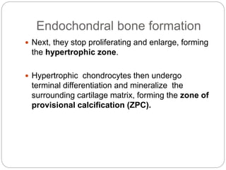
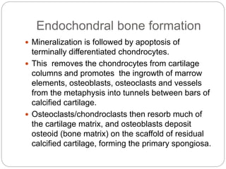
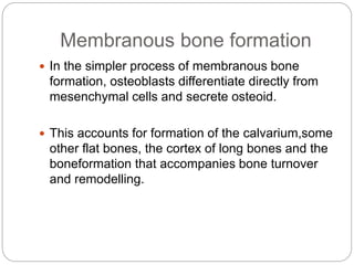
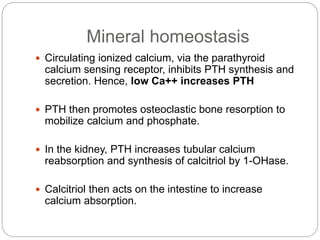
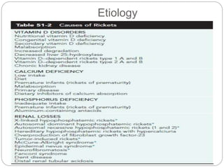
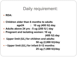
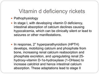
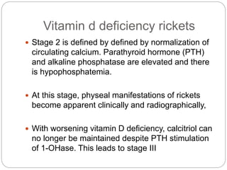
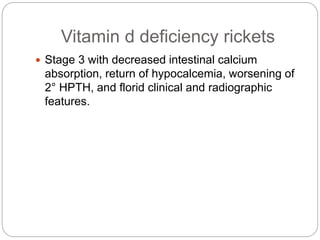
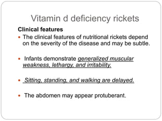
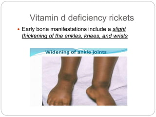
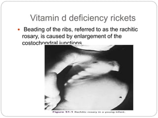
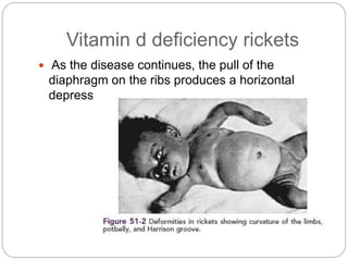
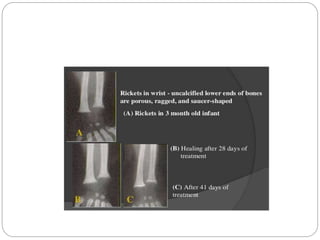
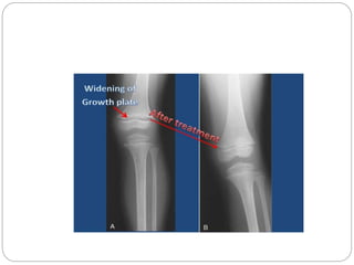
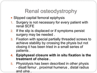
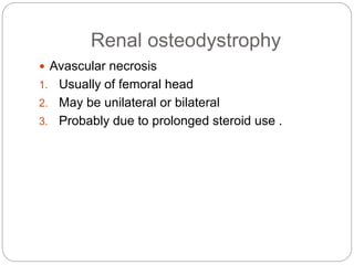
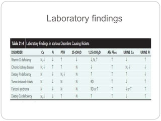
This document provides an overview of rickets and osteomalacia. It begins by defining rickets as a disease affecting the growth plates, causing deficient mineralization and impaired endochondral ossification due to failed apoptosis of hypertrophic chondrocytes. Osteomalacia is a similar disorder affecting mineralization of osteoid. Both result from insufficient calcium and phosphate levels. Vitamin D deficiency is a common cause and treatment involves vitamin D supplementation. Other causes discussed include hereditary disorders, prematurity, drugs, tumors, and renal osteodystrophy. Clinical features, radiographic findings, and management approaches are described for each condition.
1. PRESENTOR : DR VEDANT BANSAL
MODERATOR : DR KARTHIK M N
RICKETS AND OSTEOMALACIA
2. Introduction
Rickets is a syndrome of diverse etiology
characterised by failure of normal mineralisation
of bone and epiphyseal cartilage and by skeletal
deformity occuring in children.
In children, the epiphyseal ends of the bones are
the most active in osteogenesis, so the disease is
more evident there.
3. Introduction
Rickets is a disease of the physes(growth plates)
characterized not only by deficient mineralization
of cartilage and osteoid and by retarded
endochondral ossification.
Failure of normal apoptosis of hypertrophic
chondrocytes is now recognized as the key
abnormality causing this ossification defect.
4. Introduction
The abnormalities of mineralization and
ossification are caused by insufficient circulating
levels of calcium and phosphate ions.
Although mineralization depends on the calcium
phosphate (Ca × P) product, the defect in
endochondral ossification more specifically
results from hypophosphatemia, which impairs
chondrocyte apoptosis
5. Introduction
In osteomalacia, an insufficient Ca × P product
causes failure of normal mineralization of
osteoid,laid down either at sites of bone turnover
or by the periosteum in the process of
membranous bone formation.
These processes occur in both adults and
children. Hence osteomalacia can be present at
any age.
6. Introduction
Rickets involves the physes, and osteomalacia
involves other sites of bone formation
Rickets and osteomalacia result from insufficient
mineral ion concentrations, which are caused by
many disorders.
8. Endochondral bone formation
In EBF, the skeleton is pre-formed in cartilage
and then replaced by bone.
Farthest from the ossification front is the resting
zone with few and randomly distributed small
round chondrocytes.
Moving centrally, the chondrocytes proliferate
more rapidly and become flattened and arranged
in orderly columns, forming the proliferating
zone.
9. Endochondral bone formation
Next, they stop proliferating and enlarge, forming
the hypertrophic zone.
Hypertrophic chondrocytes then undergo
terminal differentiation and mineralize the
surrounding cartilage matrix, forming the zone of
provisional calcification (ZPC).
10. Endochondral bone formation
Mineralization is followed by apoptosis of
terminally differentiated chondrocytes.
This removes the chondrocytes from cartilage
columns and promotes the ingrowth of marrow
elements, osteoblasts, osteoclasts and vessels
from the metaphysis into tunnels between bars of
calcified cartilage.
Osteoclasts/chondroclasts then resorb much of
the cartilage matrix, and osteoblasts deposit
osteoid (bone matrix) on the scaffold of residual
calcified cartilage, forming the primary spongiosa.
11. Membranous bone formation
In the simpler process of membranous bone
formation, osteoblasts differentiate directly from
mesenchymal cells and secrete osteoid.
This accounts for formation of the calvarium,some
other flat bones, the cortex of long bones and the
boneformation that accompanies bone turnover
and remodelling.
12. Mineral homeostasis
Circulating ionized calcium, via the parathyroid
calcium sensing receptor, inhibits PTH synthesis and
secretion. Hence, low Ca++ increases PTH
PTH then promotes osteoclastic bone resorption to
mobilize calcium and phosphate.
In the kidney, PTH increases tubular calcium
reabsorption and synthesis of calcitriol by 1-OHase.
Calcitriol then acts on the intestine to increase
calcium absorption.
18. Daily requirement:
RDA:
Children older than 6 months to adults
age24 : 10 ug (400 IU) day.
Adults above 24 yrs : 5 ug (200 IU ) day
Pregnant and lactating women: 10 ug
(400 IU) day
Upper limit (UL) for children and adults:
50 ug (2,000 IU)/day
• Upper limit (UL) for infant 0-12 months:
25 ug (1,000 IU)/day
19. Vitamin d deficiency rickets
Pathophysiology
In stage I, with developing vitamin D deficiency,
intestinal absorption of calcium declines causing
hypocalcemia, which can be clinically silent or lead to
seizures or other manifestations.
In response, 2° hyperparathyroidism (HPTH)
develops, mobilizing calcium and phosphate from
bone, increasing renal calcium reabsorption and
phosphate excretion, and upregulating renal 25-
hydroxy-vitamin D-1α-hydroxylase (1-OHase) to
increase calcitriol and hence intestinal calcium
absorption. These adaptations lead to stage II
20. Vitamin d deficiency rickets
Stage 2 is defined by defined by normalization of
circulating calcium. Parathyroid hormone (PTH)
and alkaline phosphatase are elevated and there
is hypophosphatemia.
At this stage, physeal manifestations of rickets
become apparent clinically and radiographically,
With worsening vitamin D deficiency, calcitriol can
no longer be maintained despite PTH stimulation
of 1-OHase. This leads to stage III
21. Vitamin d deficiency rickets
Stage 3 with decreased intestinal calcium
absorption, return of hypocalcemia, worsening of
2° HPTH, and florid clinical and radiographic
features.
22. Vitamin d deficiency rickets
Clinical features
The clinical features of nutritional rickets depend
on the severity of the disease and may be subtle.
Infants demonstrate generalized muscular
weakness, lethargy, and irritability.
Sitting, standing, and walking are delayed.
The abdomen may appear protuberant.
23. Vitamin d deficiency rickets
Early bone manifestations include a slight
thickening of the ankles, knees, and wrists
24. Vitamin d deficiency rickets
Beading of the ribs, referred to as the rachitic
rosary, is caused by enlargement of the
costochondral junctions.
25. Vitamin d deficiency rickets
As the disease continues, the pull of the
diaphragm on the ribs produces a horizontal
depression known as Harrison’s groove
26. Vitamin d deficiency rickets
Short stature .
Pectus carinatum .
Closure of the fontanelles is delayed and the
sutures are thickened, which leads to a skull
appearance described as resembling hot cross
buns.
As the child begins standing and walking, the
softened long bones bow, and it is at this time
that the child is usually brought to the orthopaedic
surgeon for a diagnosis.
27. Vitamin d deficiency rickets
In toddlers,bowleg, or genu varum, is one of the
most common initial signs.
In older children, genu valgum and coxa vara
may be initial features.
28. Vitamin d deficiency rickets
Radiology
Failure of the physeal cartilage to calcify and
undergo normal endochondral ossification leads
to an increased thickness of the physis and a
hazy appearance of the provisional zone of
calcification .
The widened growth plate is particularly suspect
for rickets, which differentiatesthis rare condition
from the more common physiologic angular
deformities of the lower extremities .
29.
30. The metaphysis abutting the physis is brushlike in
appearance,with islands or columns of cartilage
persisting wellinto the metaphysis .
The metaphysis also appears cupped or flared.
The bones have an osteopenic appearance
overall, with thinning of the cortices.
31.
32. Looser’s lines, or radiolucent transverse
bands(pseudofracture lines) that extend across
the axes of the long bones, are evident on
radiographs in 20% of patients with rickets.
33. Treatment
Rickets is treated by the administration of vitamin
D under the supervision of a pediatric specialist in
metabolic bone disease.
The usual course of treatment is 6 to 10 weeks.
After 2 to 4 weeks, radiographs show
improvement in mineralization.
34. TREATMENT
SPECIFIC TREATMENT:
ADMINISTRATION OF 15000MICROGRAM OR
6LAKH UNITS OF VITAMIN D ORALLY OR
INTRAMUSCULAR
IF HEALING LINE IS NOT SEEN ON THE X RAYS
WITHIN 3-4 WEEKS OF THERAPY,THE ABOVE
DOSE IS REPEATED
ON RESPONSE , 4OO UNITS OR 10 MICROGRAM
OF VIT D/ DAY IS SUPPLEMENTED
35.
36.
37. If the child does not respond to vitamin D
therapy, vitamin D–resistant rickets should be
suspected.
Because residual deformity is rare after medical
treatment of nutritional rickets, there is no specific
orthopaedic treatment of nutritional rickets.
38. Rickets of Prematurity
Very premature infants are particularly at risk for
the development of nutritional treatment
required.Resolution of the rachitic changes and
fractures occurs as the infants gain weight.
39. Drug-Induced Rickets
Certain antiepileptic medications have been known to
produce rachitic changes in children .
Seizure medications that affect the liver may induce
the cytochrome P-450 microsomal enzyme system
and decrease levels of vitamin D.
Hypocalcemia develops, which can aggravate the
seizure disorder.
Treatment with vitamin D is very helpful.
The condition should be suspected in neurologic
patients with seizures who begin sustaining frequent
fractures
40. Vitamin D–Resistant Rickets
Vitamin D–resistant rickets, also known as
hereditary or familial hypophosphatemic rickets,
encompasses a group of disorders in which
normal dietary intake of vitamin D is insufficient to
achieve normal mineralization of bone because of
pathologic renal phosphate wasting
Hereditary hypophosphatemic rickets can be
inherited as an X-linked dominant, autosomal
dominant, or autosomal recessive form. X-linked
dominant disease is the most frequent form of
hereditary rickets
41. Vitamin D–Resistant Rickets
The disease usually becomes apparent at a slightly
older age than nutritional rickets, with most patients
becoming symptomatic between 1 and 2 years of age.
Severe hypophosphatemic rickets can be recognized
in early infancy, and when the disease is suspected
because of the family history, laboratory determination
of phosphorus concentrations can lead to the
diagnosis in infants as young as 3 months.
The usual initial complaints are delayed walking and
angular deformities of the lower extremities. In
contrast to what is seen in nutritional rickets, systemic
manifestations such as irritability and apathy are
minimal.
42. Vitamin D–Resistant Rickets
Physical findings in hypophosphatemic rickets include
skeletal deformities, which resemble those seen in
nutritional rickets but, because of the chronicity of the
disease, become much more severe.
Once affected children begin to walk, genu varum
develops, although genu valgum may occur in some
children .
Periarticular enlargement is present as a result of
widening of the physes and metaphyses.
The rachitic rosary may also occur.
Short stature is a feature of hypophosphatemic
rickets.
Height is usually 2 standard deviations (SDs) below
the mean for age in these patients.
43. Vitamin D–Resistant Rickets
The radiographic changes are the same as those
seen in
nutritional rickets and include physeal widening and
indistinct
osteopenic metaphyses.
In the lower extremities, genu
varum is obvious, and the distal femoral and
proximal tibial
physes are particularly widened medially .
44. Vitamin D–Resistant Rickets
The usual treatment consists of oral replacement
of phosphorus in large doses and the
administration of an active form of vitamin D,
calcitriol or alfacalcidol.
45. Vitamin D–Resistant Rickets
The orthotic management of vitamin D–resistant
rickets has not been efficacious.
The deformity most commonly seen in patients
with hypophosphatemic rickets is a gradual
anterolateral bowing of the femur, combined with
tibia vara.
46. Vitamin D–Resistant Rickets
Multilevel osteotomy is generally required to
satisfactorily correct the mechanical axis of the
limb.
The mechanical axis should be mildly
overcorrected at surgery
The suggested fixation varies among reports.
External fixation allows fine tuning of the
alignment postoperatively, when the patient is
able to stand
Others advocate the use of intramedullary fixation
47.
48.
49. Tumor-Related Hypophosphatemic
Rickets
An association between benign and malignant tumors and
hypophosphatemic rickets has been described, termed
oncogenic hypophosphatemic osteomalacia .
Conditions such as neurofibromatosis and fibrous
dysplasia produce rickets on rare occasion.
Osteoblastoma, hemangiopericytoma of bone, and skin
tumors have produced rachitic changes in bone by
disrupting the renal tubular resorption of phosphate.
Certain of age. The rachitic changes resolve with excision
of the tumor.
Rachitic changes resolve with excision of the tumour
50. Renal osteodystrophy
Inability of the damaged glomerulus to excrete
phosphorus ,causing hyperphospahtemia .
Increasing FGF 23 levels decreases the
production of dihydroxyvitamin D form the kidney
.
Resulting hypocalcemia triggers release of PTH
51. Renal osteodystrophy
Histology
Osteoclastic resorption of bone
Replacement of marrow with fibrous tissue
Pathcy formation of new bone leads to osteosclerosis
Clinical features
1.short for age
2.bone pain and fractures
3.genu valgum / varum
4.slipped capital femoral epiphysis
5.Trendelenburg gait
6.rachitic rosary
52. Renal osteodystrophy
Radiology
1.Osteopenia with thinning of cortices and
indistinct trabeculae
2.Skull has a salt and pepper appearance
3.Widening of physis
4.Cupping of physis not seen as in nutritional
rickets
5.Subperiosteal resorption in ulna
53. Renal osteodystrophy
Medical treatment
1. High dose pulsed iv , intraperitoneal and oral
calcitriol therapy .
2. Treament of acidosis with sodium bicarbonate
3. Phosphate binding agents .
Orthopaedic treament
1. Patients are referred for treatment of three
problems – angular deformity of lower limbs ,
SCFE, AVN .
54. Renal osteodystrophy
Angular deformity
1. Genu valgum is the most common deformity
2. If patient is symptomatic and has had optimal
medical management of the psteodystrophy
without resolution of deformity an osteotomy is
performed .
3. Distal end of femur is usually the site of greatest
deformity
4. Ilizarov device has met with success although
healing was delayed .
55. Renal osteodystrophy
Slipped capital femoral epiphysis
1. Surgery is not necessary for every patient with
renal SCFE
2. If the slip is displaced or if symptoms persist
surgery may be needed .
3. Fixation with special partially threaded screws to
achieve stability by crossing the physis but not
closing it has been tried in a small series of
patients .
4. Epiphyseal closure with in situ fixation is the
treatment of choice .
5. Physiolysis has been described in other physis
- disatl femur , proximal humerus , distal radius
and ulna .
56. Renal osteodystrophy
Avascular necrosis
1. Usually of femoral head
2. May be unilateral or bilateral
3. Probably due to prolonged steroid use .