Downloaded 2,272 times
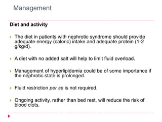
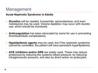
![Management
Specific treatment
In minimal-change nephropathy, glucocorticosteroids, such as prednisone,
are used. Children who relapse may be treated with rituximab]
In some lupus nephritis, prednisone and cyclophosphamide are useful
Secondary amyloidosis with nephrotic syndrome may respond to anti-inflammatory
treatment of the primary disease.
In membranous nephropathy, expectant management without
immunosuppression can be used for the first 6 months, in patients at low risk
for progression (ie, those with serum creatinine level < 1.5 mg/dL). Patients
with renal insufficiency (serum creatinine level > 1.5 mg/dL) are at greatest
risk for the development of end-stage renal disease and should receive
immunosuppressive therapy.[37]](https://image.slidesharecdn.com/nephroticsyndromeabhay-copy-140910103817-phpapp02/85/Nephrotic-syndrome-27-320.jpg)
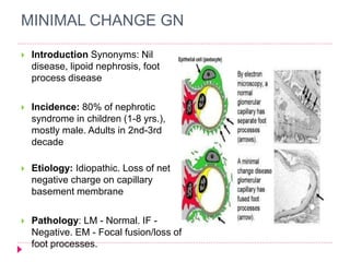
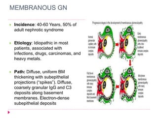
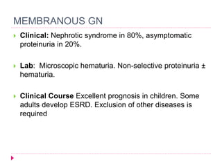


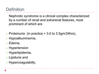
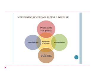
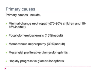
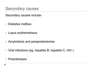
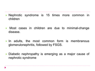
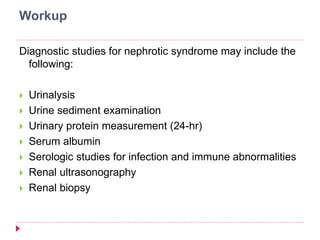
Nephrotic syndrome is characterized by proteinuria, hypoalbuminemia, edema, and hyperlipidemia. It can be primary, caused by diseases of the kidney itself, or secondary, caused by systemic illnesses that affect the kidneys. The most common primary causes are minimal-change disease in children and membranous glomerulonephritis in adults. Secondary causes include diabetes, lupus, and infections. Treatment involves controlling edema with diuretics, treating underlying conditions, and using steroids, immunosuppressants, or ACE inhibitors depending on disease type and severity.