Downloaded 1,022 times
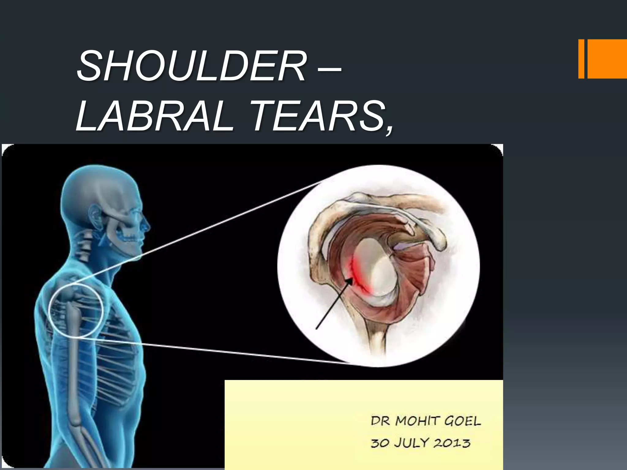
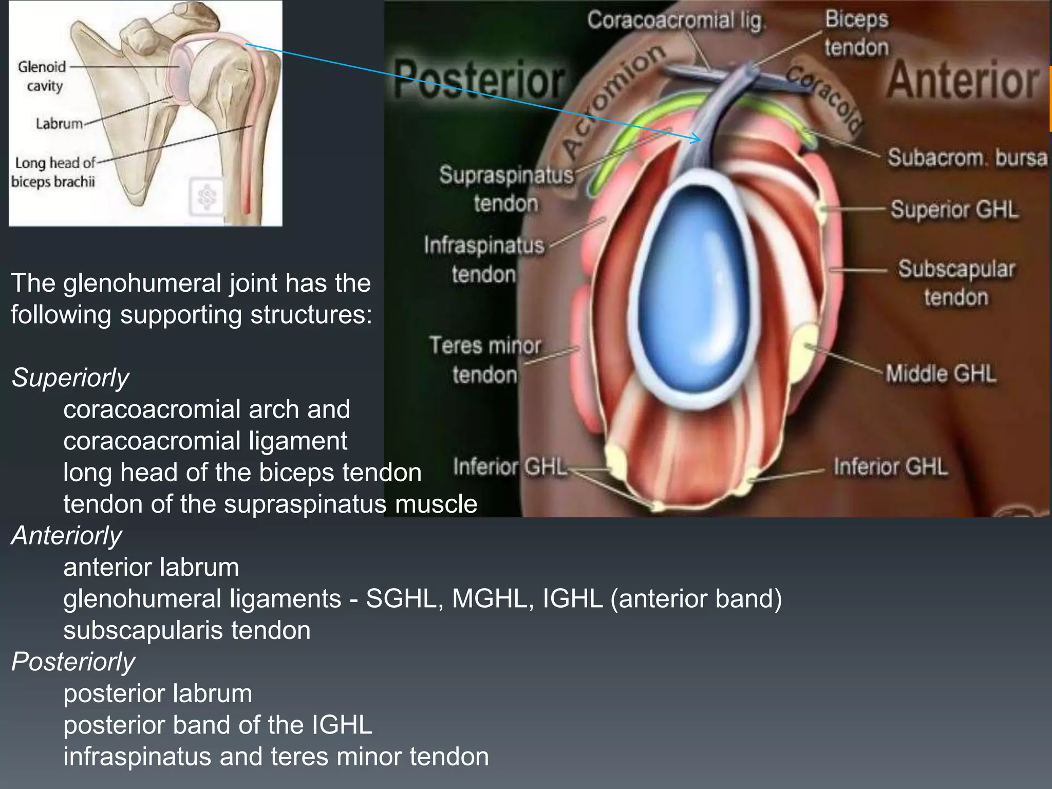
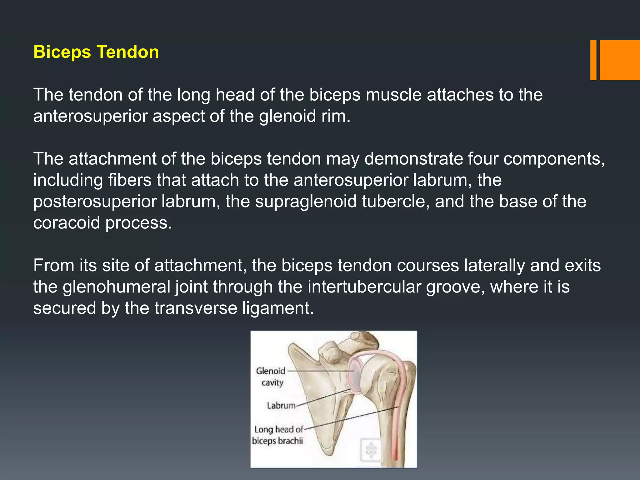


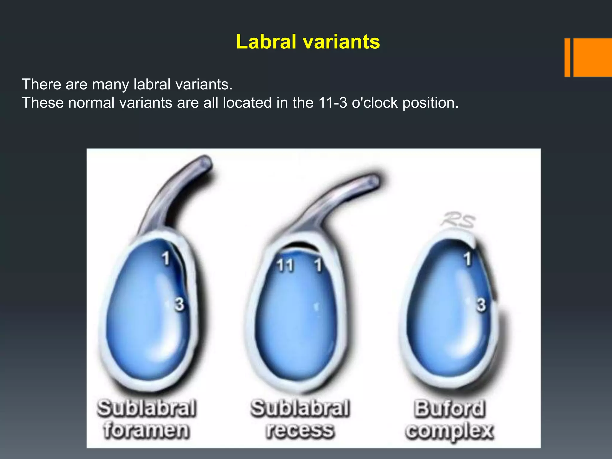

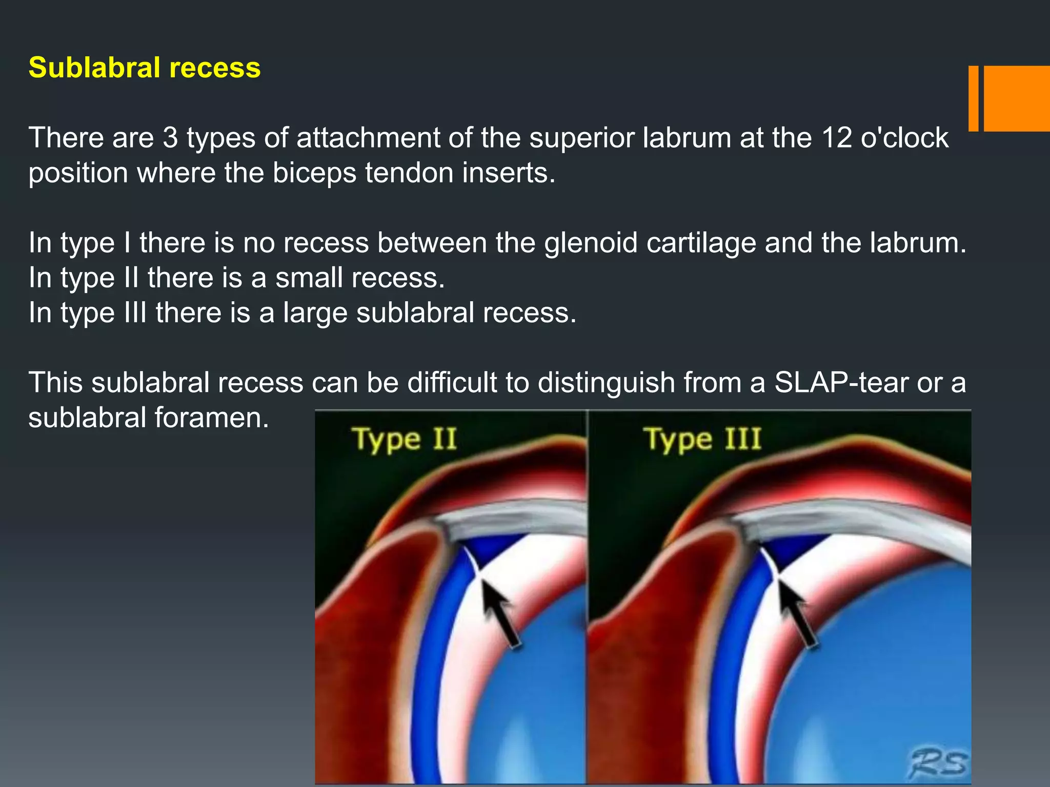
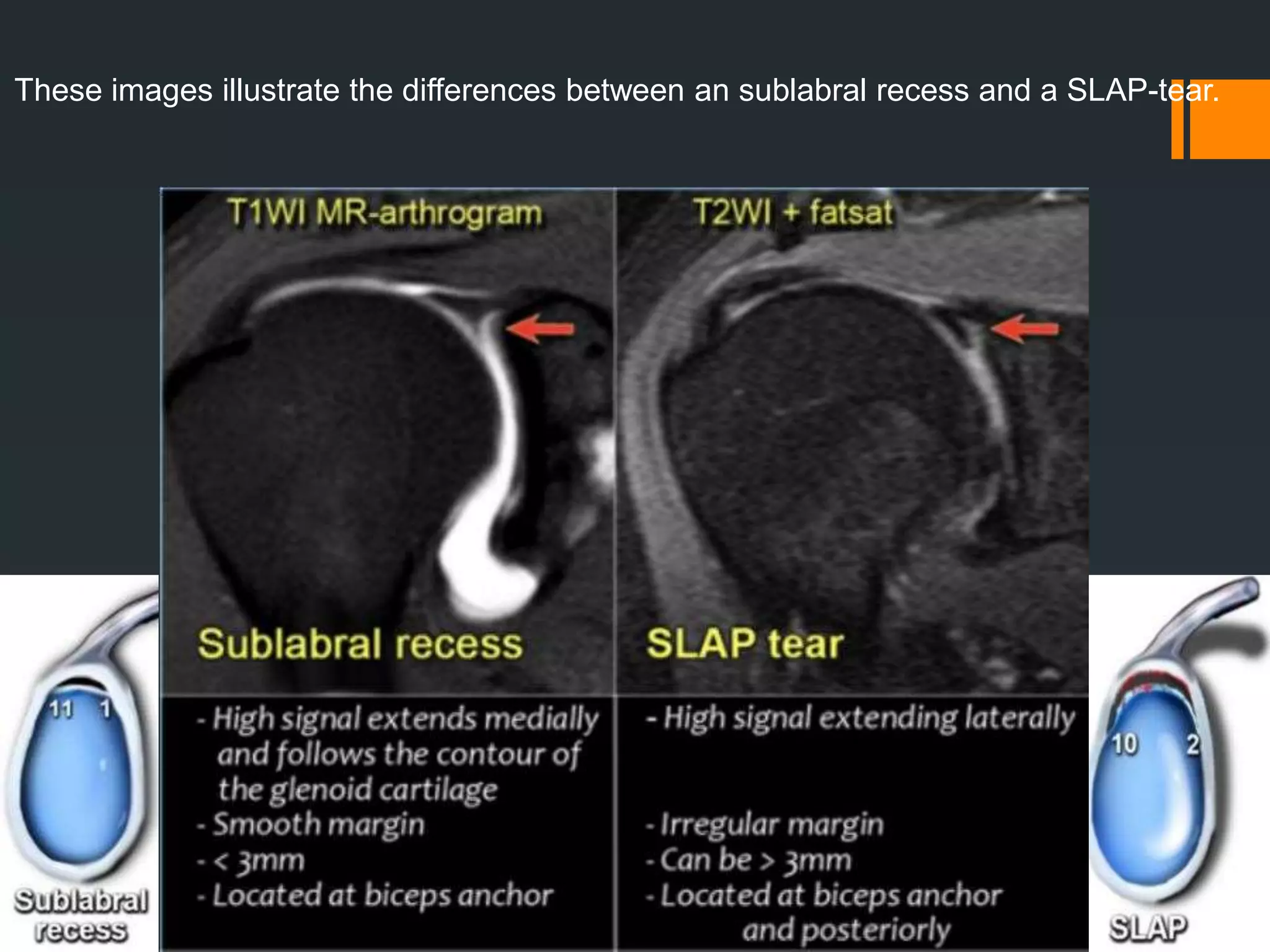


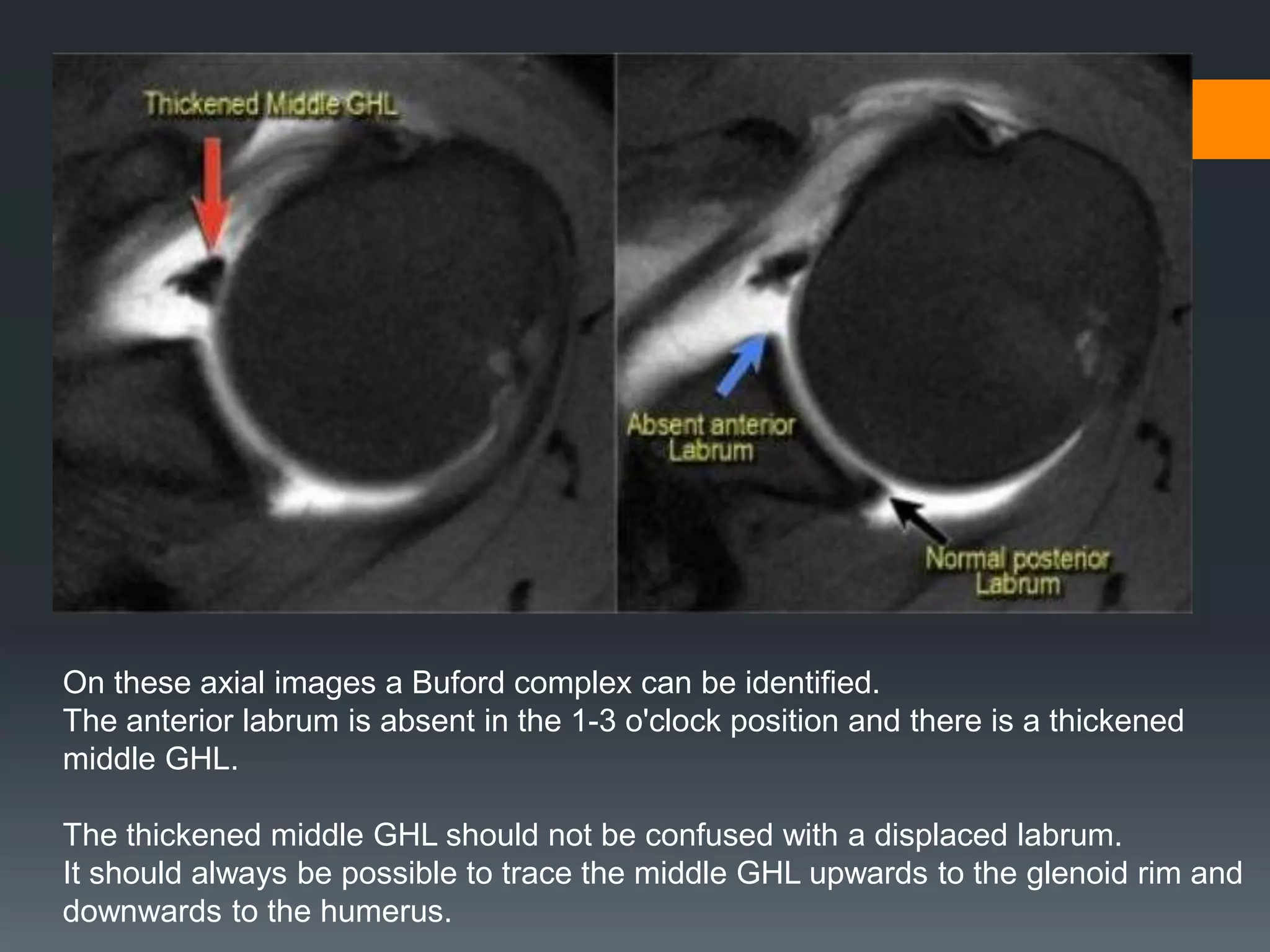

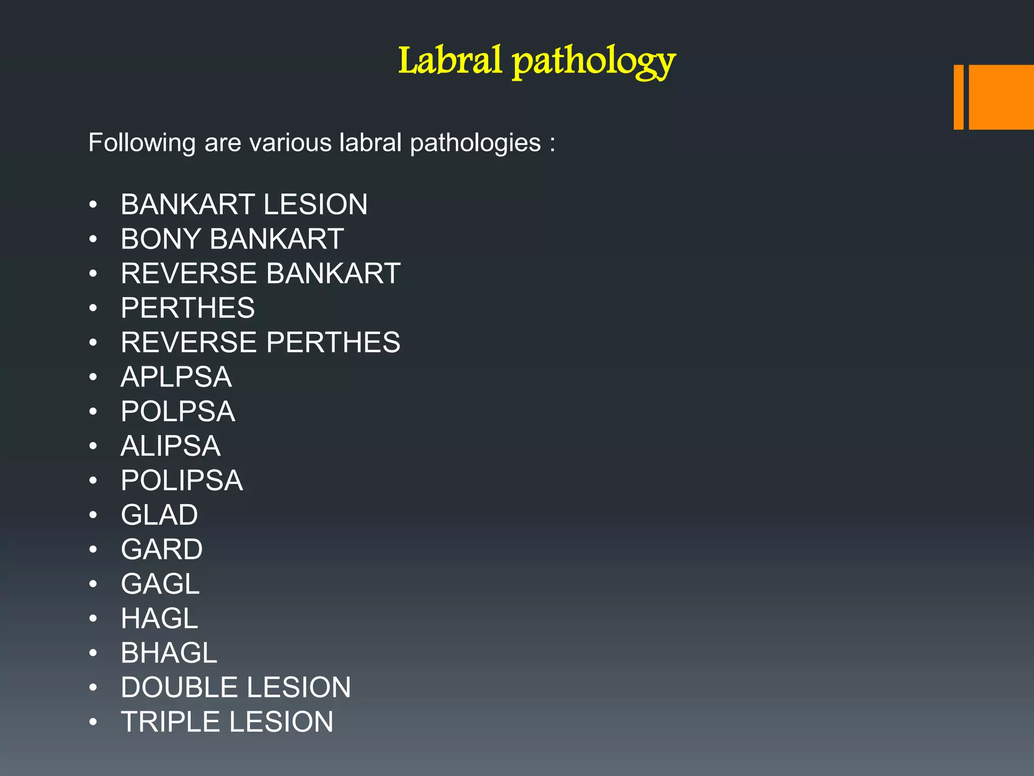
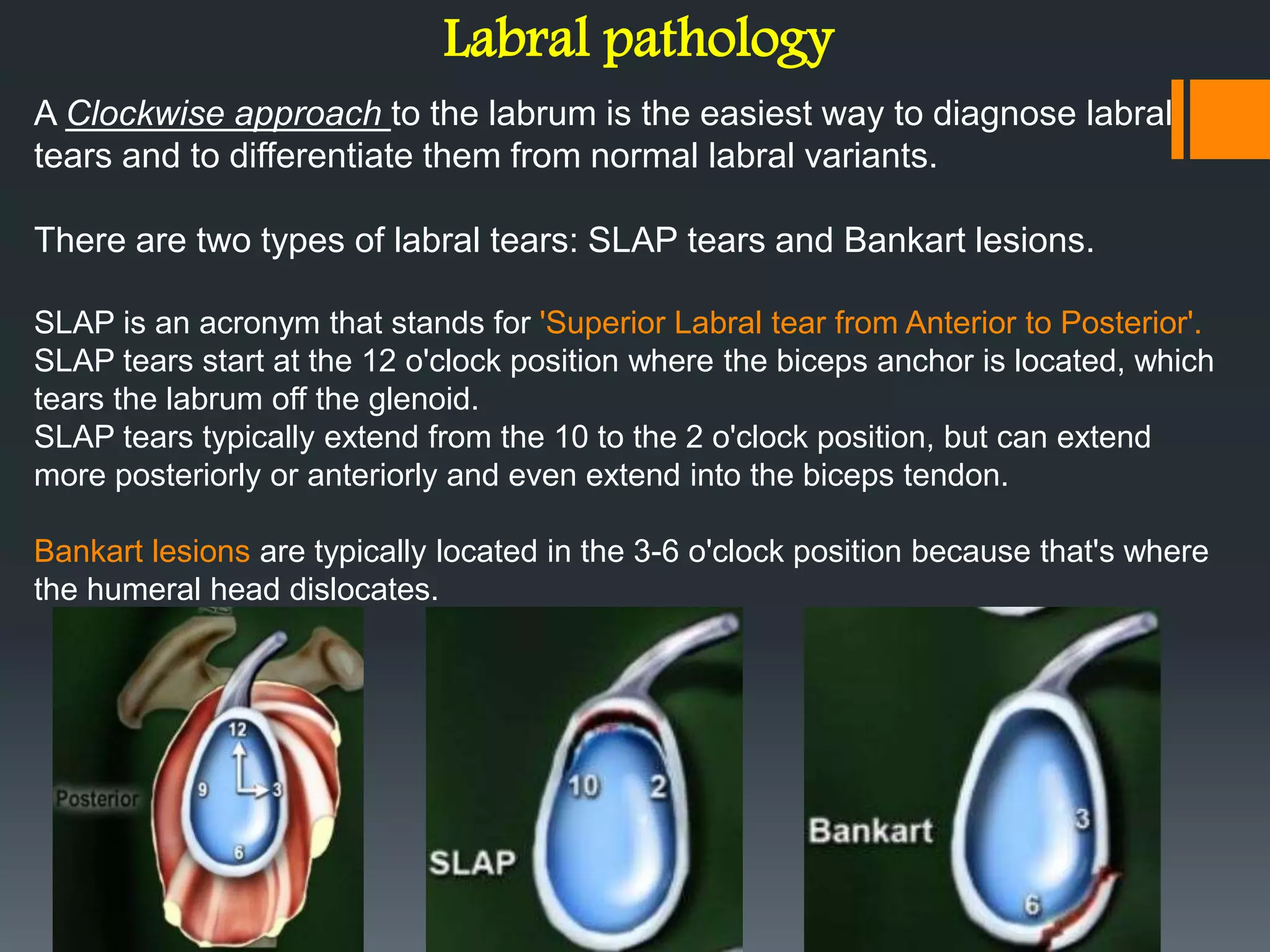
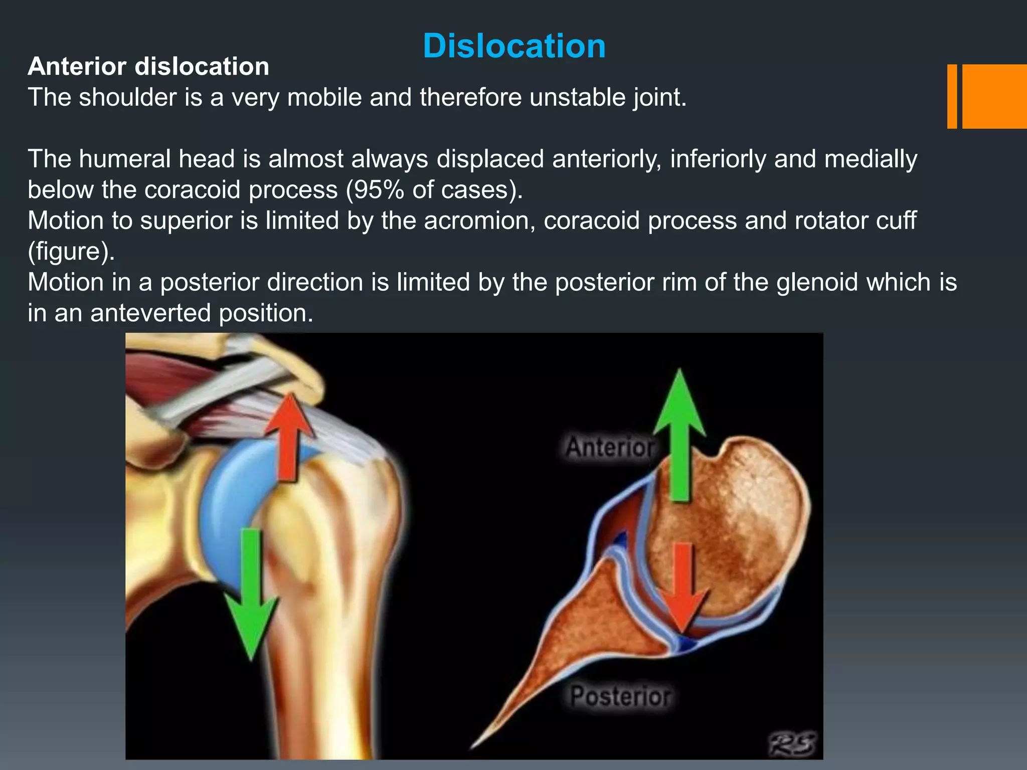
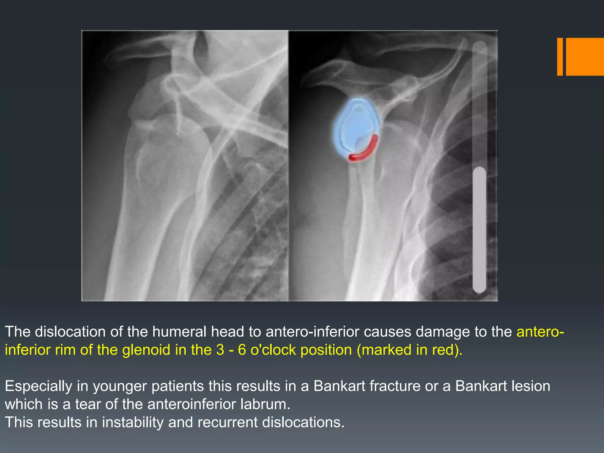

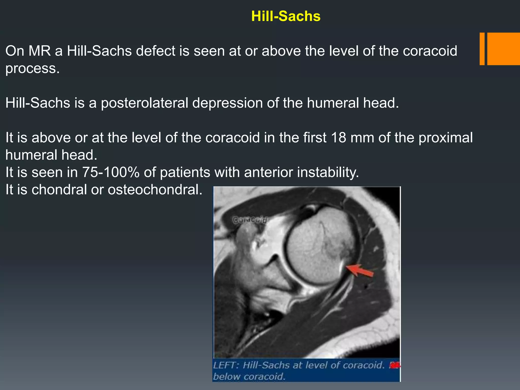
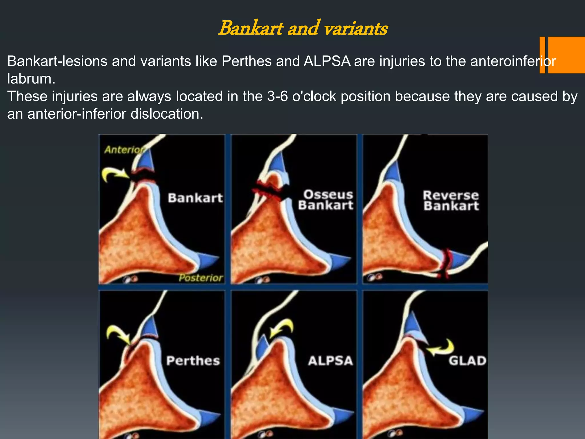

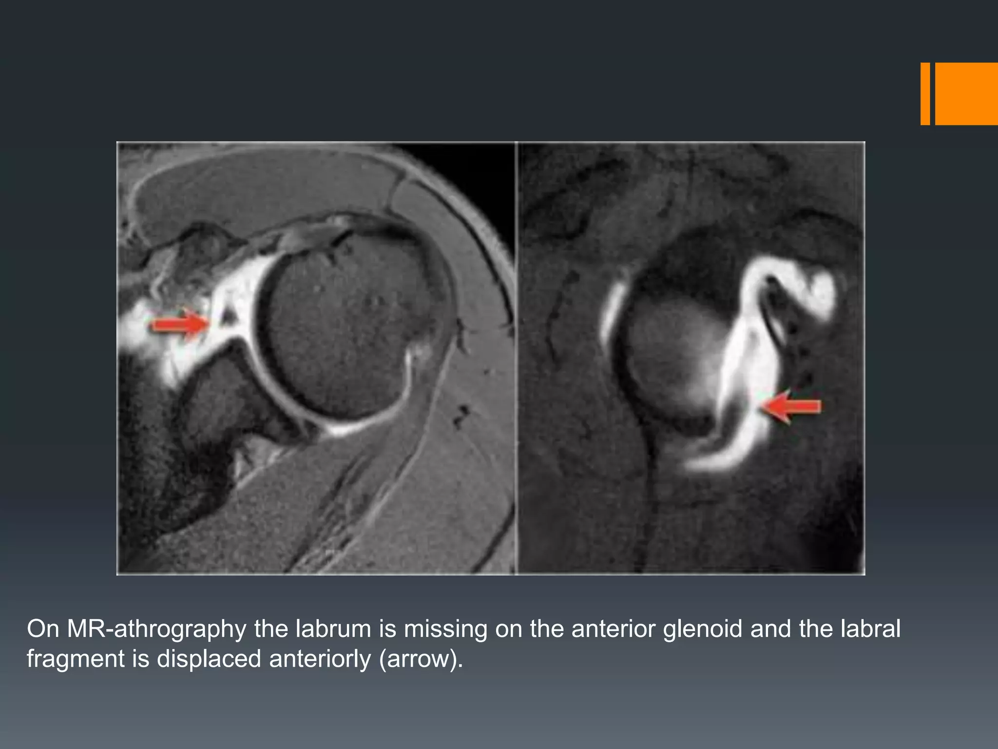


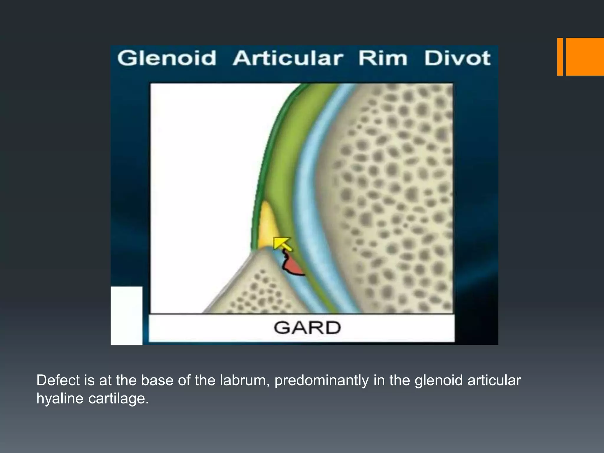

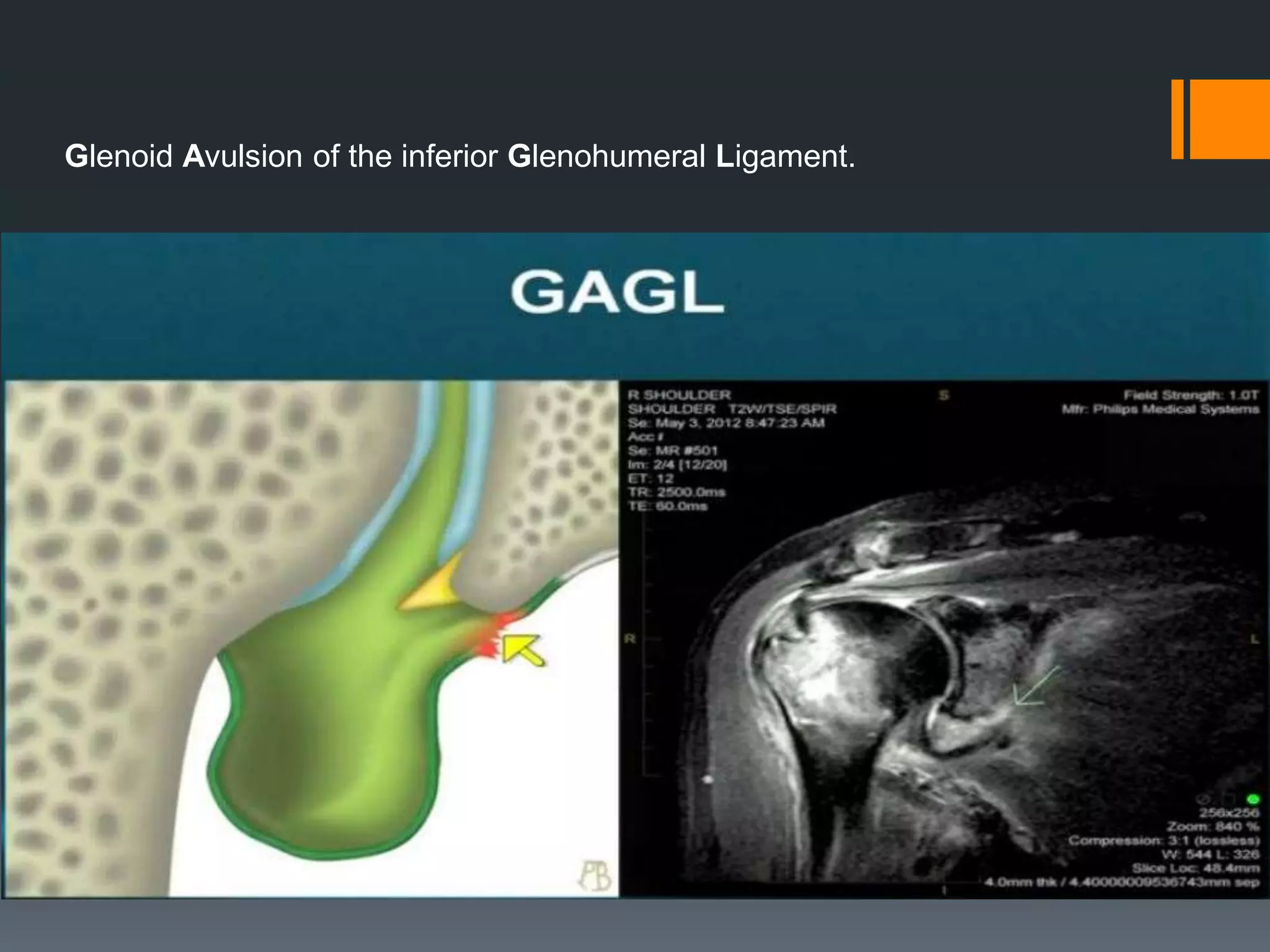


The document describes various structures of the shoulder joint that provide stability, including the labrum, biceps tendon, and glenohumeral ligaments. It discusses common labral injuries like SLAP tears and Bankart lesions caused by anterior dislocation of the humeral head. It also describes variants like Buford complex and sublabral recesses that should not be confused with pathology.


