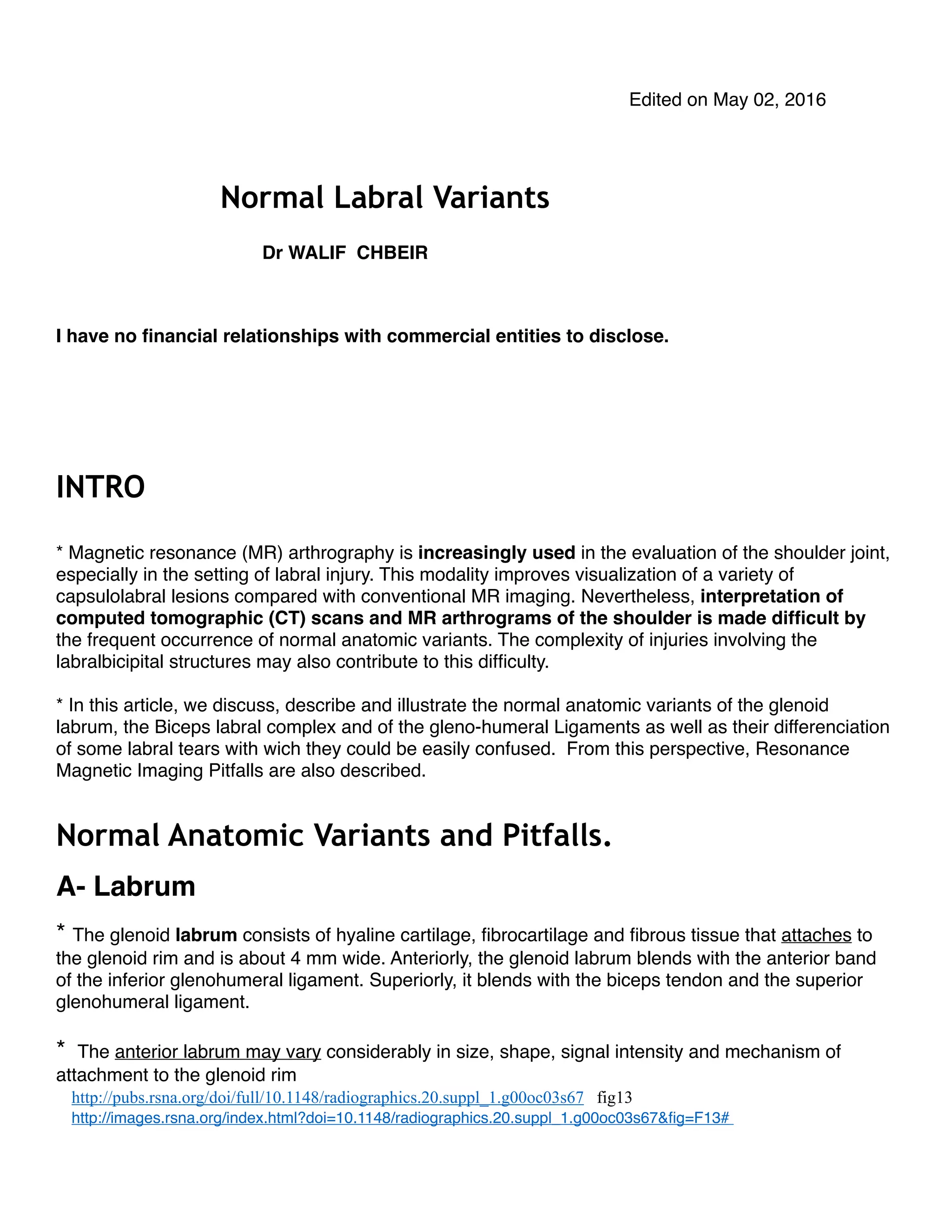
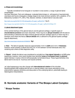
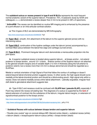
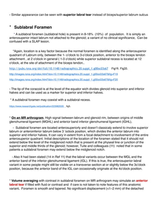
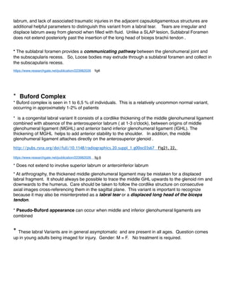
Walif Chbeir provides an in-depth look at labral variants and the analysis of CT and MRI scans on patients.
In this article, we discuss, describe and illustrate the normal anatomic variants of the glenoid labrum, the Biceps labral complex and of the gleno-humeral Ligaments as well as their differenciation of some labral tears with wich they could be easily confused. From this perspective, Resonance Magnetic Imaging Pitfalls are also described.
sauth delhi call girls in Bhajanpura 🔝 9953056974 🔝 escort Service
Normal Labral Variants - Walif Chbeir
1. Edited on May 02, 2016
Normal Labral Variants
Dr WALIF CHBEIR
I have no financial relationships with commercial entities to disclose.
INTRO
* Magnetic resonance (MR) arthrography is increasingly used in the evaluation of the shoulder joint,
especially in the setting of labral injury. This modality improves visualization of a variety of
capsulolabral lesions compared with conventional MR imaging. Nevertheless, interpretation of
computed tomographic (CT) scans and MR arthrograms of the shoulder is made difficult by
the frequent occurrence of normal anatomic variants. The complexity of injuries involving the
labralbicipital structures may also contribute to this difficulty.
* In this article, we discuss, describe and illustrate the normal anatomic variants of the glenoid
labrum, the Biceps labral complex and of the gleno-humeral Ligaments as well as their differenciation
of some labral tears with wich they could be easily confused. From this perspective, Resonance
Magnetic Imaging Pitfalls are also described.
Normal Anatomic Variants and Pitfalls.
A- Labrum
* The glenoid labrum consists of hyaline cartilage, fibrocartilage and fibrous tissue that attaches to
the glenoid rim and is about 4 mm wide. Anteriorly, the glenoid labrum blends with the anterior band
of the inferior glenohumeral ligament. Superiorly, it blends with the biceps tendon and the superior
glenohumeral ligament.
* The anterior labrum may vary considerably in size, shape, signal intensity and mechanism of
attachment to the glenoid rim ( Fig1)
a- Shape and morphology:
2. - Typically considered to be triangular or rounded in cross section, a range of glenoid labral
morphologies
has been described. Park and colleagues evaluated labral shape on arthrograms of asymptomatic
volunteers and found a triangular or wedge shape to be most common (anterior, 64%; posterior, 47%)
followed by rounded (17%, 33%). Flat, cleaved, notched, or absent labrum was also seen.
- Fig 1: Anterior labrum. Drawings representing a transverse section through the middle aspect of
the shoulder joint illustrate various appearances of the anterior labrum. This structure may be
triangular (1), undersized (2), blunttipped (arrow in 3), or crescentic or meniscoid (4) = Detached
free edge overlying glenoid articular cartilage. Alternatively, there may be a recess between the
anterior labrum and the cartilage (arrow in 5), the middle glenohumeral ligament may be located
proximal to the anterior labrum (m in 6), or the anterior labrum may appear small and be
accompanied by a thickened inferior glenohumeral ligament (i in 7).
- Labrum is not always symmetric anterior to posterior.
RadioGraphics Vol. 20, No. suppl_1: S67-S81
courtesy of : http://images.rsna.org/index.html?doi=10.1148/radiographics.20.suppl_1.g00oc03s67&fig=F15#
b-Labral attachment types
At the central interface of the glenoid labrum and the glenoid cartilage, 2 specific types of
chondrolabral junctions have been described. There may be an abrupt transition with the labrum
demonstrating a detached free edge overlying glenoid articular cartilage (type A) or there may be a
transition zone where the fibrous labrum blends with or adherent to the glenoid hyaline cartilage
(type B attachment) (fig21).
c- Size: The labrum typically measures approximately 4 mm in width and 3 mm in thickness;
however, broad variation in labral size from 2 to 14 mm between normal individuals exist, thus
rendering size criteria of little diagnostic utility.
d- Signal: Initially the labrum was considered to normally be of low signal intensity on all MR pulse
sequences; however, more recent studies have identified areas of increased linear or globular
3. signal intensity in nearly a third of arthroscopically normal labral tissue. These hyperintensities intra
labral seem to be due to mucinous and myxoid contents.
e- Labral appearance may also change with internalexternal rotation of the shoulder.
Nonarthrographic MR imaging with the arm in internal rotation may cause redundancy of the
anteroinferior capsular structures and obscuration of the underlying labrum, potentially leading to
falsenegative interpretations.
B- Normale anatomic Variants of The Biceps Labral Complex:
* Biceps Tendon
The tendon of the long head of the biceps muscle attaches to the anterosuperior aspect of the glenoid
rim ( Fig 3,4,4’,5,6,7). The attachment of the biceps tendon may demonstrate four components,
including fibers that attach to the anterosuperior labrum, the posterosuperior labrum, the supraglenoid
tubercle, and the base of the coracoid process (Fig 8). From its site of attachment, the biceps tendon
courses laterally and exits the glenohumeral joint through the intertubercular groove, where it is
secured by the transverse ligament.
* BLC and Sup SubL Recess:
* The labrum demonstrates its greatest variation in morphology and attachment above the equator
(Fig 8’). At the superior labrum, fibers from the proximal origin of the long head of the biceps tendon
blend with the labrum forming the biceps labral complex (BLC). The mechanism of attachment of
the superior labrum to the glenoid at the site of the biceps tendon insertion may show considerable
variation (Fig 9, 10, 11, 12). Three distinct types of BLC have been described:
- In a type I BLC, the labrum is firmly attached to the glenoid rim, with no intervening cartilage or
central free edge. There is no Recess. . An arthroscopic probe cannot be inserted between the deep
side of the labrum and the glenoid (fig 10). slab type.
- In type II BLC: the attachment of the glenoid labrum and biceps tendon to the glenoid occurs more
medially and there is continuation of the hyaline cartilage under the labrum accompanied by a small
synovial-lined sulcus between the labral free edge and cartilage. A small sulcus is present between
biceps/labrum and glenoid (Fig 11). May be continuous with sublabral foramen. Intermediate type .
- In a type III BLC, a prominent triangular meniscoid labrum projects into the joint space and results
in a deep recess between the labrum and the glenoid rim, allowing a probe to be inserted between
the labrum and the glenoid cartilage (Fig12a &12b) (Fig 13a,13b). Often continuous as sublabral
foramen.
4. The sublabral sulcus or recess present in type II and III BLCs represents the most frequent
normal anatomic variant of the superior labrum. Prevalence: 75%. A cadaveric study by Smith and
colleagues (10 in 2) demonstrated a recess deeper than 2 mm to be present in 39% of specimens.
* ArthroIRM: The recess can be identified on routine MR imaging and is enhanced by the presence
of a joint effusion or an intra-articular contrast solution.
a- This 3 types of BLC are demonstrated by MR Arthrography on Fig.2.
(A) Type I BLC. Oblique coronal fat-suppressed T1- weighted. MR arthrographic image demonstrates
a smooth, firm attachment of the labrum to the superior glenoid (arrow) with no intervening sulcus.
(B) Type II BLC. Oblique coronal fat-suppressed T1-weighted MR arthrographic image demonstrates
continuation of the hyaline cartilage under the labrum (arrow) accompanied by a contrast-filled sulcus
between the labral free edge and cartilage (curved arrow).
(C) Type III BLC. Oblique coronal fat-suppressed T2-weighted image shows a prominent triangular
meniscoid labrum outlined by fluid because of a deep sublabral sulcus (curved arrow) that parallels
the underlying glenoid cartilage (arrow) extending through the superior labral base. On this plan, the
labrum is triangular and demonstrates a meniscuslike projection into the joint space.
b- A superior sublabral recess is located along superior labrum , at biceps anchor , not extend
posterior to biceps tendon, around 12-1 o'clock , Anterior portion of the Superior labrum not attached
to the glenoid smooth margins and of variable depth (1-2 mm in thickness along the full anterior to
posterior extent); but a recess more than 3-5 mm is always abnormal and should be regarded as a
SLAPtear.
Medial or vertical orientation of high Signal of fluid that follow the contour of cartilage or dosen’t
extend beyond labral-chondral junction suggests recess. In other words, the high signal should curve
medially at the labral-chondral junction and should be a blind-ending pouch. High signal only within a
recess: Does not extend medially across entire labral attachment. Laterally angulated, irregular fluid
cleft distal to glenolabral attachment suggests tear.
c- Type III BLC and recessus could be confused with SLAPII tear ( pseudo-SLAP), especially if
Fluid may extend into recess simulating tear. The diagnosis of a sulcus is supported by the lack of
lateral extension of contrast into the substance of the labrum, irregular labral margins, or extension of
signal/contrast posterior to the biceps anchor. (Fig 13a &13b).
* Sublabral Recess with sulcus between biceps tendon and superior labrum
- "Double oreo cookie" sign on coronal oblique MR: Glenoid cortex (black) + sublabral recess (white)
+ labrum (black) + biceps/superior labrum sulcus (white) + biceps tendon (black)
- Similar appearance can be seen with superior labral tear instead of biceps/superior labrum sulcus
5. * Sublabral Foramen
* A sublabral foramen (sublabral hole) is present in 8-18% (10%) of population. It is simply an
anterosuperior intact labrum not attached to the glenoid; a variant of no clinical significance, Can be
confused with a SLAP lesion.
*Again, location is a key factor because the normal foramen is identified along the anterosuperior
quadrant of Labrum only, between the 1- o’clock to 3-o’clock position, anterior to the biceps tendon
attachment , at 2 o'clock in general ( 1-3 o'clock) while superior sublabral recess is located at 12
o'clock, at the site of attachment of the biceps tendon. ( Fig.14a & 14b,15).
- The tip of the coracoid is at the level of the equator wich divides glenoid into superior and inferior
halves and can be used as a maeker for superior and inferior halves.
* A sublabral foramen may coexist with a sublabral recess. Fig 16.
* On an MR arthrogram, High signal between labrum and glenoid rim, between origins of middle
glenohumeral ligament (MGHL) and anterior band inferior glenohumeral ligament (IGHL),
- Sublabral foramen are located anterosuperiorly and doesn’t classicaly extend to involve superior
labrum or anteroinferior labrum below 3 ‘oclock position, which divides the anterior labrum into
superior and inferior halves. It can vary in extent from a focal detachment to involvement of the entire
anterosuperior quadrant. Initial descriptions of the location of the foramen stated that it should not
extend below the level of the midglenoid notch that is present at the physeal line or junction of the
superior and middle thirds of the glenoid; however, Tuite and colleagues (11) noted that in some
patients a sublabral foramen may extend below the midglenoid notch.
- Also It had been stated (14 in Ref 11) that the labral variants occur between the MGL and the
anterior band of the inferior glenohumeral ligament (IGL). If this is true, the anterosuperior labral
variant in some people might still be visible on a transverse section at or slightly below the 3o’clock
position, because the anterior band of the IGL can occasionally originate at the 4o’clock position.
* Volume averaging with contrast in sublabral foramen on MR arthrogram may simulate an anterior
labral tear if filled with fluid or contrast and if care is not taken to note features of this anatomic
variant. Foramen is smooth and tapered. No significant displacement (<1–2 mm) of the detached
labrum, and lack of associated traumatic injuries in the adjacent capsuloligamentous structures are
additional helpful parameters to distinguish this variant from a labral tear. Tears are irregular and
displace labrum away from glenoid when filled with fluid. Unlike a SLAP lesion, Sublabral Foramen
does not extend posteriorly past the insertion of the long head of biceps brachii tendon .
6. * The sublabral foramen provides a communicating pathway between the glenohumeral joint and
the subscapularis recess (Fig.17). So, Loose bodies may extrude through a sublabral foramen and
collect in the subscapularis recess.
* Buford Complex
* Buford complex is seen in 1 to 6,5 % of individuals. This is a relatively uncommon normal variant,
occurring in approximately 1-2% of patients
* is a congenital labral variant It consists of a cordlike thickening of the middle glenohumeral ligament
combined with absence of the anterosuperior labrum ( at 1-3 o'clock), between origins of middle
glenohumeral ligament (MGHL) and anterior band inferior glenohumeral ligament (IGHL). The
thickening of MGHL helps to add anterior stability to the shoulder. In addition, the middle
glenohumeral ligament attaches directly on the anterosuperior glenoid (Fig 18,19, 20).
* Does not extend to involve superior labrum or anteroinferior labrum
* At arthrography, the thickened middle glenohumeral ligament may be mistaken for a displaced
labral fragment. It should always be possible to trace the middle GHL upwards to the glenoid rim and
downwards to the humerus. Care should be taken to follow the cordlike structure on consecutive
axial images cross-referencing them in the sagittal plane. This variant is important to recognize
because it may also be misinterpreted as a labral tear or a displaced long head of the biceps
tendon.
* Pseudo-Buford appearance can occur when middle and inferior glenohumeral ligaments are
combined
* These labral Variants are in general asymptomatic and are present in all ages. Question comes
up in young adults being imaged for injury. Gender: M = F. No treatment is required.
C- Imaging Pitfalls
* Magic angle artifact
- Anteroinferior and posterosuperior labrum
* Intra-articular biceps tendon dislocation
- Dislocated tendon lies adjacent to labrum. May simulate tear
* Hyaline cartilage undercutting
7. - Cartilage lying beneath labrum may simulate tear. The Cartilage signal intensity is higher than
fibrous labrum. The cartilage is smooth and tears tend to be irregular. Cartilage does not extend
through to opposite labral surface. This aspect of undercutting cartilage is found mostly in type B
attachments where intermediate signal intensity may be noted at the chondrolabral junction
corresponding to the transitional zone of fibrocartilage, which should not be misinterpreted as a labral
tear. fig 21B..
* Confusion with middle glenohumeral ligament
- On axial images, middle glenohumeral ligament lies adjacent to anterior labrum and then, may
appear to be fragment of anterior labrum if there is a crescent of fluid between labrum and middle
glenohumeral ligament wich can simulate tear. A "pseudo-sublabral foramen" appearance may occur
when oblique sagittal images are improperly oriented. Follow oblique course of middle glenohumeral
ligament on consecutive images to confirm that it is a separate structure from labrum and Confirm
normal signal in the underlying labrum.
D-GlenoHumeral Ligaments
* Ligaments have low signal intensity on all MR imaging sequences
* Superior Glenohumeral Ligament : The SGH ligament is the most consistently identified
capsular ligament. May origin from biceps tendon, anterior labrum, or in common with MGHL. The
superior glenohumeral ligament courses in a plane nearly perpendicular to the middle glenohumeral
ligament (Fig.6, 22) and parallel to the coracoid process.
* Middle Glenohumeral Ligament. typically has an oblique orientation from superomedial to
inferolateral, extending along deep surface of subcapsularis to lesser tuberosity. Blends anteriorly
with joint capsule and labrum. Can fuse with anterior band of IGHL. The middle glenohumeral
ligament varies most in size and site of attachment to the glenoid. It may be absent or small in
30%. (Fig23) or may appear thick and cordlike (as, for example, in Buford complex).
Originate from superoanterior labrum (Fig 24) or more frequently from glenoid neck (Fig 25).
The appearance of the middle glenohumeral ligament may also change significantly with
internalexternal rotation of the shoulder . With the arm in external rotation, the ligament is stretched
and is located adjacent to the capsule. With internal rotation, the ligament may appear redundant.
* Inferior Glenohumeral Ligament: More accurately termed IGHL complex. Extends from inferior
glenoid labrum to inferior humeral anatomic neck. The inferior glenohumeral ligament is an
important stabilizer of the anterior shoulder joint and consists of the axillary pouch and anterior and
posterior bands (Fig 26 and 3).
8. The anterior band inserts along the inferior two thirds of the anterior glenoid labrum. It presents a
vertical orientation. When redundant, it may overlap the anterior edge of the glenoid cartilage. The
anterior band is usually larger and prominent, although in approximately 25% of cases it is very thin
(fig 27).
The posterior band is usually thinner than the anterior band but has also vertical orientation.
*
- Because the middle and inferior glenohumeral ligaments may be located adjacent to the anterior
labrum, they may give the erroneous impression on transverse CT or MR arthrograms that a
portion of the labrum is detached or torn.
- Most often, the labrum is firmly fixed to the glenoid cartilage, but occasionally a recess of variable
size may be present between the anterior labrum and the glenoid rim. Such a recess may also be
present between the sites of attachment of the middle and inferior glenohumeral ligaments and may
simulate labral detachment. Occasionally, a small recess is evident along the entire labrum.
- The anterior labrum may be very thin and mostly replaced by a thick inferior glenohumeral
ligament.
- Folds of synovial tissue may form along the anterior capsule due to a chronic inflammatory
condition. Such
folds may be misinterpreted as labral fragments or fraying of the labrum.
- When the MGHL origins from scapular neck, it may simulate stripping of anterior capsule.
MR Imaging Technique
* MR arthrography: . Best imaging study for biceps labral complex and for glenohumeral ligaments
(Fibrocartilaginous labrum and glenoHumeral Ligaments outlined by contrast).
* Standards MR Sequences for Shoulder MR Arthrography
- T1-weighted sequences: 1-Axial without fat saturation. Evaluates bony Bankart and
cartilage injuries
2- Oblique coronal with fat saturation
- PD with fat-saturation sequence: Oblique sagittal
- T2-weighted with fat-saturation sequence: Oblique coronal
* The labrum is routinely evaluated in all 3 planes on MR imaging, with the axial plane providing the
most diagnostic information and the coronal plane serving an adjunctive role for evaluation of the
9. superior labrum (biceps labral complex) and inferior glenohumeral capsulolabral complex. MGHL and
IGHL are best evaluated by Sagital Oblique Plane. SGHL and MGHL by Axial Plane.
* Several reports have detailed the importance of proper shoulder positioning to optimize
evaluation of its complex anatomy and specifically to aid in detection of subtle abnormalities of the
glenoid labrum and GHLs.
* The shoulder is routinely imaged in neutral or slight external rotation. The degree of rotation
can be assessed by noting the position of the bicipital grove on axial images.
- Significant internal rotation should be avoided for conventional MR imaging of the shoulder
because it may cause redundancy of the anteroinferior capsular structures medial displacement of
the joint capsule and contraction of the subscapularis tendon, and obscuration of the underlying
anteroinferior labrum, potentially leading to falsenegative interpretations. With the administration of
intra-articular contrast and distension of the joint, capsular apposition becomes less problematic.
* MR imaging of the shoulder with the arm in alternate positions has been advocated to better
assess the integrity of specific labroligamentous structures.
- Abduction and External Rotation: Images acquired only in the ABER position if there was
only a clinical concern for anteroinferior labroligamentous pathology (Bankart lesion, anterior
labroligamentous periosteal sleeve avulsion (ALPSA), Perthes lesion)
- Imaging of the shoulder in flexion, adduction, and internal rotation (FADIR) has been
advocated to better evaluate the posterior capsulolabral complex.
* MDCT arthrography with multiplanar reformations can be useful in patients with contraindication to
MR. Employ oblique coronal and sagittal to correspond to standard MR planes
Differential Diagnostic
It is important when interpreting a shoulder MR study to distinguish between the anterosuperior labral
variants and a labral tear. Patients with pain, instability, and a labral tear benefit from surgery, while
surgery is unnecessary for the anterosuperior labral variants. It has been reported (7 in 11 ) that if the
MGL of a Buford complex is mistakenly surgically attached to the glenoid rim patients can have
painful restriction of humeral rotation and elevation. Fixing a SLF or superior recess can have also
the same consequences.
* Anterior Labral Tears:
- SubLabral Foramen may be confused with an anterior labral tear if filled with fluid or contrast and
if care is not taken to note features of this anatomic variant. Foramen is smooth and tapered and
unlike a SLAP lesion it does not extend posteriorly past the insertion of the long head of biceps
brachii tendon .
10. - Tears are irregular and displace labrum away from glenoid when filled with fluid (Fig 28, 29, 30).
- They most commonly involve the anteroinferior aspect of the labrum (Bankart lesion, anterior
labroligamentous periosteal sleeve avulsion (ALPSA), Perthes lesion). Bankart tears can extend into
anterosuperior labrum Positioning the arm in abduction and external rotation at MR arthrography
has been used for better evaluation of these lesions.
- The second most common type of labral tear involves the entire anterior labrum .
- Isolated tears of the anterosuperior labrum are uncommon, and in the absence of other
pathologic findings, the possibility that they represent normal anatomic variants should always be
considered.
- Microinstability, Shoulder Can have isolated tear of anterosuperior labrum
* SLAP Lesions
* The Superior SubLabral recess must be distinguished from SLAP tear wich correspond to
Anteriortoposterior lesions of the superior labrum. SLAP lesions are centered at the attachment of
the biceps tendon. However, the portions of the labrum situated anterior and posterior to the site of
attachment of the biceps tendon may be involved to a variable extent. High Signal extends posterior
to biceps anchor in SLAP: Only 75% specificity at conventional MR (T2WI), 55% at MR arthrography.
This is controversial. Indeed, some believe that superior recess can extend posterior to biceps.
-Above type II, SLAP tears require Surgery. Type II SLAP lesions consist of detachment of the superior
labralbicipital complex from the superior glenoid rim (Fig 31,32,33,34,35). It may be difficult to
differentiate this type of SLAP lesion from a deep superior sublabral recess at CT and MR
arthrography ( Pseudo-SLAP).
High signal of SLAP (fluid on T2WI or arthrographic contrast on T1WI) is seen extending into the superior
labrum, and tracking into the labrum and sometimes into the biceps tendon is the characteristic finding. The
investigation of choice is MR arthrogram, which is variably reported as having accuracies of 75- 90%,
although distinguishing between subtypes of SLAP can be difficult. Increased distance between the labrum
and the glenoid, an irregular appearance, or lateral extension may suggest a SLAP lesion rather than
a normal anatomic variant (Fig 36). In short, 3 signs are suggestive of tear: 1 - Lateral curving high
T2 signal in tear is the most useful sign 2 - "Double Oreo" sign in setting of SLAP tear associated
with subL Recess: two high signal T2 lines; one is Variant superior recess and other is superior
labrum anterior-posterior (SLAP) tear.
11. 3 - High signal too wide on oblique coronal images: > 2 mm on conventional MR or / and > 3 mm
on direct MR arthrography: suggests tear rather than recess
- Again the High signal of the recess curves medially at labral-chondral junction and should be a
blind-ending pouch without extension medially across entire labral attachment. The diagnostic is
supported by the lack of lateral extension of contrast into the substance of the labrum, the regular
labral margins, the absence of extension of signal/contrast posterior to the biceps anchor and the
parallelisme between hyaline cartilage and the sulcus (fig.37).
The Sublabral recess typically extends only to the most posterior attachment point of the biceps
tendon to the labrum and glenoid. It does not extend posterior to the Biceps . Beyond , there is tear.
* A SLAP tear may, however, extend into anterosuperior labrum ( 1-3 o'clock) : in 40% of SLAP tears
and 90% specificity (10% of people have sublabral foramen). Indeed, SLAP tear type II, in which
the labrum is avulsed from the underlying glenoid can look similar to a sublabral foramen but can be
distinguished from the latter by observing high signal extending between the glenoid and labrum posterior to the
attachment of the biceps tendon. Scroll through the images and notice the unattached labrum at the
1-2-3 o'clock position at the site of the sublabral foramen. Notice the smooth borders unlike the
margins of a SLAP tear.
- SubL Foramen : Unlike a SLAP lesion, it does not extend posteriorly past the insertion of the long head of
biceps brachii tendon. It neither extends inferior to the equator into the anterior inferior labrum. If there
is extension beyond these regions, then it is a tear.
* Constricted joint fluid access to the subscapularis recess through the sublabral foramen
should not be misconstrued as a type II SLAP tear with associated paralabral cyst. High origin of
the middle and inferior
GHLs (MGHL and IGHL) with narrowing of Weitbrecht foramen may promote extension of fluid and/or
debris through the sublabral foramen as an alternative path to the subscapularis recess.
* SLAP tears occur in setting of fall on outstretched arm, sports activity involving overhead arm
motion and acute or repetitive biceps traction injury. Other abnormalities are usually associated:
Other labral or ligamentous tears, Bankart lesion, Rotator cut in 40%, Spinoglenoid notch paralabral
cyst associated with traumatic posterior SLAP tear.
12. * - Anatomic Variants are asymptomatic when isolated but may escort labral tears wich clinically
include mostly pain and a clicking sensation. Biceps weakness. Positive O'Brien test and
microinstability.
- In short, if the the signal between the labrum and glenoid extends into the anterior inferior
labrum or posterior to the biceps insertion, it is a tear. Fig 38-42.
CONCLUSION
* MR Imaging and MR Arthrography remain the primary imaging modalities for evaluation of
patients with suspected internal derangement of the shoulder. Accurate interpretation of these
studies, and of the CT Arthrography, requires an understanding of the complex anatomy of the
shoulder joint, imaging pitfalls, the normal variants of the L. and the complexity of injuries involving
the labralbicipital complex. - It remains difficult, somehow, to differentiate normal variants from
pathologic conditions and to distinguish between various types of SLAP lesions.
* Consider
- A Variant if labral "abnormality" is between 12-3 o'clock. However If the abnormality goes posterior
to or into the biceps tendon insertion or extends below the equator into the anterior inferior quadrant,
it is NOT a variant.
- That the Sublabral recess does not extend posterior to the Biceps insertion and the Sublabral
Foramen does not extend inferior to the equator into the anterior inferior labrum. There is a tear, if
extension is beyond these regions. However, in 6% of patients with sublabral foramen and Bufford
Complex, the labrum is abnormal just below the midpoint of the glenoid. In this setting, other MR
findings may be helpful in distinguishing a variant from an extensive chronic tear of the anterior
labrum.
BIBLIOGRAPHY
1-CT and MR Arthrography of the Normal and Pathologic Anterosuperior Labrum and
LabralBicipital Complex. Michel De Maeseneer & co.
Radiographics October 2000, Volume 20, Issue suppl 1, Special Issue
http://pubs.rsna.org/doi/full/10.1148/radiographics.20.suppl_1.g00oc03s67
13. 2-Anatomic Variants and Pitfalls of the Labrum, Glenoid Cartilage, and Glenohumeral
Ligaments. Kevin S. Dunham & co.
Magn Reson Imaging Clin N Am 20 (2012) 213–228
https://www.researchgate.net/publication/223982026
3- Shoulder MR Anatomy: Normal anatomy, Variants and Checklist
Robin Smithuis and Henk Jan van der Woude
http://www.radiologyassistant.nl/en/p4f49ef79818c2/shouldermranatomy. html
4-Glenoid labrum variants
Dr Henry Knipe ◉ and A.Prof Frank Gaillard ◉ et al.
http://radiopaedia.org/articles/glenoidlabrumvariants
5-GLENOID LABRUM MRI SIMPLIFIED 1,2,3 &4.
http://radedasia.com/glenoidlabrummrisimplified
6-Normal Labral Variants
Michael J. Tuite, MD
In STATdx: Musculoskeletal > Diagnosis > Trauma > Shoulder and Humerus > Ligamentous Pathology > Instability and
Labrum
https://my.statdx.com/document/normallabralvariants/ ba8a04a082f14791812cada37a410416/ print
7- SLAP Tear
Michael J. Tuite, MD
In STATdx: Musculoskeletal > Diagnosis > Trauma > Shoulder and Humerus > Ligamentous Pathology > Superior
Labrum
https://my.statdx.com/document/slaptear/
12c9219761fd4cee9da20774ce481479/ print 1/8
8- Shoulder Arthrography
Julia R. Crim, MD
In STATdx
https://my.statdx.com/document/shoulderarthrography/
d3ed4f3015fb4bd3a1f48c494d6f49cc/ print 1/5
9- Imaging Anatomy, MusculoSkeletal, Shoulder,
Labrum (Catherine C. Roberts, MD & co.) and Ligaments (Catherine C. Roberts, MD & co.)
2005-2013, Amirsys,Inc.
https://ebooks.amirsys.com/EbookAdvantageMain.jsp?rc=false#1
10- Glenoid labrum
Glenoid labrum Wikipedia,
the free encyclopedia https://en.wikipedia.org/wiki/Glenoid_labrum
14. 11-Sublabral Foramen and Buford Complex: Inferior Extent of the Unattached or Absent
Labrum in 50 Patients. Tuite et co Radiology April 2002 Volume 223, Issue 1 : p 137-42
http://pubs.rsna.org/doi/full/10.1148/radiol.2231010896