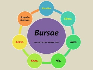
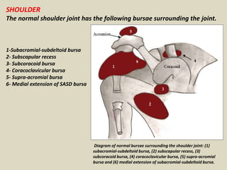
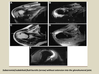
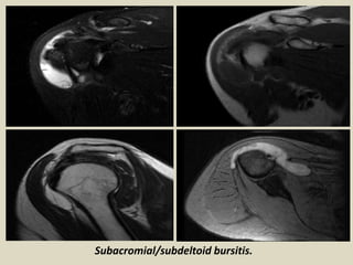
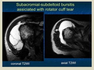
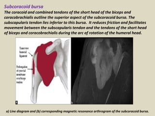
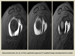
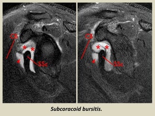
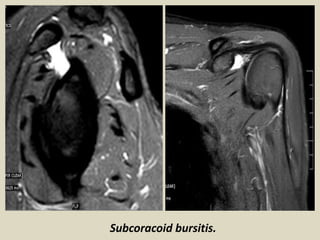
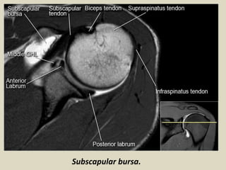
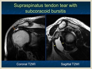
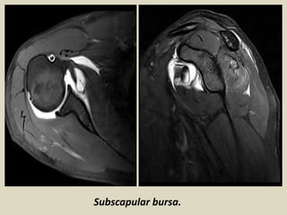
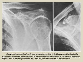
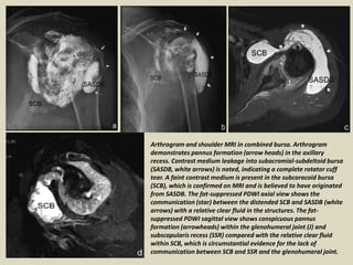
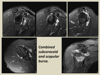
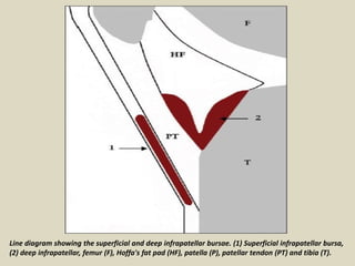
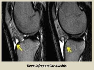
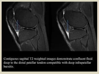
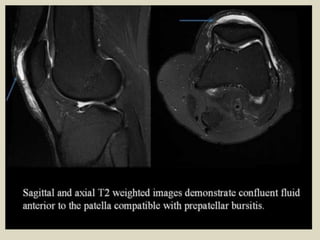
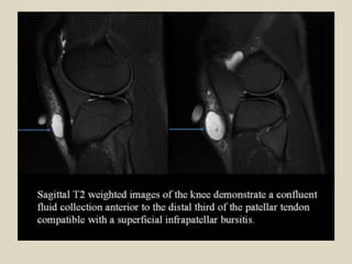
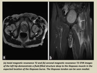
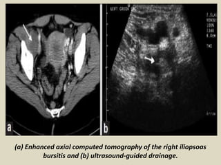
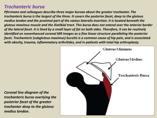
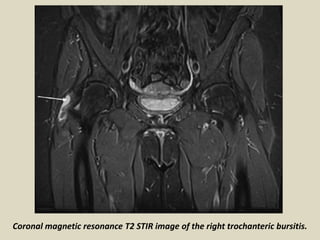
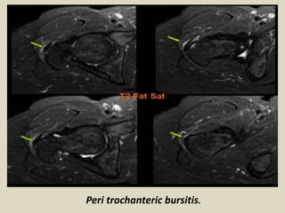
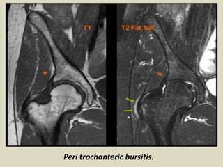
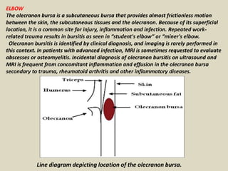
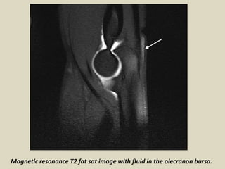
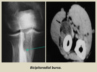
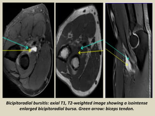
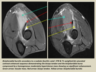
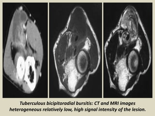
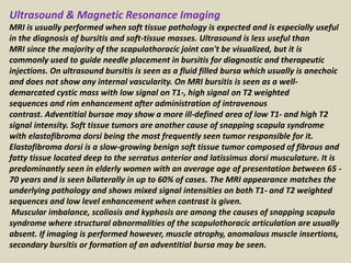
This document discusses various bursae in the body, including those around the shoulder, knee, and other joints. It provides details on the anatomy and locations of bursae and describes imaging findings of normal and pathological bursae. Key points include:
- Bursae are fluid-filled sacs that reduce friction between tissues like tendons and bones. They can communicate with joints or be separate.
- Common shoulder bursae include the subacromial-subdeltoid bursa and subcoracoid bursa. MRI is useful for identifying bursal inflammation or fluid from rotator cuff tears.
- Around the knee, the superficial and deep infrapatellar burs
Anatomy and imaging of wrist joint (MRI AND XRAY)Kajal Jha
Anatomy and imaging of wrist joint (xray and MRI).
this ppt was made as the class presentation by Kajal Jha as the part of the course of BSC MIT at BPKIHS,Dharan . It covers the part of syllabus of third year of BSC MIT of this institution.
Anatomy and imaging of wrist joint (MRI AND XRAY)Kajal Jha
Anatomy and imaging of wrist joint (xray and MRI).
this ppt was made as the class presentation by Kajal Jha as the part of the course of BSC MIT at BPKIHS,Dharan . It covers the part of syllabus of third year of BSC MIT of this institution.
Direct MR Arthrography of Hip joint in Children for Acetabular Labrum- Techni...scrappmd
Direct MR Arthrography of Hip joint in Children for Acetabular Labrum- Techniques, Findings and Pitfalls *Zahir U Sarwar, MBBS, CAQ, Pediatric Radiology, *Nemours Clinic and Wolfson Children’s Hospital, Jacksonville, FL, zsawar@nemors.org; **Seth J Crapp, MD, *Kevin Neal, MD, *Inbal Cohen MD, Chief Pediatric Radiology- **University of Florida College of Medicine Shands Hospital-Jacksonville, FL
Presented at the Society for Pediatric Radiology Conference, Scottsdale, Arizona 2008
A number of periarticular disorders have become increasingly common over the past two to three decades, due in part to greater participation in recreational sports by individuals of a wide range of ages. Periarticular disorders most commonly affect the knee or shoulder. With the exception of bursitis, hip pain is most often articular or is being referred from disease affecting another structure.
Patient-based learning made simple
Understanding the anatomy of a sports injury is the key to unlocking the diagnosis for most clinicians. Unfortunately, anatomy is often poorly taught, is not clinically focused and many anatomy textbooks are so complicated that searching for clinically useful information is difficult. In addition, multiple pathologies can present in an overlapping fashion, making the differentiation of the various possible causes of injury problematic.
Clinical Sports Anatomy classifies structures according to their anatomical reference points to form a diagnostic triangle. Discriminant questions are coupled with the more useful clinical tests and diagnostic manoeuvres to direct the reader toward a definitive clinical diagnosis. This approach is firmly rooted in evidence-based medicine and includes a list of the most appropriate investigations required to confirm diagnosis.
step by step presentation on ultrasound evaluation of shoulder and knee joints with illustrations of probe positioning.multiple examples of pathologies also added.
Shoulder joint (Biomechanics, Anatomy, Kinesiology) by Muhammad Arslan Yasin,
Anatomy Of Shoulder Joint,
Muscles Of Shoulder Joint,
Biomechanics Of Shoulder Joint,
Common Injuries Of Shoulder Joint.
Knee anatomy and clinical tests 2024.pdfvimalpl1234
This includes all relevant anatomy and clinical tests compiled from standard textbooks, Campbell,netter etc..It is comprehensive and best suited for orthopaedicians and orthopaedic residents.
Title: Sense of Smell
Presenter: Dr. Faiza, Assistant Professor of Physiology
Qualifications:
MBBS (Best Graduate, AIMC Lahore)
FCPS Physiology
ICMT, CHPE, DHPE (STMU)
MPH (GC University, Faisalabad)
MBA (Virtual University of Pakistan)
Learning Objectives:
Describe the primary categories of smells and the concept of odor blindness.
Explain the structure and location of the olfactory membrane and mucosa, including the types and roles of cells involved in olfaction.
Describe the pathway and mechanisms of olfactory signal transmission from the olfactory receptors to the brain.
Illustrate the biochemical cascade triggered by odorant binding to olfactory receptors, including the role of G-proteins and second messengers in generating an action potential.
Identify different types of olfactory disorders such as anosmia, hyposmia, hyperosmia, and dysosmia, including their potential causes.
Key Topics:
Olfactory Genes:
3% of the human genome accounts for olfactory genes.
400 genes for odorant receptors.
Olfactory Membrane:
Located in the superior part of the nasal cavity.
Medially: Folds downward along the superior septum.
Laterally: Folds over the superior turbinate and upper surface of the middle turbinate.
Total surface area: 5-10 square centimeters.
Olfactory Mucosa:
Olfactory Cells: Bipolar nerve cells derived from the CNS (100 million), with 4-25 olfactory cilia per cell.
Sustentacular Cells: Produce mucus and maintain ionic and molecular environment.
Basal Cells: Replace worn-out olfactory cells with an average lifespan of 1-2 months.
Bowman’s Gland: Secretes mucus.
Stimulation of Olfactory Cells:
Odorant dissolves in mucus and attaches to receptors on olfactory cilia.
Involves a cascade effect through G-proteins and second messengers, leading to depolarization and action potential generation in the olfactory nerve.
Quality of a Good Odorant:
Small (3-20 Carbon atoms), volatile, water-soluble, and lipid-soluble.
Facilitated by odorant-binding proteins in mucus.
Membrane Potential and Action Potential:
Resting membrane potential: -55mV.
Action potential frequency in the olfactory nerve increases with odorant strength.
Adaptation Towards the Sense of Smell:
Rapid adaptation within the first second, with further slow adaptation.
Psychological adaptation greater than receptor adaptation, involving feedback inhibition from the central nervous system.
Primary Sensations of Smell:
Camphoraceous, Musky, Floral, Pepperminty, Ethereal, Pungent, Putrid.
Odor Detection Threshold:
Examples: Hydrogen sulfide (0.0005 ppm), Methyl-mercaptan (0.002 ppm).
Some toxic substances are odorless at lethal concentrations.
Characteristics of Smell:
Odor blindness for single substances due to lack of appropriate receptor protein.
Behavioral and emotional influences of smell.
Transmission of Olfactory Signals:
From olfactory cells to glomeruli in the olfactory bulb, involving lateral inhibition.
Primitive, less old, and new olfactory systems with different path
Explore natural remedies for syphilis treatment in Singapore. Discover alternative therapies, herbal remedies, and lifestyle changes that may complement conventional treatments. Learn about holistic approaches to managing syphilis symptoms and supporting overall health.
ARTIFICIAL INTELLIGENCE IN HEALTHCARE.pdfAnujkumaranit
Artificial intelligence (AI) refers to the simulation of human intelligence processes by machines, especially computer systems. It encompasses tasks such as learning, reasoning, problem-solving, perception, and language understanding. AI technologies are revolutionizing various fields, from healthcare to finance, by enabling machines to perform tasks that typically require human intelligence.
Flu Vaccine Alert in Bangalore Karnatakaaddon Scans
As flu season approaches, health officials in Bangalore, Karnataka, are urging residents to get their flu vaccinations. The seasonal flu, while common, can lead to severe health complications, particularly for vulnerable populations such as young children, the elderly, and those with underlying health conditions.
Dr. Vidisha Kumari, a leading epidemiologist in Bangalore, emphasizes the importance of getting vaccinated. "The flu vaccine is our best defense against the influenza virus. It not only protects individuals but also helps prevent the spread of the virus in our communities," he says.
This year, the flu season is expected to coincide with a potential increase in other respiratory illnesses. The Karnataka Health Department has launched an awareness campaign highlighting the significance of flu vaccinations. They have set up multiple vaccination centers across Bangalore, making it convenient for residents to receive their shots.
To encourage widespread vaccination, the government is also collaborating with local schools, workplaces, and community centers to facilitate vaccination drives. Special attention is being given to ensuring that the vaccine is accessible to all, including marginalized communities who may have limited access to healthcare.
Residents are reminded that the flu vaccine is safe and effective. Common side effects are mild and may include soreness at the injection site, mild fever, or muscle aches. These side effects are generally short-lived and far less severe than the flu itself.
Healthcare providers are also stressing the importance of continuing COVID-19 precautions. Wearing masks, practicing good hand hygiene, and maintaining social distancing are still crucial, especially in crowded places.
Protect yourself and your loved ones by getting vaccinated. Together, we can help keep Bangalore healthy and safe this flu season. For more information on vaccination centers and schedules, residents can visit the Karnataka Health Department’s official website or follow their social media pages.
Stay informed, stay safe, and get your flu shot today!
HOT NEW PRODUCT! BIG SALES FAST SHIPPING NOW FROM CHINA!! EU KU DB BK substit...GL Anaacs
Contact us if you are interested:
Email / Skype : kefaya1771@gmail.com
Threema: PXHY5PDH
New BATCH Ku !!! MUCH IN DEMAND FAST SALE EVERY BATCH HAPPY GOOD EFFECT BIG BATCH !
Contact me on Threema or skype to start big business!!
Hot-sale products:
NEW HOT EUTYLONE WHITE CRYSTAL!!
5cl-adba precursor (semi finished )
5cl-adba raw materials
ADBB precursor (semi finished )
ADBB raw materials
APVP powder
5fadb/4f-adb
Jwh018 / Jwh210
Eutylone crystal
Protonitazene (hydrochloride) CAS: 119276-01-6
Flubrotizolam CAS: 57801-95-3
Metonitazene CAS: 14680-51-4
Payment terms: Western Union,MoneyGram,Bitcoin or USDT.
Deliver Time: Usually 7-15days
Shipping method: FedEx, TNT, DHL,UPS etc.Our deliveries are 100% safe, fast, reliable and discreet.
Samples will be sent for your evaluation!If you are interested in, please contact me, let's talk details.
We specializes in exporting high quality Research chemical, medical intermediate, Pharmaceutical chemicals and so on. Products are exported to USA, Canada, France, Korea, Japan,Russia, Southeast Asia and other countries.
Recomendações da OMS sobre cuidados maternos e neonatais para uma experiência pós-natal positiva.
Em consonância com os ODS – Objetivos do Desenvolvimento Sustentável e a Estratégia Global para a Saúde das Mulheres, Crianças e Adolescentes, e aplicando uma abordagem baseada nos direitos humanos, os esforços de cuidados pós-natais devem expandir-se para além da cobertura e da simples sobrevivência, de modo a incluir cuidados de qualidade.
Estas diretrizes visam melhorar a qualidade dos cuidados pós-natais essenciais e de rotina prestados às mulheres e aos recém-nascidos, com o objetivo final de melhorar a saúde e o bem-estar materno e neonatal.
Uma “experiência pós-natal positiva” é um resultado importante para todas as mulheres que dão à luz e para os seus recém-nascidos, estabelecendo as bases para a melhoria da saúde e do bem-estar a curto e longo prazo. Uma experiência pós-natal positiva é definida como aquela em que as mulheres, pessoas que gestam, os recém-nascidos, os casais, os pais, os cuidadores e as famílias recebem informação consistente, garantia e apoio de profissionais de saúde motivados; e onde um sistema de saúde flexível e com recursos reconheça as necessidades das mulheres e dos bebês e respeite o seu contexto cultural.
Estas diretrizes consolidadas apresentam algumas recomendações novas e já bem fundamentadas sobre cuidados pós-natais de rotina para mulheres e neonatos que recebem cuidados no pós-parto em unidades de saúde ou na comunidade, independentemente dos recursos disponíveis.
É fornecido um conjunto abrangente de recomendações para cuidados durante o período puerperal, com ênfase nos cuidados essenciais que todas as mulheres e recém-nascidos devem receber, e com a devida atenção à qualidade dos cuidados; isto é, a entrega e a experiência do cuidado recebido. Estas diretrizes atualizam e ampliam as recomendações da OMS de 2014 sobre cuidados pós-natais da mãe e do recém-nascido e complementam as atuais diretrizes da OMS sobre a gestão de complicações pós-natais.
O estabelecimento da amamentação e o manejo das principais intercorrências é contemplada.
Recomendamos muito.
Vamos discutir essas recomendações no nosso curso de pós-graduação em Aleitamento no Instituto Ciclos.
Esta publicação só está disponível em inglês até o momento.
Prof. Marcus Renato de Carvalho
www.agostodourado.com
Presentation1.pptx, radiological imaging of bursae.
1. Bursae
Dr/ ABD ALLAH NAZEER. MD.
Shoulder.
Elbow.
Wrist.
Hip.Knee.
Ankle.
Scapulo-
thoracic.
2. A bursa is a synovial-lined sac overlying the bony surfaces at
areas of tendon friction. Bursae are located where tendons
move against each other or glide over a bony surface. They are
classified according to their location: subcutaneous, subfascial,
subtendinous, and submucosal. Bursae can also be classified as
communicating or non communicating. When a bursa is
located adjacent to a joint, the synovial membrane of the
bursae may communicate with the joint. This bursa is termed a
communicating bursa. Some examples are the iliopsoas bursa
lateral to the hip and the gastrocnemius-semimembranosus
bursa posteromedial to the knee. In certain locations,
communication between the joint and the bursa is abnormal.
An example is the subacromial-subdeltoid (SASD) bursa that
lies superior to the rotator cuff and inferior to the acromion.
Most of the bursae are potential spaces and are not normally
visualized on imaging.
3. In pathological conditions such as excessive local friction,
infection, arthritides or direct trauma, fluid and debris collect
within the bursa or fluid extends into the bursa from the adjacent
joint. The walls of the bursa thicken as the bursal inflammation
becomes longstanding. The term bursitis refers to pathological
enlargement of the bursa. If the abnormally distended bursa is
superficially located, it can be visualized by ultrasound as the
ultrasound beam is able to penetrate through this region. The
bursa is seen as a fluid-filled anechoic structure lined by a
hyperechoic wall. Deep-seated bursae are depicted on magnetic
resonance imaging (MRI) or computed tomography (CT). On MRI,
the bursa is seen as a high T2 fluid-filled structure. CT shows the
inflamed bursa as hypodense with an enhancing wall. Clinically,
bursitis mimics several peripheral joint and muscle abnormalities.
Therefore, it is important for the radiologist to identify bursal
pathology and direct management geared toward bursitis.
4. SHOULDER
The normal shoulder joint has the following bursae surrounding the joint.
1-Subacromial-subdeltoid bursa
2- Subscapular recess
3- Subcoracoid bursa
4- Coracoclavicular bursa
5- Supra-acromial bursa
6- Medial extension of SASD bursa
Diagram of normal bursae surrounding the shoulder joint: (1)
subacromial-subdeltoid bursa, (2) subscapular recess, (3)
subcoracoid bursa, (4) coracoclavicular bursa, (5) supra-acromial
bursa and (6) medial extension of subacromial-subdeltoid bursa.
5. Subacromial-subdeltoid bursa
The subacromial-subdeltoid (SASD) bursa comprise of two bursae that lie
between the rotator cuff tendons and the undersurface of the acromion.
They are located deep in the deltoid muscle and acromioclavicular joint
(AC joint) and overlie the bicipital groove and rotator cuff.
The causes of SASD bursal fluid include: rotator cuff tears, impingement,
septic bursitis, and reactive bursitis from glenohumeral joint disease,
such as calcium deposition arthropathy. In full-thickness tears of the
rotator cuff, fluid from within the glenohumeral joint tracks through the
tear into the bursa. Communication with the joint is abnormal in this
location. This abnormal communication is best depicted on an MR
arthrogram. Intraarticular gadolinium outlines the tear and extends into
the bursa. On MRI, the abnormal collection of fluid is seen as a low T1,
high T2 signal within the bursa, either due to bursal inflammation or
from a full-thickness rotator cuff tear. On ultrasound, the fluid-filled
bursa overlies the rotator cuff and is clearly depicted as a fluid-filled
anechoic structure both on longitudinal and transverse planes.
12. Subcoracoid bursa
The coracoid and combined tendons of the short head of the biceps and
coracobrachialis outline the superior aspect of the subcoracoid bursa. The
subscapularis tendon lies inferior to this bursa. It reduces friction and facilitates
movement between the subscapularis tendon and the tendons of the short head
of biceps and coracobrachialis during the arc of rotation of the humeral head.
a) Line diagram and (b) corresponding magnetic resonance arthrogram of the subcoracoid bursa.
13. Subcoracoid bursitis, (1a, 1b, 1c) Three sagittal fat-suppressed T1-weighted images extending lateral to medial.
19. Inadvertent bursogram (a. b.) in a 44-year-old male. The tip of the needle is adjusted to a
deeper position to complete the arthrogram immediately. This burso-arthrogram
demonstrates the anterior location of the SCB relative to the glenohumeral joint (J).
20. X-ray photographs in chronic supracoracoid bursitis. Left: Cloudy calcification in the
coracoclavicular region while the arm is in zero position and the direction of the x-rays is horizontal.
Right: Arm is in 900 anteflexion and the x-rays are from anterocaudal to posterocranial.
21. Arthrogram and shoulder MRI in combined bursa. Arthrogram
demonstrates pannus formation (arrow heads) in the axillary
recess. Contrast medium leakage into subacromial-subdeltoid bursa
(SASDB, white arrows) is noted, indicating a complete rotator cuff
tear. A faint contrast medium is present in the subcoracoid bursa
(SCB), which is confirmed on MRI and is believed to have originated
from SASDB. The fat-suppressed PDWI axial view shows the
communication (star) between the distended SCB and SASDB (white
arrows) with a relative clear fluid in the structures. The fat-
suppressed PDWI sagittal view shows conspicuous pannus
formation (arrowheads) within the glenohumeral joint (J) and
subscapularis recess (SSR) compared with the relative clear fluid
within SCB, which is circumstantial evidence for the lack of
communication between SCB and SSR and the glenohumeral joint.
23. Bursae around the knee joints:
Infrapatellar Bursitis
Infrapatellar bursae can be superficial or deep. The superficial infrapatellar bursa is
located between the tibial tubercle and the overlying skin, whereas the deep
infrapatellar bursa is located between the posterior aspect of the patellar tendon and
the tibia.
Superficial Infrapatellar Bursitis
Superficial infrapatellar bursitis, also called clergyman's knee, is due to inflammation
and fluid accumulation resulting from chronic stress. Clinically, there is a palpable
swelling inferior to the patella. On MRI, it appears as a loculated collection that
projects exophytically, anterior to the patellar tendon, forming a swelling. On imaging,
it should be differentiated from subcutaneous edema: edema is seen as a diffuse fluid
collection seen all over the anterior aspect of the knee, whereas bursitis appears as a
localized collection with well-defined borders.
Deep Infrapatellar Bursitis
On MRI, deep infrapatellar bursitis appears as a triangular fluid collection posterior to
the patellar tendon. When found in adolescents, it should be differentiated from
Osgood–Schlatter disease. Osgood–Schlatter disease is associated with thickening and
hyperintense signal intensity of the patellar tendon, but these findings are absent in
deep infrapatellar bursitis. In addition, cases of Osgood–Schlatter disease, on clinical
examination, display pain and tenderness at the insertion of the patellar tendon.
24. Line diagram showing the superficial and deep infrapatellar bursae. (1) Superficial infrapatellar bursa,
(2) deep infrapatellar, femur (F), Hoffa's fat pad (HF), patella (P), patellar tendon (PT) and tibia (T).
26. Superficial infrapatellar bursitis. A 42-year-old female presented with an anterior
knee swelling. Sagittal proton-density fat saturated (A) and sagittal gradient-echo
T2W (B) images show a distended superficial infrapatellar bursa (arrows)
30. Prepatellar bursa
The prepatellar bursa is a tricompartmental structure. The superficial
compartment lies between the subcutaneous tissue and an extension
of the fascia lata. The intermediate compartment is situated between
the transverse superficial fascia and an intermediate oblique fascia
formed by fascial extension of the vastus lateralis and vastus medialis
muscles. The deep compartment lies between the intermediate oblique
fascia and the deep longitudinal fibers of the rectus femoris tendon.
Inflammation of this bursa occurs from repetitive trauma from
kneeling, as seen with housemaids, carpet-layers, and wrestlers. It is
also seen in inflammatory conditions such as gout.
Chronic trauma in the form of prolonged or repeated kneeling leads to
inflammation and hemorrhagic bursitis. Clinically, patients may present
with pain and swelling over the patella.
On MRI, prepatellar bursitis appears as an oval fluid-signal-intensity
lesion between the subcutaneous tissue and the patella. Hemorrhagic
lesions show T2 shortening on gradient-echo (GRE) images.
31. Line diagram showing
compartmentalization of
the prepatellar bursa.
SCCT - subcutaneous
cellular tissues; QT -
quadriceps tendon; PT -
patellar tendon; F -
femur; P, patella;
(1) superficial
compartment;
(2) intermediate
compartment;
(3) deep compartment.
32. Sagittal magnetic resonance T2 fat sat image showing high-signal
fluid intensity within the prepatellar bursitis.
33. Axial magnetic resonance T2 fat sat image depicting Baker's cyst with its neck between the
semimembranosus and the medial gastrocnemius tendons. Image also depicts prepatellar bursitis.
34. Prepatellar bursitis. A 44-year-old female presented with anterior knee pain. Axial (A) and
coronal (B) T2W fat saturated images showing a distended pre patellar bursa (arrows)
38. Suprapatellar bursa
The suprapatellar bursa lies between the distal rectus femoris tendon and the femur. In
the fifth fetal month, a septum (suprapatellar plica) that lies between the knee joint and
the bursa perforates and results in a communication. This situation occurs in 85% of the
adults. The suprapatellar bursa is an example of bursae normally communicating with
joints. On routine ultrasound and MR imaging, fluid in the suprapatellar bursa is a
common finding in individuals with joint effusion. In patients with a non communicating
bursa and bursitis, fluid is localized only to the suprapatellar bursa.
Longitudinal ultrasound image demonstrating fluid within the suprapatellar bursa.
40. Suprapatellar bursitis. A 29-year-old male presented with internal derangement of the knee: sagittal proton
density fat saturated (A) and axial T2W gradient-echo (B) images show a distended suprapatellar bursa
(arrows) and in addition, a partial tear of the anterior cruciate ligament (thick arrow in A)
41. Pes Anserine Bursitis
The Pes anserine bursa separates the Pes anserine tendons,
consisting of the distal sartorius, gracilis, and semitendinosus
tendons, from the subjacent distal portion of the tibial collateral
ligament and the bony surface of the medial tibial condyle.
Anserine bursitis results from overuse, especially in runners.
On MRI, Pes anserine bursitis appears as an oblong multiloculated
fluid collection seen along the anserine tendons on the
posteromedial aspect of the knee. This is best appreciated on T2W
axial images. It is commonly confused with a popliteal cyst; the Pes
anserine bursa is located posteriorly and medially along the
semitendinosus, whereas the popliteal cyst is located more often in
the midline posteriorly. Also, Pes anserine bursae are small in size;
they do not extend into the thigh and never show communication
with the knee joint, whereas a popliteal cyst can extend into the
thigh and may or may not communicate with the knee joint.
42. a) Axial line diagram and (b) axial magnetic resonance image showing Pes anserine bursitis.
43. Pes anserine bursitis. A 32-year-old female presented with pain along the posteromedial aspect
of the knee: Coronal proton density (A) and axial T2W (B) images show a distended Pes anserine
bursa (arrows). The Pes anserine tendons (arrowhead in B) are seen inferior to the bursa.
46. Medial Collateral Ligament Bursitis
The medial collateral ligament bursa is located between the superficial and deep layers of the
medial collateral ligament. Clinically, fluid collection in this bursa mimics a medial meniscus tear
and/or medial collateral ligament tear. On MRI, medial collateral ligament bursitis appears as a
T2 hyperintensity between the superficial and deep fibers of the medial collateral ligament. It
must be differentiated from meniscocapsular separation. In meniscocapsular separation, in
addition to fluid collection between the superficial and deep fibers of the medial collateral
ligament, there is a tear of the peripheral corner of the medial meniscus and meniscal
displacement from the outer cortical margin of the tibia.
Medial collateral ligament bursitis. A 54-year-old female presented with knee swelling: sagittal true inversion recovery
magnitude (TIRM) (A) and axial T2W (B) images show a distended medial collateral ligament burse (arrows)
48. Medial collateral ligament bursitis located between the deep MCL
(yellow arrow) and the superficial MCL (green arrow).
49. Axial magnetic resonance T2 fat sat image showing fluid within the medial collateral ligament bursa.
50. Ilio tibial Bursitis
The ilio tibial bursa is located between the distal part of the ilio tibial band near its insertion
on Gerdy tubercle and the adjacent tibial surface. Ilio tibial bursitis and tendinitis are usually
due to overuse and varus stress of the knee.
On MRI ilio tibial bursitis appears as a fluid collection near the insertion of the ilio tibial tract
in its distal part, close to the lateral aspect of the tibia. It must be differentiated from iliotibial
tendinitis. On MRI, ilio tibial tendinitis appears as a fluid collection encircling the tendon,
whereas bursitis appears as a localized oblong fluid collection adjacent to the tendon.
Iliotibial bursitis. A 16-year-old female presented with lateral knee swelling: sagittal
STIR (A) and axial T2W (B) images show a distended iliotibial bursa (arrows)
51. Baker's Cyst
This is also called a popliteal cyst and typically involves the gastrocnemius- semimembranosus bursa and
is located between the medial femoral condyle, semimembranosus tendon and the medial head of the
gastrocnemius. It may or may not communicate with the knee joint. It may rupture and extend inferiorly
along the gastrocnemius muscle into the calf or extend superiorly into thigh along the semimembranosus.
On MRI they appear as well-defined hyperintense lesions on T2W images at the characteristic location.
Axial images are the best to demonstrate the communication with the knee joint and also the
characteristic location between the semimembranosus tendon and medial head of gastrocnemius.
Baker's cyst. A 48-year-old male presented with swelling in the popliteal fossa: sagittal (A) and axial (B)
T2W gradient-echo images show a distended gastrocnemius--semimembranosus bursa (arrows)
52.
53. Fibular bursa. Sequential coronal fat suppressed T2WI (A) from anterior to posterior show a
multilocular cystic mass posterolateral to the iliotibial band (solid arrow), insinuating between
the fibular collateral ligament (dashed arrow) and the distal biceps femoris tendon (arrow
head), consistent with fibular bursitis. Sagittal ultrasound of the lateral knee (B) at the level of
the popliteal notch (*) demonstrates a loculated hypoechoic focus between the fibular collateral
ligament (dashed arrow) and the biceps femoris tendon (arrow heads).
54.
55. Lateral Gastrocnemius Bursa. Axial PD (A) and coronal fat sat T2WI (B)
demonstrating a distended multiloculated lateral gastrocnemius bursa.
56.
57. ANKLE
Retrocalcaneal bursa
The retrocalcaneal bursa is a saddle-shaped bursa situated between the
calcaneous and the Achilles tendon. It assists in decreasing friction during plantar
flexion. Retrocalcaneal bursitis is common and is caused by Reiter disease,
psoriasis, ankylosing spondylitis and, often, with calcaneal fractures.
Sagittal magnetic resonance T2 fat sat image showing retrocalcaneal bursitis with a thick synovial wall.
58. MRI shows effusion (green arrow), Stieda’s
process (white arrow), bony overgrowth
(short white arrow), tendinopathy (thin
yellow arrow), retrocalcaneal bursitis (open
arrow), bone marrow edema (orange
arrow) and chronic bursitis (orange arrow).
MRI shows the Stieda's process
(arrowhead), Haglund’s deformity (open
arrow) and retrocalcaneal bursitis (yellow
arrow) and retroachilles chronic bursitis
with thickening of the skin (white arrow).
59. Superficial retrocalcaneal bursa
A second more superficial bursa lies between the Achilles tendon and the
overlying subcutaneous fat. Inflammation results from poorly fitting shoes. When
superficial retrocalcaneal bursitis occurs in the setting of Achilles tendinopathy, a
diagnosis of Haglund syndrome is made.
Longitudinal ultrasound image showing superficial retrocalcaneal bursa.
61. 1. Left ankle, lateral view shows equivocal
prominence of the posterior superior calcaneal
tuberosity.
2. Sagittal T1-weighted image demonstrates
focal decreased T1 signal within pre-Achilles fat
and prominent posterior superior calcaneal
tuberosity.
3. Sagittal proton density image shows
increased signal intensity in pre-Achilles
fat/retrocalcaneal bursa.
4. Sagittal T2-weighted image reveals mildly
increased signal intensity within distal Achilles
tendon without evidence of full-thickness tear.
5. Axial T2-weighted image better demonstrates
increased signal intensity within the Achilles
tendon with associated retrocalcaneal
inflammation.
62. MRI of left ankle. Axial proton density spectral presaturation inversion recovery (SPIR) images (A, B, and C) show a
bright signal intensity lesion (*), extending from the retrocalcaneal region (A) to over the distal tibial metaphysis (C).
It surrounds the Achilles tendon (white arrows), which appears enlarged, not homogeneous and subtotally torn (B,
curved white arrows). Sagittal T1-weighted image (D) and DP SPIR image (E) after IV administration of gadolinium
contrast material show a localized fluid collection with intermediate to low signal intensity (*), which enhances
peripherally and lacks enhancement centrally, found in soft tissue abscess likely originating from the subcutaneous
bursa. There is clear partition between the fluid collection and Kager’s fat pad (black arrow), which appears spared by
the septic process (D). White arrows show distal and proximal ends of Achilles tendon (E).
64. Tibialis anterior subtendinous bursitis - Note fluid distension only partially involving the
tendon (white arrows), in its distal portion, compared to the contralateral normal tendon
(asterisk). This partial involvement is necessary to differentiate from tenosynovitis.
65. Surgically proven discrete adventitial bursal sac in 22-year-old female ice skater. Axial T1-weighted (A)
(TR/TE, 450/15) and axial fat-saturated T2-weighted fast spin-echo (B) (2,500/90) images show mass
with well-defined borders and fluid signal characteristics (arrow) in subcutaneous soft-tissues.
The medial malleolar bursa, an adventitial bursa, may develop over the medial
malleolus in response to abnormal pressure, usually from footwear that closely
approximates the ankle, such as boots, skates, and high-top shoes. The rise in
popularity of sporting activities that require tight-fitting boots, such as figure skating
and ice hockey, has led to an increased incidence of painful medial malleolar bursitis.
66. Surgically-proven discrete adventitial bursal sac in 27-year-old male ice hockey player. Axial fat-
suppressed image (TR/TE, 3,400/43) depicts mass (arrow) with fluid signal characteristics in
subcutaneous fat posteromedial to medial malleolus. Soft-tissue edema is noted adjacent to mass
67. Partly scarred down bursal sac in 15-year-old female ice skater. Patient's symptoms
improved with modification of skates. Axial STIR image (6,300/60) depicts heterogeneous
increased signal of mass (arrow), which is compatible with fibrosis of a bursal sac.
68. HIP
Iliopsoas bursa
The iliopsoas bursa is the largest bursa around the hip joint (average
size, 6 cm × 3 cm) and is situated beneath the musculotendinous portion
of the iliopsoas muscle, anterior to the hip joint capsule and lateral to
the femoral vessels. It is found in about 98% of the subjects, and
communicates with the cavity of the hip joint in about 15% of the cases.
Normally collapsed, distension of the bursa is usually caused by
overproduction of synovial fluid in an arthritic hip leading to increased
intraarticular pressure and extension of fluid into the potential space of
the bursa.
Iliopsoas bursa distension in association with disease has been
described in trauma, osteoarthritis, avascular necrosis, rheumatoid
arthritis, synovial chondromatosis, pigmented villonodular synovitis,
gout, and pyogenic infection. The differential diagnosis of an enlarged
iliopsoas bursa includes inguinal or femoral hernia, neoplasm,
lymphadenopathy, undescended testis, hematoma, psoas abscess,
femoral aneurysm, and arteriovenous fistula.
69. (a) Axial magnetic resonance T2 and (b) coronal magnetic resonance T2 STIR images
of the left hip demonstrate a fluid-filled structure deep to the iliopsoas muscle in the
expected location of the iliopsoas bursa. The iliopsoas tendon can be seen medial.
70. (a) Enhanced axial computed tomography of the right iliopsoas
bursitis and (b) ultrasound-guided drainage.
71. Trochanteric bursa
Pfirrmann and colleagues describe three major bursae about the greater trochanter. The
trochanteric bursa is the largest of the three. It covers the posterior facet, deep to the gluteus
medius tendon and the proximal part of the vastus lateralis insertion. It is located beneath the
gluteus maximus muscle and the iliotibial tract. This bursa does not extend over the anterior border
of the lateral facet. It is lined by a small layer of fat on both sides. Therefore, it can be routinely
identified on nonenhanced coronal MR images as a fine linear structure paralleling the posterior
facet. Trochanteric (subgluteus maximus) bursitis is a common cause of hip pain, and is associated
with obesity, trauma, inflammatory arthritides, and in patients with total hip arthroplasty.
Coronal line diagram of the
trochanteric bursa overlying the
posterior facet of the greater
trochanter deep to the gluteus
medius tendon.
74. Bursitis (green arrows) appears as a fluid collection(decreased T1 signal, increased
(T2signal). Iliotibial band (thin green arrows), greater trochanter (blue arrows).
75. Subgluteus medius bursa
The subgluteus medius bursa is deep in the distal gluteus medius tendon. This bursa
covers an area of the superior part of the lateral facet. Its superior extent is marked
by the tip of the trochanter; its anterior extent, by the lateral facet; and its posterior
and inferior extent, by the tendinous insertion of the gluteus medius muscle.
Coronal magnetic resonance T2 STIR image of the bilateral subgluteus medius bursitis, larger on the right side.
76. Subgluteus minimus bursa
The subgluteus minimus bursa is in the area of the anterior facet. It lies
beneath the gluteus minimus tendon, medial and superior to its insertion.
Coronal magnetic resonance STIR image of the right subgluteus minimus bursitis.
79. ELBOW
The olecranon bursa is a subcutaneous bursa that provides almost frictionless motion
between the skin, the subcutaneous tissues and the olecranon. Because of its superficial
location, it is a common site for injury, inflammation and infection. Repeated work-
related trauma results in bursitis as seen in “student's elbow” or “miner's elbow.
Olecranon bursitis is identified by clinical diagnosis, and imaging is rarely performed in
this context. In patients with advanced infection, MRI is sometimes requested to evaluate
abscesses or osteomyelitis. Incidental diagnosis of olecranon bursitis on ultrasound and
MRI is frequent from concomitant inflammation and effusion in the olecranon bursa
secondary to trauma, rheumatoid arthritis and other inflammatory diseases.
Line diagram depicting location of the olecranon bursa.
82. 61-year-old man with surgically confirmed rupture of triceps tendon at insertion of olecranon
and concomitant non septic effusion of olecranon bursa. Sagittal T2-weighted fat-suppressed
image (4,100/100) reveals tear of triceps tendon (white arrowhead). Note concomitant elbow
joint effusion (arrow). Olecranon bursa is marked with black arrowhead.
86. Bicipitoradial bursitis refers to inflammation of the bicipitoradial bursa.
The bicipitoradial bursa surrounds the biceps tendon in supination. In pronation, the radial
tuberosity rotates posteriorly, which compresses the bicipitoradial bursa between the biceps
tendon and the radial cortex which consequently increases the pressure within the bursa.
Epidemiology
It typically presents in adults and may be more common in males
Clinical presentation
Patients often presents with elbow swelling, pain, tenderness, redness and limited movement.
Radiographic features
Ultrasonography
May show evidence of distention of the bicipitoradial bursa by fluid which appear anechoic or
hypoechoic soft tissue. The distal biceps tendon should be evaluated for injury. Nodular soft-
tissue debris and small calcifications may be seen within the fluid. Power Doppler imaging may
show hyperaemia and suggests active inflammation.
MRI
Features observed on different sequences include
T1: homogeneous hypointense signal mass in characteristic location; associated tendinosis of
biceps tendon, thickening and intermediate signal intensity at insertion
T2: hyperintense flattened, oval, or round shaped cystic appearing mass that shows fluid signal
intensity on all pulse sequences unless containing inflammatory debris or calcification +/- rice
bodies in patients with rheumatoid arthritis
STIR: as with T2 with more homogeneous fat saturation
T1 C+ (Gd): may demonstrate thin rim enhancement after gadolinium administration and
homogeneous low central signal intensity.
90. Bicipitoradial bursitis: axial T1, T2-weighted image showing a isointense
enlarged bicipitoradial bursa. Green arrow: biceps tendon.
91. Paget's disease of the proximal ulna and bicipitoradial bursitis. Yellow arrow: osseous
hypertrophy and deformation associated with cortical thickening of the ulna.
92. Bicipitoradial bursitis secondary to a nodular fasciitis: axial STIR & T1-weighted fat saturated
contrast-enhanced sequence demonstrating the biceps tendon and the bicipitoradial bursa
compressed by a homogeneous moderately hyperintense mass showing no significant enhancement.
Green arrow: tissular mass. Red arrow: biceps tendon. Yellow arrow: bicipitoradial bursitis
94. Palmar Bursae of the Wrist and Hand
Bursae are fluid-filled sacs lined by a synovial membrane, acting to decrease friction
between adjacent structures, allowing smooth, gliding movement. They are located
throughout the body in locations where tendons are closely apposed to bones and
rigid fibrous structures. The flexor tendons of the carpal tunnel are enveloped in two
palmar bursae: the ulnar bursa and the radial bursa. The ulnar and radial bursae
begin proximal to the carpal tunnel, usually near the distal margin of the pronator
quadratus muscle, and they extend distally to the level of the mid-palm, allowing for
longitudinal excursion of the tendons during normal wrist movement. These bursae
extend over a longer distance than the extensor tendon sheaths, a difference likely
related to the greater range of wrist motion that takes place in flexion compared with
extension The smaller radial bursa surrounds the flexor pollicis longus tendon. The
larger ulnar bursa surrounds the eight flexor digitorum tendons, doing so by means of
three invaginations: a deep invagination between the carpal bones and the profundus
tendons, a middle invagination between the profundus and superficialis tendons, and
a superficial invagination between the superficialis tendons and the flexor
retinaculum. These invaginations arise from a common space at the ulnar side of the
tendons adjacent to the FD5 tendon (7a). Although there are several anatomic
variations in the communication patterns between the ulnar bursa, the radial bursa,
and the digital flexor tendon sheaths, a typical anatomic arrangement found in the
majority of persons has been established through anatomic dissection as well as
tenography in cadavers.
95. A fat-suppressed T1-weighted post-contrast axial image at the level of the
metacarpal shafts reveals avid synovial enhancement of the ulnar (long arrows) and
radial (short arrows) bursae. Note that the low signal foci seen in (A) do not enhance.
Observe the two-tiered arrangement of the flexor digitorum profundus tendons
(above dotted line) and flexor digitorum superficialis tendons (below dotted line).
98. On fat-suppressed T2-weighted image of the right hand, the coronal view (A) shows a partial tear of the fifth finger
flexor tendon (arrow), a fluid collection in the tendon sheath (open arrow), contiguous with the flexor compartment
along the ulnar side (arrowheads). The axial views were obtained at the levels of the proximal phalanx (B), mid-palm
(C) and proximal palm (D-1). The contrast-enhanced T1-weighted axial image of the proximal palm (D-2) shows a
thickened synovium of the ulnar bursa (open arrowheads).
99.
100. A fat-suppressed proton density axial image just proximal to the carpal tunnel demonstrates increased
tenosynovial fluid (short arrows) and synovial proliferation (arrowheads) in the radial and ulnar bursae
as well as multiple extensor tendon sheaths in this 45-year-old female with rheumatoid arthritis.
101. Scapulothoracic bursitis and snapping scapula:
Snapping scapula syndrome usually is a result of abnormal anatomy, abnormal
scapulothoracic motion, space-occupying bony or soft-tissue lesions or a combination of
these. Imaging is commonly used to determine the nature and location of snapping scapula
syndrome and is essential in treatment planning. Conventional radiographs normally are
the first diagnostic step, but since the anterior aspect of the scapula is difficult to visualize
and soft tissue masses are not well visualized on plain films, additional cross-sectional
imaging is commonly performed.
Computed Tomography
CT is indicated when an underlying osseous abnormality is suspected and plain films do not
reveal any abnormalities. Anatomical variations comprise the most common subgroup of
osseous abnormalities causing snapping scapula syndrome and include (a) an excessive
anterior curvature of the superomedial scapular angle rubbing across the ribs, (b) a
Luschka’s tubercle being a bony prominence at the supermedial angle of the scapula and
(c) a teres major process representing an anterior curvature towards the chest wall at the
inferior aspect of the scapula. It is important to realize that these anatomic variants are
commonly seen and increase the risk of snapping scapula, but do not cause symptoms in
the majority of people. Other common causes of snapping scapula include mal united
scapula- or rib fractures, reactive spurs and osteochondromas arising from the rib or
anterior surface of the scapula. Chondrosarcoma are a rare cause of snapping scapula and
are usually seen in an older age group
102. Ultrasound & Magnetic Resonance Imaging
MRI is usually performed when soft tissue pathology is expected and is especially useful
in the diagnosis of bursitis and soft-tissue masses. Ultrasound is less useful than
MRI since the majority of the scapulothoracic joint can't be visualized, but it is
commonly used to guide needle placement in bursitis for diagnostic and therapeutic
injections. On ultrasound bursitis is seen as a fluid filled bursa which usually is anechoic
and does not show any internal vascularity. On MRI bursitis is seen as a well-
demarcated cystic mass with low signal on T1-, high signal on T2 weighted
sequences and rim enhancement after administration of intravenous
contrast. Adventitial bursae may show a more ill-defined area of low T1- and high T2
signal intensity. Soft tissue tumors are another cause of snapping scapula syndrome
with elastofibroma dorsi being the most frequently seen tumor responsible for it.
Elastofibroma dorsi is a slow-growing benign soft tissue tumor composed of fibrous and
fatty tissue located deep to the serratus anterior and latissimus dorsi musculature. It is
predominantly seen in elderly women with an average age of presentation between 65 -
70 years and is seen bilaterally in up to 60% of cases. The MRI appearance matches the
underlying pathology and shows mixed signal intensities on both T1- and T2 weighted
sequences and low level enhancement when contrast is given.
Muscular imbalance, scoliosis and kyphosis are among the causes of snapping scapula
syndrome where structural abnormalities of the scapulothoracic articulation are usually
absent. If imaging is performed however, muscle atrophy, anomalous muscle insertions,
secondary bursitis or formation of an adventitial bursa may be seen.
103. (a) Axial CT image of a 29-year old man showing an osteochondroma (arrow)
arising from the posterior aspect of the 8th rib. (b) T2 FS weighted image of the
same patient showing the osteochondroma (arrow) with an associated bleeding
in the supraserratus bursa (arrowheads) due to an acute injury.
104. Longitudinal sonographic and axial T1 FS weighted image after administration of intravenous contrast image
in a 53-year old woman presenting with pain at the inferior aspect of her left scapula and associated
snapping showing a fluid filled infra-serratus bursa (arrows) abutting the 7th posterior rib (arrowhead).
107. (A) Coronal and (B) axial T1-weighted images) demonstrate a well-demarcated low signal intensity lenticular mass.
(C) Coronal and (D) axial T2-weighted images shows high signal intensity mass situated at the subscapular region
between the serratus anterior and the rib cage. SC: subscapularis muscle, LD: latissimus dorsi muscle, SA: serratus
anterior muscle. Gadolinium enhanced (E) Coronal and (F) axial T1-weighted image shows rim-like enhancement.
108. 74-year-old woman with scapulothoracic bursitis associated with thoracoplasty. A, Anteroposterior chest
radiograph shows deformity of left upper thorax and mal-positioned left scapula as result of
thoracoplasty. B, Axial CT image shows mass adjacent to deformed rib at level of inferior tip of left scapula
and evidence of abnormal articulation between rib and scapula with flattening and sclerosis indicating
chronic change. C, Coronal multiplanar reformatted CT image shows ellipsoidal mass along left upper
chest wall. D, Axial T2-weighted image shows cystic nature of mass corresponding to lesion in B.