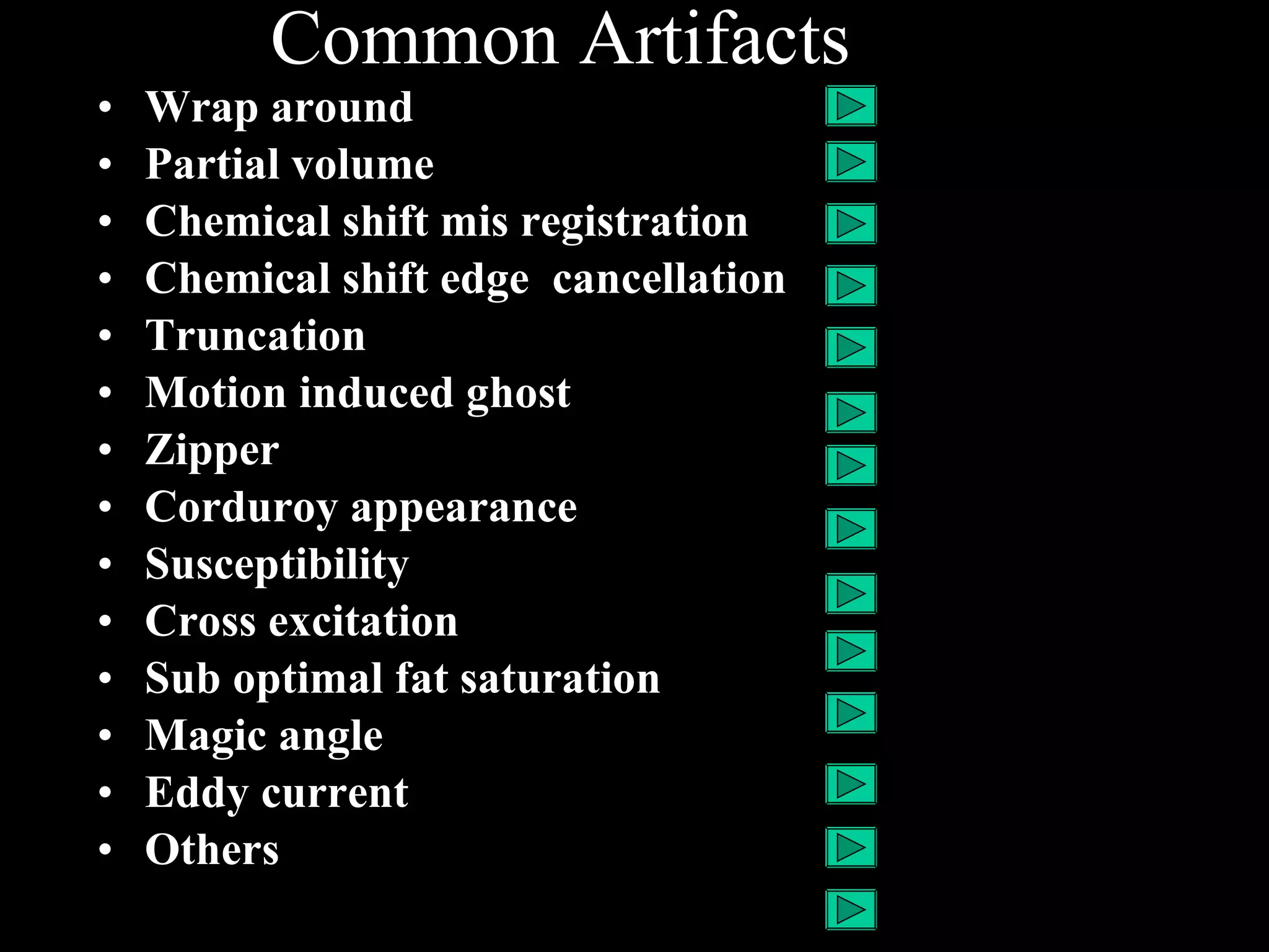
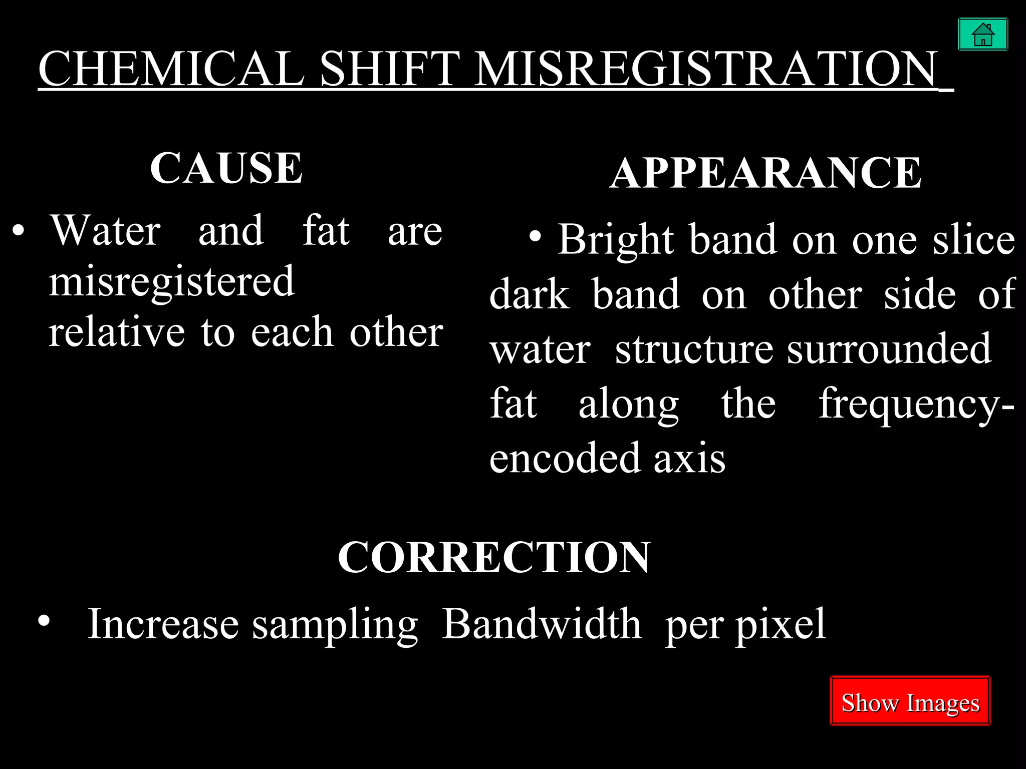
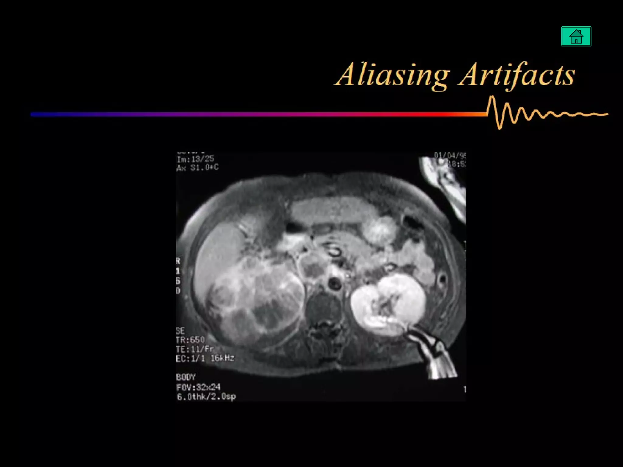
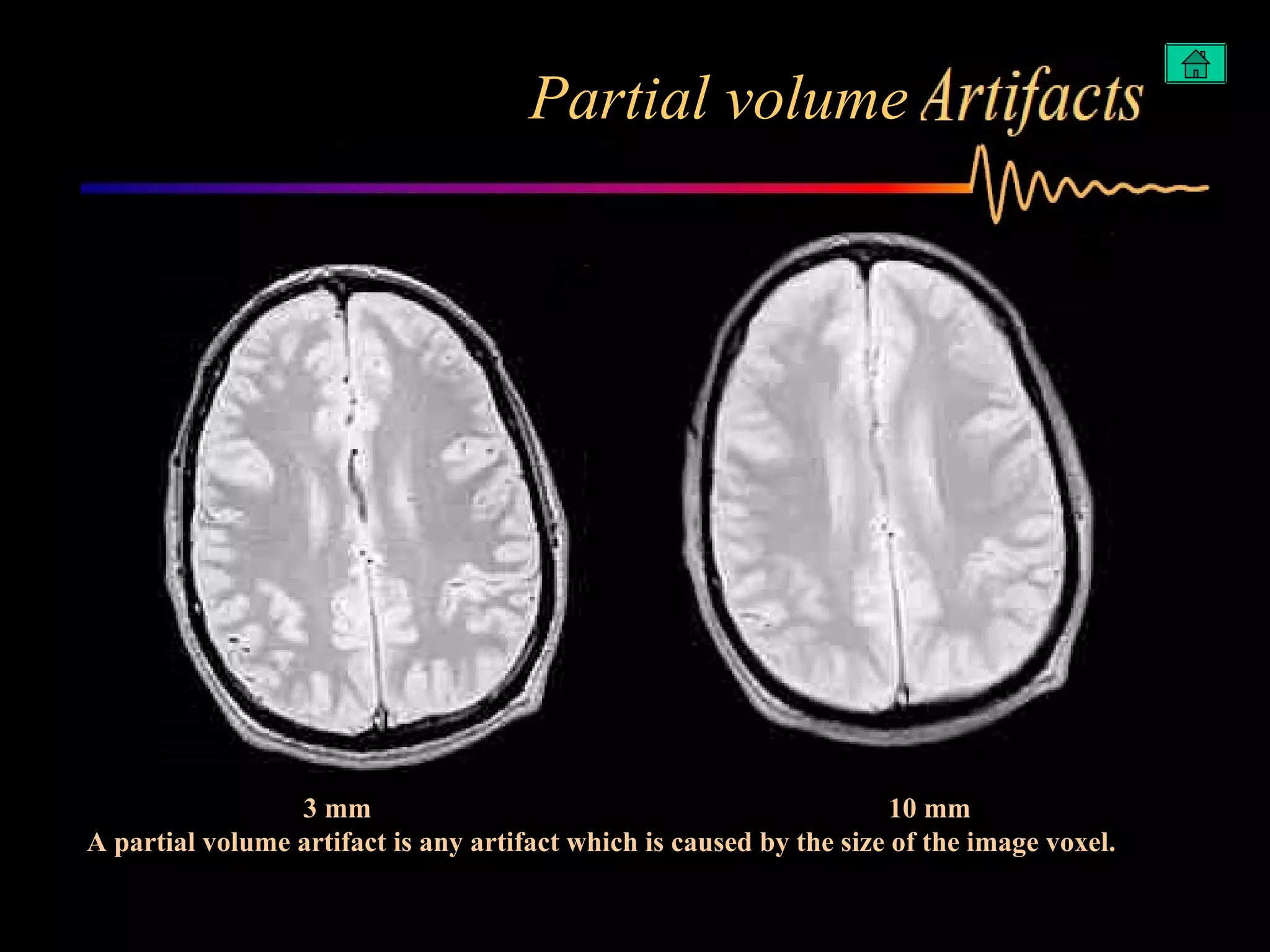
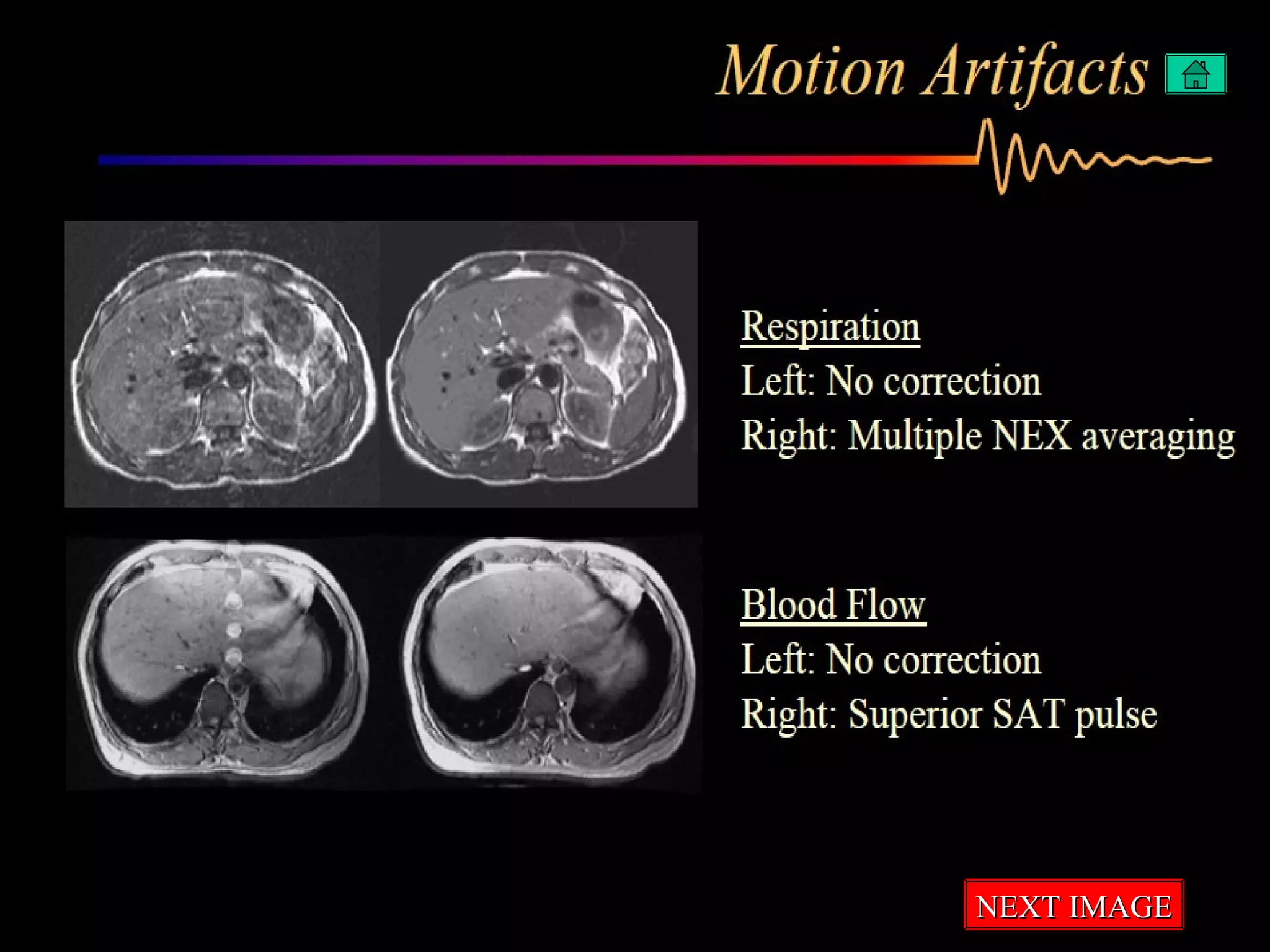
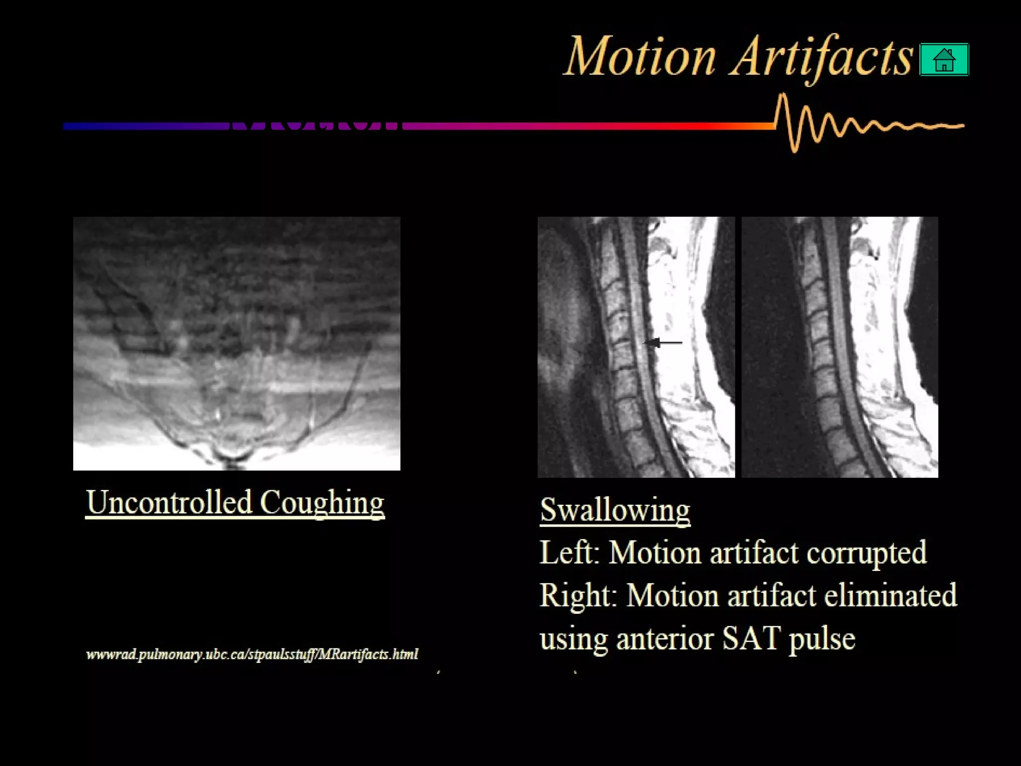
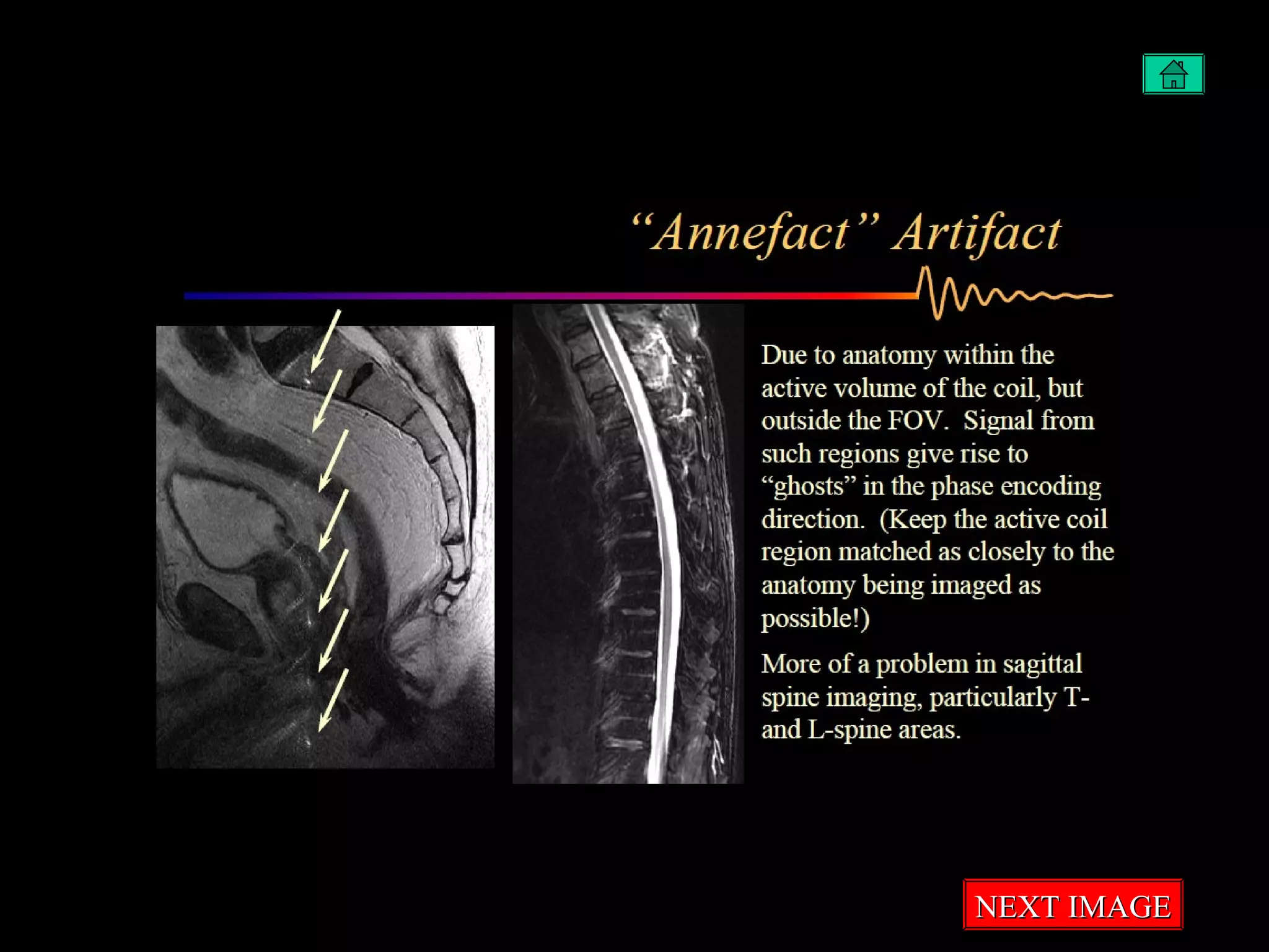
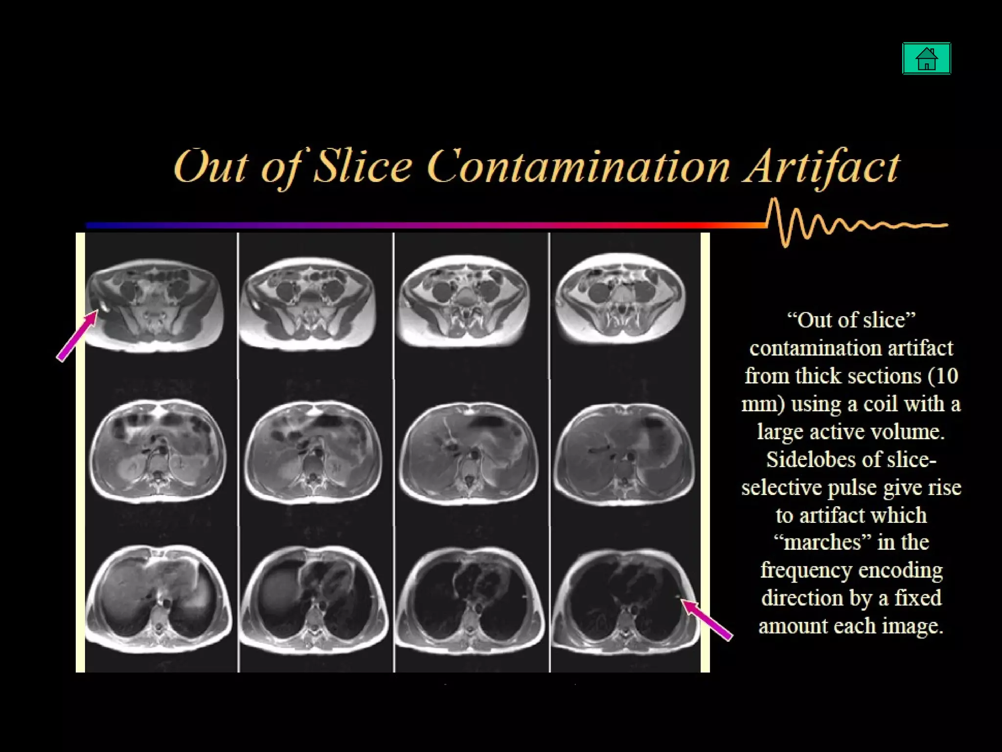
Artifacts in MRI BY  Arun A Dr.Shaji’s MRI Perinthalmanna
Common Artifacts Wrap around Partial volume  Chemical shift mis registration Chemical shift edge  cancellation Truncation  Motion induced ghost Zipper  Corduroy appearance  Susceptibility Cross excitation Sub optimal fat saturation Magic angle Eddy current Others
WRAPAROUND CAUSE FOV too small for body part   CORRECTION   Sample data sufficient  for a larger FOV Use of local coil to reduce  sensitivity of tissue outside image APPEARANCE   Body parts extends beyond  one edge of  image  and is projected at the opposite side   Show Images
PARTIAL VOLUME  CAUSE More than one tissue is included with in a voxel   Usually because of thick slices  CORRECTION   Increase spatial resolution  (particularly in the slice direction ) APPEARANCE   Edge of structure contain tissues from two adjacent slices  Show Images
CHEMICAL SHIFT MISREGISTRATION   CAUSE Water and fat are misregistered relative to each other  CORRECTION   Increase sampling  Bandwidth  per pixel APPEARANCE   Bright band on one slice dark band on other side of water  structure surrounded  fat along the frequency- encoded axis  Show Images
CHEMICAL SHIFT  EDGE CANCELLATION  CAUSE Water and lipid signals in a voxel at fat – water interfaces  destructively  CORRECTION   Use spin echo or In phase gradient echo  technique Suppress lipid signal intensity  APPEARANCE   Black line at fat–water interfaces  Show Images
TRUNCATION  CAUSE Abrupt ,high contrast boundaries cannot be accurately represented by Fourier Transform  CORRECTION   Increase spatial resolution  APPEARANCE   Ring –down artifact at high contrast interfaces  Show Images
MOTION INDUCED GHOST  CAUSE Motion causes phases changes during signal sampling  Or intensity changes  b/w signals  Both result in phase encoding errors  CORRECTION   Average multiple signals  Complete image acquisition rapidly  Trigger acquisitions so the image anatomy is constant from view to view  Balance gradients  to eliminate gradient moment  Reduce signal of moving tissue  APPEARANCE   Repetitive patterned noise that resembles the source structure  Repeated at regular intervals  along phase encoding axis   Show Images
ZIPPER  CAUSE Nondecayed transverse magnetization  RF feed through  Or extraneous noise  CORRECTION   Improve spoiling of transverse magnetization  Alternate polarity of excitation pulses  Eliminate source of noise  APPEARANCE   Band of alternative white and black dots  Show Images
CORDUROY APPEARANCE  CAUSE Bad data points  CORRECTION   Increase sapling of k-space  APPEARANCE   Corduroy appearance across some images
SUSCEPTIBILITY  CAUSE Magnetic field altered by heterogeneous magnetic properties  in body part  CORRECTION   Eliminate source of heterogeneous  susceptibility Maximize sampling bandwidth  Minimize TE Refocus echoes  APPEARANCE   Signal loss  Distortion  Show Images
CROSS EXITATION  CAUSE RF pulse targeted to one slice particularly excites an adjacent  slice  CORRECTION   Use gap b/t slices in 2D multislice acquisitions Allow sufficient time for recovery b/w radial overlapping slice acquisitions  APPEARANCE   Reduced signal intensity
SUBOPTIMAL FAT SATURATION  CAUSE Magnetic field heterogeneity causes the lipid resonance to vary within the field of view  so the fat suppression RF pulse doesn't  correspond to all lipid  CORRECTION   Improve magnetic field shimming  Improve tuning of fat suppression  pulse  APPEARANCE   increased signal intensity on lipid  Possibly suppression of  water signal
MAGIC ANGLE  CAUSE T2 of oriented fibers depends on angle relative to magnetic field  CORRECTION   Use sufficiently long TE for structure to have long signal intensity regardless of angle  APPEARANCE   variable signal intensity on of tendons and other structures  Show Images
EDDY CURRENT CAUSE Electrical currents caused by rapidly  switching gradients  CORRECTION   Improve precision of gradient pulses  Use eddy current compensation APPEARANCE   Reduced peripheral signal intensity  Show Images
OTHERS  In homogeneity on fat sat  Annefect Gradient amplifier failure Spike noise Spatial saturation Out of slice contamination
Wrap around NEXT IMAGE
wraparound
Partial volume 3 mm  Partial volume 3 mm  10 mm A partial volume artifact is any artifact which is caused by the size of the image voxel.
Chemicals shift  NEXT IMAGE
Chemicals shift
Motion NEXT IMAGE
Motion
Metal object/susceptibility NEXT IMAGE
Metal object/susceptibility
Eddy current  Metal object/susceptibility
ZIPPER NEXT IMAGE
ZIPPER
Truncation NEXT IMAGE
Truncation Truncation
Magic Angle Truncation
In homogeneity on fat sat NEXT IMAGE Truncation
Annefect NEXT IMAGE
Gradient amplifier failure NEXT IMAGE
Spike noise NEXT IMAGE Annefect
Spatial saturation NEXT IMAGE Annefect
Out of slice contamination

Artifacts in MRI

  • 1.
    Artifacts in MRIBY Arun A Dr.Shaji’s MRI Perinthalmanna
  • 2.
    Common Artifacts Wraparound Partial volume Chemical shift mis registration Chemical shift edge cancellation Truncation Motion induced ghost Zipper Corduroy appearance Susceptibility Cross excitation Sub optimal fat saturation Magic angle Eddy current Others
  • 3.
    WRAPAROUND CAUSE FOVtoo small for body part CORRECTION Sample data sufficient for a larger FOV Use of local coil to reduce sensitivity of tissue outside image APPEARANCE Body parts extends beyond one edge of image and is projected at the opposite side Show Images
  • 4.
    PARTIAL VOLUME CAUSE More than one tissue is included with in a voxel Usually because of thick slices CORRECTION Increase spatial resolution (particularly in the slice direction ) APPEARANCE Edge of structure contain tissues from two adjacent slices Show Images
  • 5.
    CHEMICAL SHIFT MISREGISTRATION CAUSE Water and fat are misregistered relative to each other CORRECTION Increase sampling Bandwidth per pixel APPEARANCE Bright band on one slice dark band on other side of water structure surrounded fat along the frequency- encoded axis Show Images
  • 6.
    CHEMICAL SHIFT EDGE CANCELLATION CAUSE Water and lipid signals in a voxel at fat – water interfaces destructively CORRECTION Use spin echo or In phase gradient echo technique Suppress lipid signal intensity APPEARANCE Black line at fat–water interfaces Show Images
  • 7.
    TRUNCATION CAUSEAbrupt ,high contrast boundaries cannot be accurately represented by Fourier Transform CORRECTION Increase spatial resolution APPEARANCE Ring –down artifact at high contrast interfaces Show Images
  • 8.
    MOTION INDUCED GHOST CAUSE Motion causes phases changes during signal sampling Or intensity changes b/w signals Both result in phase encoding errors CORRECTION Average multiple signals Complete image acquisition rapidly Trigger acquisitions so the image anatomy is constant from view to view Balance gradients to eliminate gradient moment Reduce signal of moving tissue APPEARANCE Repetitive patterned noise that resembles the source structure Repeated at regular intervals along phase encoding axis Show Images
  • 9.
    ZIPPER CAUSENondecayed transverse magnetization RF feed through Or extraneous noise CORRECTION Improve spoiling of transverse magnetization Alternate polarity of excitation pulses Eliminate source of noise APPEARANCE Band of alternative white and black dots Show Images
  • 10.
    CORDUROY APPEARANCE CAUSE Bad data points CORRECTION Increase sapling of k-space APPEARANCE Corduroy appearance across some images
  • 11.
    SUSCEPTIBILITY CAUSEMagnetic field altered by heterogeneous magnetic properties in body part CORRECTION Eliminate source of heterogeneous susceptibility Maximize sampling bandwidth Minimize TE Refocus echoes APPEARANCE Signal loss Distortion Show Images
  • 12.
    CROSS EXITATION CAUSE RF pulse targeted to one slice particularly excites an adjacent slice CORRECTION Use gap b/t slices in 2D multislice acquisitions Allow sufficient time for recovery b/w radial overlapping slice acquisitions APPEARANCE Reduced signal intensity
  • 13.
    SUBOPTIMAL FAT SATURATION CAUSE Magnetic field heterogeneity causes the lipid resonance to vary within the field of view so the fat suppression RF pulse doesn't correspond to all lipid CORRECTION Improve magnetic field shimming Improve tuning of fat suppression pulse APPEARANCE increased signal intensity on lipid Possibly suppression of water signal
  • 14.
    MAGIC ANGLE CAUSE T2 of oriented fibers depends on angle relative to magnetic field CORRECTION Use sufficiently long TE for structure to have long signal intensity regardless of angle APPEARANCE variable signal intensity on of tendons and other structures Show Images
  • 15.
    EDDY CURRENT CAUSEElectrical currents caused by rapidly switching gradients CORRECTION Improve precision of gradient pulses Use eddy current compensation APPEARANCE Reduced peripheral signal intensity Show Images
  • 16.
    OTHERS Inhomogeneity on fat sat Annefect Gradient amplifier failure Spike noise Spatial saturation Out of slice contamination
  • 17.
  • 18.
  • 19.
    Partial volume 3mm Partial volume 3 mm 10 mm A partial volume artifact is any artifact which is caused by the size of the image voxel.
  • 20.
    Chemicals shift NEXT IMAGE
  • 21.
  • 22.
  • 23.
  • 24.
  • 25.
  • 26.
    Eddy current Metal object/susceptibility
  • 27.
  • 28.
  • 29.
  • 30.
  • 31.
  • 32.
    In homogeneity onfat sat NEXT IMAGE Truncation
  • 33.
  • 34.
  • 35.
    Spike noise NEXTIMAGE Annefect
  • 36.
    Spike noise NEXTIMAGE Annefect
  • 37.
  • 38.
    Out of slicecontamination